. 2019 Jan:73:55-63.
doi: 10.1016/j.dnarep.2018.11.002. Epub 2018 Nov 13.

The Ataxia telangiectasia-mutated and Rad3-related protein kinase regulates cellular hydrogen sulfide concentrations

Affiliations

The Ataxia telangiectasia-mutated and Rad3-related protein kinase regulates cellular hydrogen sulfide concentrations

Jie Chen et al. DNA Repair (Amst). 2019 Jan.

Abstract

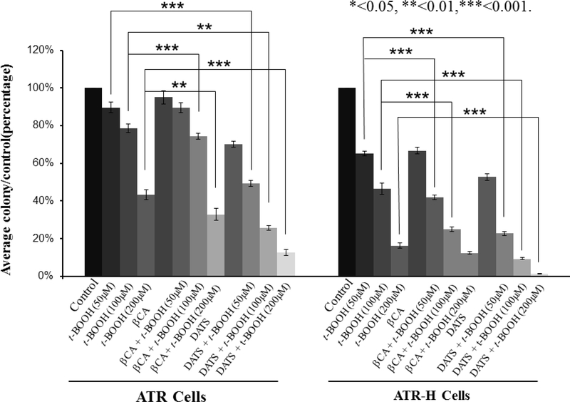
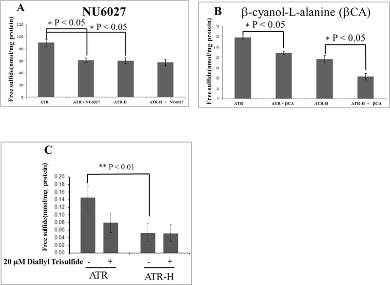
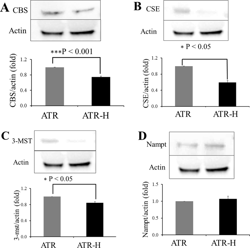
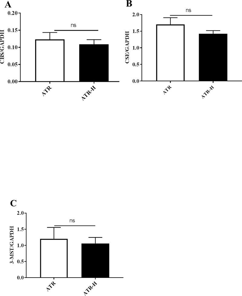
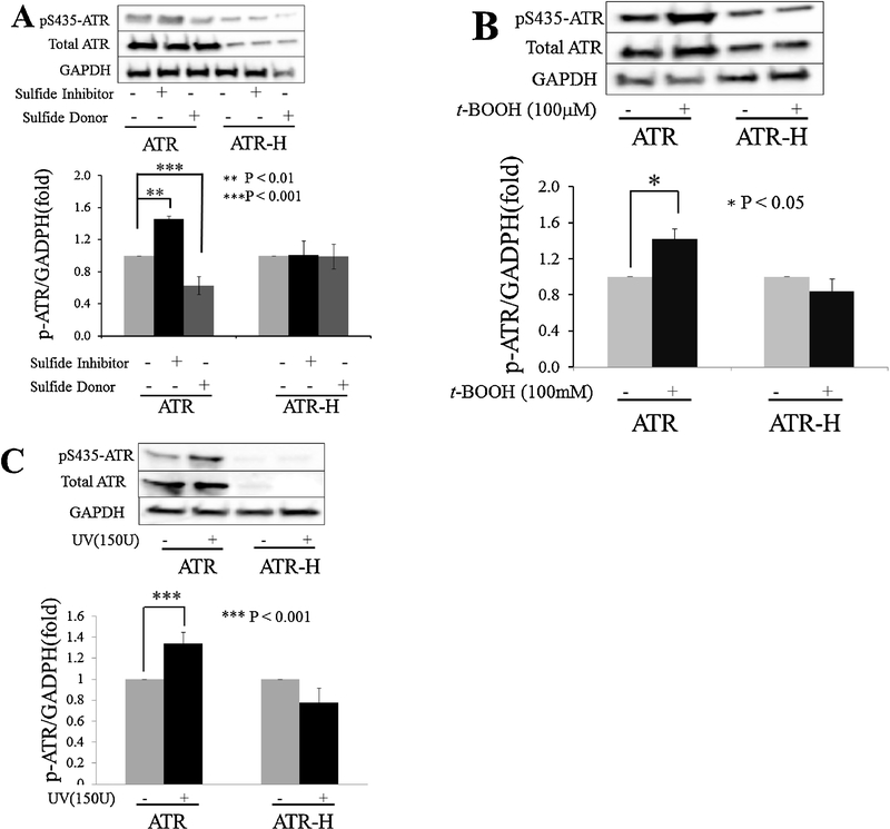
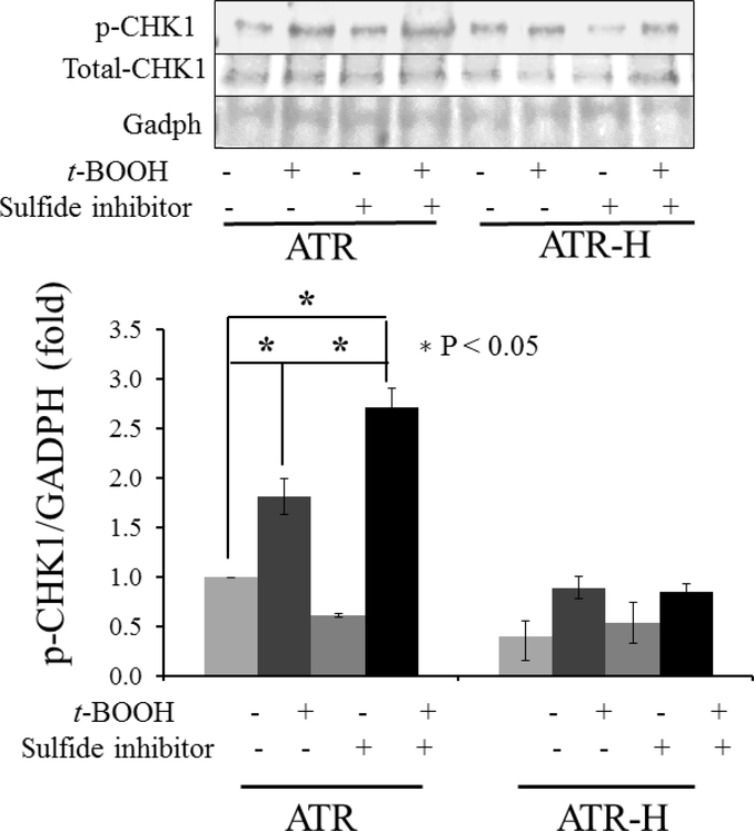
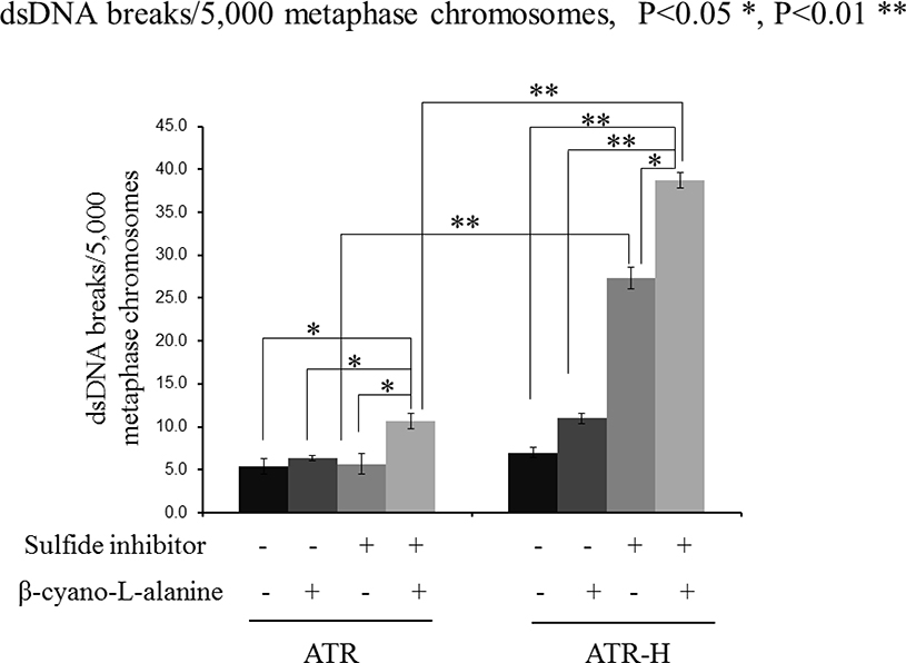
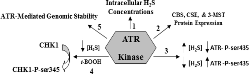
The ataxia telangiectasia-mutated and Rad3-related (ATR) serine/threonine kinase plays a central role in the repair of replication-associated DNA damage, the maintenance of S and G2/M-phase genomic stability, and the promotion of faithful mitotic chromosomal segregation. A number of stimuli activate ATR, including persistent single-stranded DNA at stalled replication folks, R loop formation, hypoxia, ultraviolet light, and oxidative stress, leading to ATR-mediated protein phosphorylation. Recently, hydrogen sulfide (H2S), an endogenous gasotransmitter, has been found to regulate multiple cellular processes through complex redox reactions under similar cell stress environments. Three enzymes synthesize H2S: cystathionine-β-synthase, cystathionine γ-lyase, and 3-mercaptopyruvate sulfurtransferase. Since H2S can under some conditions cause DNA damage, we hypothesized that ATR activity may regulate cellular H2S concentrations and H2S-syntheszing enzymes. Here we show that human colorectal cancer cells carrying biallelic knock-in hypomorphic ATR mutations have lower cellular H2S concentrations than do syngeneic ATR wild-type cells, and all three H2S-synthesizing enzymes show lower protein expression in the ATR hypomorphic mutant cells. Additionally, ATR serine 428 phosphorylation is altered by H2S donor and H2S synthesis enzyme inhibition, while the oxidative-stress induced phosphorylation of the ATR-regulated protein CHK1 on serine 345 is increased by H2S synthesis enzyme inhibition. Lastly, inhibition of H2S production potentiated oxidative stress-induced double-stranded DNA breaks in the ATR hypomorphic mutant compared to ATR wild-type cells. Our findings demonstrate that the ATR kinase regulates and is regulated by H2S.

Keywords: 3-Mercaptopyruvate sulfurtransferase; ATR; CHK1; Cystathionine γ-lyase; Cystathionine-β-synthase; H(2)S; Hydrogen sulfide; Nicotinamide; phosphoribosyltransferase.


Figures

Figure 1.
Figure 1.
The effects of t-BOOH and H2S inhibitor and donor treatments in the colony-forming efficiency assay with ATR and ATR-H cells. Twelve hours after plating in appropriate media, exponentially growing ATR and ATR-H cells were treated for two hours with either an H2S inhibitor (1 mM β-cyano-L-alanine, βCA) or an H2S donor (20 μM diallyl trisulfide, DATS), and subjected to 15 minutes 50, 100, or 200 μM t-BOOH oxidative stress. After 11 days, the cells were fixed, stained, and the colonies counted. Data indicates survival as a percentage of untreated cells.
Figure 2.
Figure 2.
Free cellular H2S concentrations in the ATR wild-type cells compared to the ATR-H mutant cells. Figure 2A, H2S concentrations were compared in ATR and ATR-H, with and without the ATR kinase inhibitor NU6027. The cells were treated for two hours with 12 μM NU6027 in standard media and harvested (2A). Figure 2B, H2S concentrations were compared in ATR and ATR-H, with and without the CBS and CSE inhibitor β-cyanol-L-alanine. The cells were treated with 1 mM β-cyanol-L-alanine for two hours in standard media and harvested (2B). Figure 2C, H2S concentrations were compared in ATR and ATR-H, with and without 20μM diallyl trisulfide for two hours in standard media and harvested (2C). “βCA” = 1 mM β-cyanol-L-alanine. Free H2S is in nmol/mg protein.
Figure 3.
Figure 3.
Representative western blots for CBS (3A), CSE (3B), 3-MST (3C), and Nampt (3D) comparing ATR and ATR -H cells protein expression with β-actin as a control protein. All western blots were performed at least in triplicate.
Figure 4.
Figure 4.
Quantitative real-time polymerase chain reaction analyses of CBS, CSE, and 3-MST mRNA levels in the ATR and ATR-H cell lines. Statistical analyses revealed no significant differences between CBS, CSE, and 3-MST mRNA levels between the two cell lines. The primers used in the quantitative real-time polymerase chain reactions are given in Table 1. GADPH was used as a control.
Figure 5.
Figure 5.
ATR protein phosphorylation on serine-435 with an H2S donor and inhibitor, or t-BOOH treatments. ATR and ATR-H cells were treated with 1mM β-cyanol-L-alanine or 20 μM diallyl trisulfide for two hours and harvested (5A). In Figure 5B, ATR and ATR-H cells were treated with 100 μM t-BOOH for 15 minutes, incubated in standard media for 45 minutes, and harvested. In Figure 5C, ATR and ATR-H cells were treated with 15,000 μJ/cm2 UV light and the cells were harvested 45 minutes later.
Figure 6.
Figure 6.
CHK1 serine 345 phosphorylation in ATR and ATR-H cells with H2S synthesis inhibition followed by t-BOOH treatment was examined. ATR and ATR-H cells were pretreated with 1mM β-cyanol-L-alanine for two hours, then 15 minutes with 10μM t-BOOH, and harvested following a 45-minute incubation in standard media.
Figure 7.
Figure 7.
dsDNA break formation in ATR and ATR-H cells following H2S synthesis inhibition. ATR and ATR-H cells were pretreated with 1mM β-cyanol-L-alanine for two hours, then 15 minutes with t-BOOH, cultured one hour in standard media, treated with colcemid for four hours, and harvested. dsDNA breaks in Giemsa stained, Colcemid-treated cells, were counted under oil immersion microscopy. The t-BOOH concentration was 10μM.
Figure 8.
Figure 8.
A summary of the findings presented in this paper. The ATR kinase regulates intracellular H2S concentrations (arrow 1) and the levels of the three H2S-synthesizing enzymes (arrow 2). Increased in intracellular H2S concentrations decreased ATR serine 435 phosphorylation, while decreased in intracellular H2S concentrations increased this phosphorylation (arrow 3). Attenuation of intracellular H2S synthesis also potentiates CHK1 serine 345 phosphorylation following ATR cell exposure to low t-BOOH concentration, an event not seen in ATR-H cells (arrow 4). Lastly, low cellular H2S concentrations in the hypomorphic ATR-H cells increase genomic instability by itself, and when combined with a low dose of t-BOOH. Taken together, our data suggests that the ATR kinase regulates and is in turn regulated by H2S.

References

    1. Yazinski SA, Zou L. Functions, Regulation, and Therapeutic Implications of the ATR Checkpoint Pathway. Annu Rev Genet. 50 (2016) 155–173. - PubMed
    1. Kabeche L, Nguyen HD, Buisson R, Zou L. A mitosis-specific and R loop-driven ATR pathway promotes faithful chromosome segregation. Science. 359 (2018) 108–114. - PMC - PubMed
    1. Buisson R, L Boisvert J, H Benes C, Zou L. Distinct but Concerted Roles of ATR, DNA-PK, and Chk1 in Countering Replication Stress during S Phase. Mol Cell. 59 (2015) 1011–1024. - PMC - PubMed
    1. Durocher D, Jackson JP, DNA-PK ATM and ATR as sensors of DNA damage: variations on a theme? Curr Opin Cell Biol. 13 (2001) 225–231. - PubMed
    1. Hanawalt PC, Historical perspective on the DNA damage response. DNA Repair. 36 (2015) 2–7. - PMC - PubMed

Publication types

MeSH terms