Review
. 2019 Jul:25:101047.
doi: 10.1016/j.redox.2018.11.005. Epub 2018 Nov 14.

Redox signaling and unfolded protein response coordinate cell fate decisions under ER stress

Affiliations
Review

Redox signaling and unfolded protein response coordinate cell fate decisions under ER stress

Zhe Zhang et al. Redox Biol. 2019 Jul.

Abstract

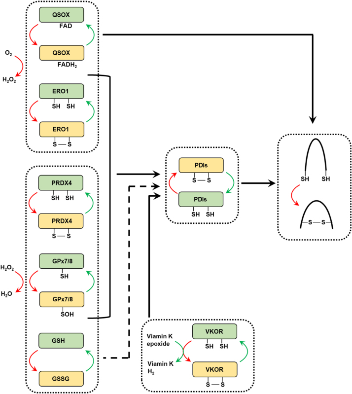
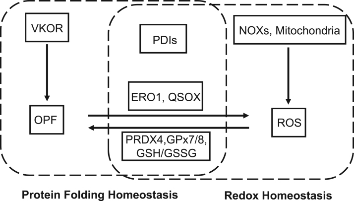
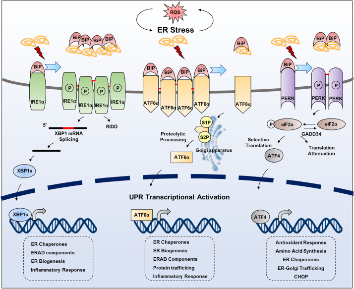
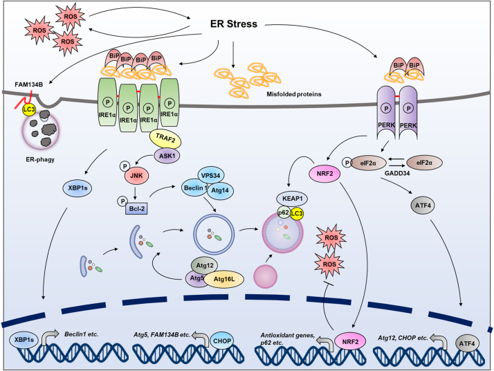
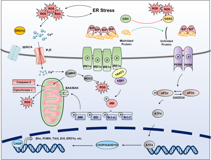
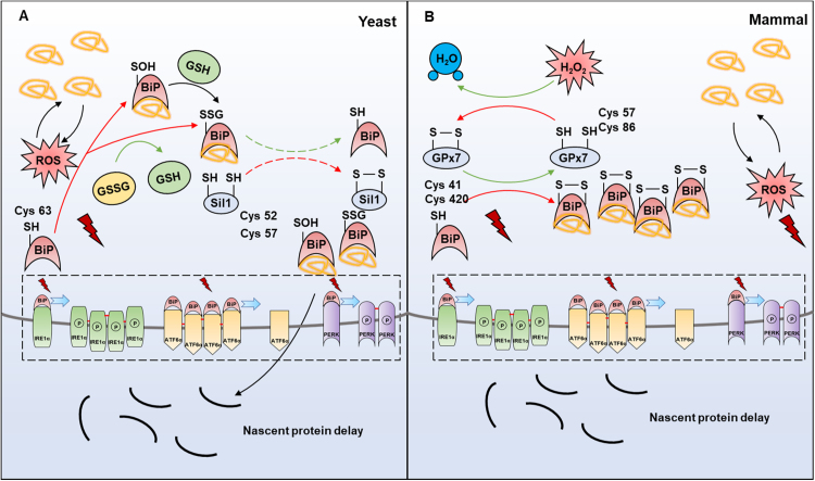
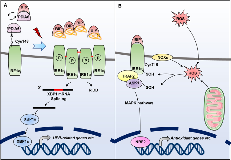
Endoplasmic reticulum (ER) is a dynamic organelle orchestrating the folding and post-translational maturation of almost all membrane proteins and most secreted proteins. These proteins synthesized in the ER, need to form disulfide bridge to acquire specific three-dimensional structures for function. The formation of disulfide bridge is mediated via protein disulfide isomerase (PDI) family and other oxidoreductases, which contribute to reactive oxygen species (ROS) generation and consumption in the ER. Therefore, redox regulation of ER is delicate and sensitive to perturbation. Deregulation in ER homeostasis, usually called ER stress, can provoke unfolded protein response (UPR) pathways with an aim to initially restore homeostasis by activating genes involved in protein folding and antioxidative machinery. Over time, however, activated UPR involves a variety of cellular signaling pathways which determine the state and fate of cell in large part (like autophagy, apoptosis, ferroptosis, inflammation, senescence, stemness, and cell cycle, etc.). This review will describe the regulation of UPR from the redox perspective in controlling the cell survival or death, emphasizing the redox modifications of UPR sensors/transducers in the ER.

Keywords: Cell fate; ER stress; Redox regulation; UPR.


Figures

Fig. 1
Fig. 1
Protein disulfide bond formation in the ER. Formation of disulfide bond in substrate proteins is mainly mediated by PDI family members. PDIs are oxidized to form a disulfide bond, then the disulfide bond is introduced to substrates, and PDIs are simultaneously reduced. ERO1, PRDX4, GPX7/8 and VKOR contribute to the re-oxidization and reactivation of PDIs. ERO1 is able to catalyze oxidation reaction by coupling de novo disulfide formation to the reduction of oxygen to H2O2. PRDX4 and GPX7/8 utilize H2O2 as oxidant, then oxidized PRDX4 and GPX7/8 is able to reactive PDIs. VKOR can accept electrons from PDIs to regenerate vitamin K. QSOX uses oxygen to contribute to disulfide formation of substrate proteins directly and H2O2 production from oxygen. The oxidized glutathione (GSSG) also plays an important role in maintaining ER redox homeostasis. Red arrows indicate the flow of oxidizing equivalents, while green arrows indicate the flow of reducing equivalents. Dashed-lined arrows indicate the potential function in biochemistry, but the function of these molecular still lack solid cell biological evidence in mammalian cells so far.
Fig. 2
Fig. 2
Crosstalk between redox homeostasis and protein folding homeostasis in the ER. OPF is deeply associated with redox balance in the ER. ROS (mainly H2O2) can be generated during OPF process through ERO1 and QSOX, and from NOXs in the ER membrane, as well as ER-associated mitochondria. ER-resident PRDX4, GPx7/8 are oxidized by H2O2 and further oxidize PDIs, simultaneously consume H2O2. VKOR facilitate OPF without affecting ROS.
Fig. 3
Fig. 3
UPR pathway under ER stress. Under physiological conditions, BiP binds with and inhibits IRE1α, ATF6α and PERK. When ER stress occurs, the misfolded peptides sequester BiP to release the three UPR transducers. IRE1α undergoes homodimerization and autophosphorylation, subsequently activating the endoribonuclease activity to splice XBP1 mRNA. The encoded XBP1s from spiced mRNA acts as a transcription factor to induce transcription of various target genes. ATF6 oligomer that exists under normal condition is reduced and exported from the ER to the Golgi, where it is proteolysed by Golgi-resident proteases, S1P and S2P. The truncated ATF6 translocates to the nucleus and induces transcription of target genes. PERK activation requires oligerization and autophosphorylation, which obtains the ability to phosphorylate eIF2α. Phosphorylated eIF2α transiently downregulates protein synthesis to decreasing ER protein load, or selectively translates ATF4, which is a transcription factor for a series of UPR target genes.
Fig. 4
Fig. 4
Redox regulation of autophagy under ER stress. In certain conditions, the damage of ER and the disturbance of ER homeostasis caused by ER stress or oxidative stress are the main causes of autophagy (including ER-phagy). Autophagy is activated by JNK, which is regulated by IRE1α during ER stress. JNK mediates phosphorylation of Bcl-2, which results in the disruption of the Beclin1/Bcl-2 complex, thus releasing free Beclin1 to form the Vps34-Beclin1 complex. Vps34-Beclin1 complex then drives the nucleation of the isolated membrane to form autophagosome. In addition, XBP1, the transcription factor which is mediated by IRE1α RNase domain, also triggers autophagy through transcriptional activation of Beclin1. PERK signaling cascade can regulate autophagy through ATF4, CHOP and NRF2. ATF4 and CHOP transcriptionally upregulate various autophagy-related genes, such as Atg5, Atg12, and FAM134B (ER-phagy-related gene). NRF2 can be activated by oxidative stress, which induces p62/SQSTM1 binding to KEAP1 and promotes NRF2 release.
Fig. 5
Fig. 5
Redox regulation of apoptosis under ER stress. PERK can provoke apoptotic signaling by phosphorylating eIF2α, which stimulates the translation of ATF4. Acting as a transcription factor, ATF4 then promotes the transcription of CHOP/GADD153, which downregulates the anti-apoptotic protein Bcl-2 and upregulates the pro-apoptotic proteins BIM and PUMA. CHOP/GADD153 also transcriptionally induces ERO1 to produce ROS. Another apoptotic pathway is the JNK, which is activated by the IRE1α-TRAF2-ASK1 complex. Phosphorylated JNK activates the downstream apoptotic pathways, and induces oxidative stress related to mitochondria. In addition, the accumulation of misfolded proteins induced by ER stress will consume a large amount of GSH to induce oxidative stress. Moreover, calcium channel proteins IP3R can be activated by SERCA, leading to calcium outflow which induces apoptosis.
Fig. 6
Fig. 6
Redox regulation of BiP to control UPR. (A) BiP is an ER chaperone that interacts with downstream transducers in the ER. This interaction can be disrupted by the accumulation of misfolded proteins. In yeast, the Cys63 of BiP can be sulfenylated by H2O2, or glutathionylated by GSSG, which enhances its ability to bind to misfolded proteins. Meanwhile, its ATPase activity is downregulated, resulting in the blockage of nascent proteins’ transportation. Sil1 and BiP exchange disulfide, thereby allowing BiP to restore its ATPase activity. (B) In mammals, the redox modification of BiP is mediated by disulfide-bonded GPx7, which oxidizes BiP to form a disulfide bond between its Cys41 and Cys420 residues.
Fig. 7
Fig. 7
Redox regulation of IRE1α to control UPR. (A) Under physiological condition, the Cyst148 of IRE1α forms an intermolecular disulfide bond with PDIA6 and is therefore kept in an inactive state. PDIA6 can also form a complex with BiP by non-covalent bonding to catalyze the oxidative protein folding. Under ER stress, IRE1α forms tetramers through disulfide bonds and is auto-phosphorylated, activating downstream signals. (B) IRE1α is able to sense the level of ROS in the ER or cytoplasm. Under oxidative stress, Cys715 that locates in the cytoplasmic region of IRE1α is sulfenylated by ROS (generated from NOXs, ER as well as mitochondria), thus activating the MAPK pathway to initiate NRF2-mediated antioxidant system, meanwhile inhibiting the UPR-initiating function of IRE1α.

References

    1. Kosuri P., Alegre-Cebollada J., Feng J., Kaplan A., Ingles-Prieto A., Badilla C.L., Stockwell B.R., Sanchez-Ruiz J.M., Holmgren A., Fernandez J.M. Protein folding drives disulfide formation. Cell. 2012;151(4):794–806. - PMC - PubMed
    1. Qin M., Wang W., Thirumalai D. Protein folding guides disulfide bond formation. Proc. Natl. Acad. Sci. USA. 2015;112(36):11241–11246. - PMC - PubMed
    1. Malhotra J.D., Kaufman R.J. Endoplasmic reticulum stress and oxidative stress: a vicious cycle or a double-edged sword? Antioxid. Redox Signal. 2007;9(12):2277–2293. - PubMed
    1. Araki K., Iemura S., Kamiya Y., Ron D., Kato K., Natsume T., Nagata K. Ero1-alpha and PDIs constitute a hierarchical electron transfer network of endoplasmic reticulum oxidoreductases. J. Cell Biol. 2013;202(6):861–874. - PMC - PubMed
    1. Enyedi B., Varnai P., Geiszt M. Redox state of the endoplasmic reticulum is controlled by Ero1L-alpha and intraluminal calcium. Antioxid. Redox Signal. 2010;13(6):721–729. - PubMed

Publication types

LinkOut - more resources