Review
. 2019 Apr;37(4):428-441.
doi: 10.1016/j.tibtech.2018.10.009. Epub 2018 Nov 21.

Zinc-Based Biomaterials for Regeneration and Therapy

Affiliations
Review

Zinc-Based Biomaterials for Regeneration and Therapy

Yingchao Su et al. Trends Biotechnol. 2019 Apr.

Abstract

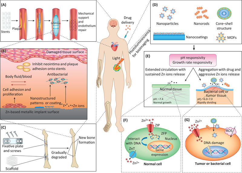
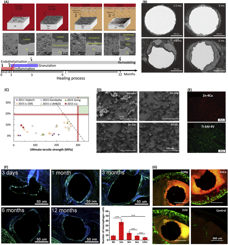
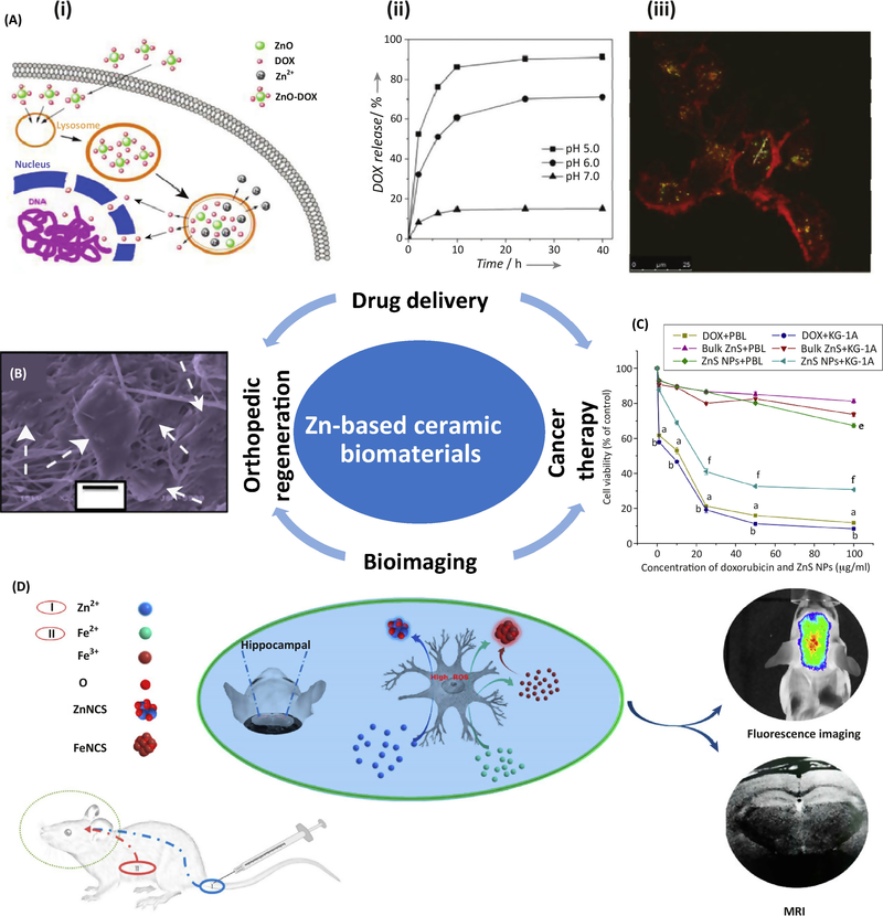
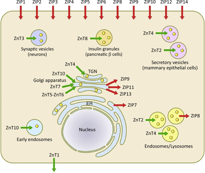
Zinc has been described as the 'calcium of the twenty-first century'. Zinc-based degradable biomaterials have recently emerged thanks to their intrinsic physiological relevance, biocompatibility, biodegradability, and pro-regeneration properties. Zinc-based biomaterials mainly include: metallic zinc alloys, zinc ceramic nanomaterials, and zinc metal-organic frameworks (MOFs). Metallic zinc implants degrade at a desirable rate, matching the healing pace of local tissues, and stimulating remodeling and formation of new tissues. Zinc ceramic nanomaterials are also beneficial for tissue engineering and therapy thanks to their nanostructures and antibacterial properties. MOFs have large surface areas and are easily functionalized, making them ideal for drug delivery and cancer therapy. This review highlights recent developments in zinc-based biomaterials, discusses obstacles to overcome, and pinpoints directions for future research.

Keywords: biodegradable; biometal; bioresorbable; nanomaterials; tissue engineering; zinc.


Figures

Figure 1.
Figure 1.
Key Figure: Zn-based biomaterials and their in vivo interactions with different tissues and cells for tissue regeneration and therapy. (A) Metallic Zn-based coronary stents provide mechanical support to the vascular wall and help repair the endothelium by removing plaque to avoid thrombosis and stent restenosis. (B) In vivo interactions of a Zn-based metallic implant surface with damaged tissues: an ideal nanostructured pattern or coating on the surface, and the release of Zn ions from the degradation process, may promote cellular adhesion and proliferation while inhibiting the adhesion of bacterial cells or other subjects (such as smooth muscle cells and plaque for a stent). (C) Metallic Zn-based orthopedic implants (fixative plates, screws and porous scaffolds) provide temporary mechanical support for bone tissue regeneration in a parallel process of implant biodegradation and new bone formation. (D) Nano-structured Zn-based ceramic and organic biomaterials provide high surface/volume ratios for drug delivery and excellent photoluminescence for in vivo bioimaging. (E) Nanostructured Zn-based ceramic and organic biomaterials have sensitive responses to pH and cell growth rate, differentiating their circulation or aggregation behaviors on normal tissues, bacterial cell or tumor tissues. (F) Relatively low concentrations of Zn ions have no adverse, and sometime beneficial, effects on normal cells. (G) High concentrations of Zn ions, aggressive Zn ion release, cellular surface aggregation of Zn-based nanomaterials, and induced ROS can damage the cell surface and DNA in tumors or bacterial cells.
Figure 2.
Figure 2.
In vitro and in vivo performance and potential clinical applications of Zn-based biodegradable metals. (A) Schematic diagrams showing the degradation mechanism of zinc stents associated with the conversion of degradation microenvironments during the healing process, including the formation of zinc phosphate under dynamic flow conditions in blood fluid and its conversion to ZnO and calcium phosphate [31]. (B) Representative backscattered electron images of cross-sectional areas from a pure Zn wire explant after 1.5, 3, 4.5, and 6 months in the abdominal aorta of an adult male Sprague-Dawley rat [3]. (C) A comparison of experimental Zn-based biodegradable metals with approximate mechanical benchmarks (red lines) [10]. (D) Cell morphologies adhered on pure Zn and Zn-1X (X = Mg, Ca, Sr) alloy after one day of culture [41]. (E) Biofilm formation on Zn-4Cu alloy compared to Ti-6Al-4V after incubation with S. aureus for 1 day, illustrated by live/dead staining [36]. (F) Representative immunofluorescence staining images of macrophage antibody during implantation and the corresponding number of macrophages per strut [31]. (G) Representative histology of cross-sections of a mouse distal femoral shaft from Zn-1Mg, Zn-1Ca, and Zn-1Sr implanted pins and a sham control group observed under fluorescent microscopy at week 8 post-implant, with green fluorescence indicating new bone formation [41]. Images reproduced with permission from the indicated references.
Figure 3.
Figure 3.
Potential clinical applications of Zn-based ceramic biomaterials. (A) Multifunctional ZnO@polymer–DOX composites for drug delivery: (i) schematic delivery process into the cells, (ii) drug release profiles at different pH values, and (iii) CLSM images of U251 cells after 3 h incubation with the composites and lysotracker. The green emission is from the lysotracker; the red emission is from the composites [58]. (B) SEM images of the cellular spread pattern (white arrows) of C2C12 cells after 72h incubation on electrospun ZnO/TiO2 nanofibers [57]. (C) Cell viability assay of peripheral blood lymphocytes (PBL) and KG-1A cell (leukemic cells) when cultured with DOX, bulk zinc sulfide powder, and ZnS nanoparticles for 24 h [69]. (D) Scheme illustration of in vivo dual-modality bioimaging of modeled Alzheimer’s mice brains through biosynthesized zinc and iron oxide via the combined injection of ferrous chloride solution post-stomach and zinc gluconate solution post-tail vein [63]. Images reproduced with permission from the indicated references
Figure I.
Figure I.
Cellular Receptors and Channels of Zn. Image reproduced with permission from [11].

References

    1. Chen Q and Thouas GA (2015) Metallic implant biomaterials. Materials Science and Engineering: R: Reports 87, 1–57
    1. Zheng YF, et al. (2014) Biodegradable metals. Materials Science and Engineering R: Reports 77, 1–34
    1. Bowen PK, et al. (2013) Zinc exhibits ideal physiological corrosion behavior for bioabsorbable stents. Adv. Mater 25, 2577–2582 - PubMed
    1. Frederickson CJ, et al. (2005) The neurobiology of zinc in health and disease. Nat. Rev. Neurosci 6, 449–462 - PubMed
    1. Xiong HM (2013) ZnO Nanoparticles Applied to Bioimaging and Drug Delivery. Adv. Mater 25, 5329–5335 - PubMed

Publication types

MeSH terms