. 2019 Jan 2;27(1):59-75.
doi: 10.1016/j.ymthe.2018.10.014. Epub 2018 Oct 24.

Non-viral, Tumor-free Induction of Transient Cell Reprogramming in Mouse Skeletal Muscle to Enhance Tissue Regeneration

Affiliations

Non-viral, Tumor-free Induction of Transient Cell Reprogramming in Mouse Skeletal Muscle to Enhance Tissue Regeneration

Irene de Lázaro et al. Mol Ther. .

Abstract

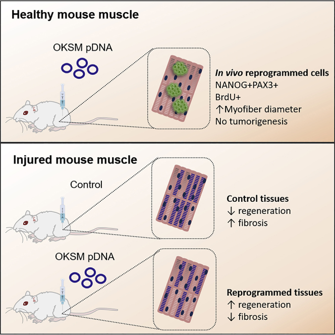
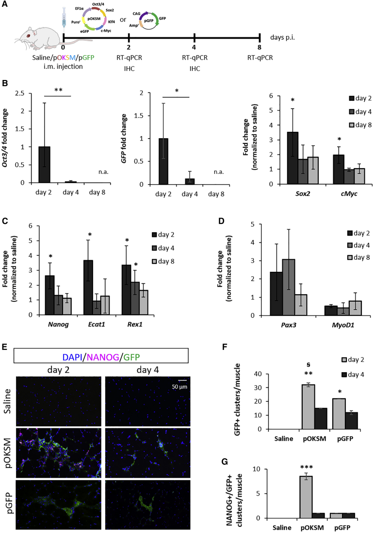
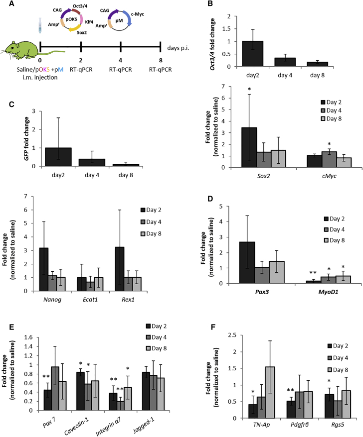
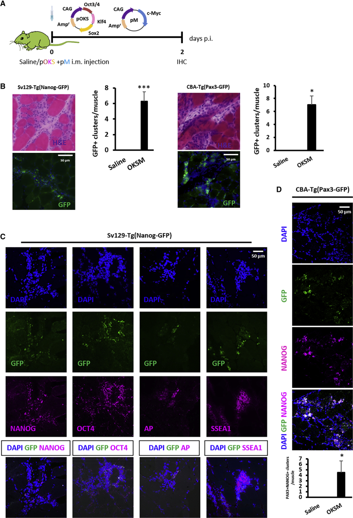
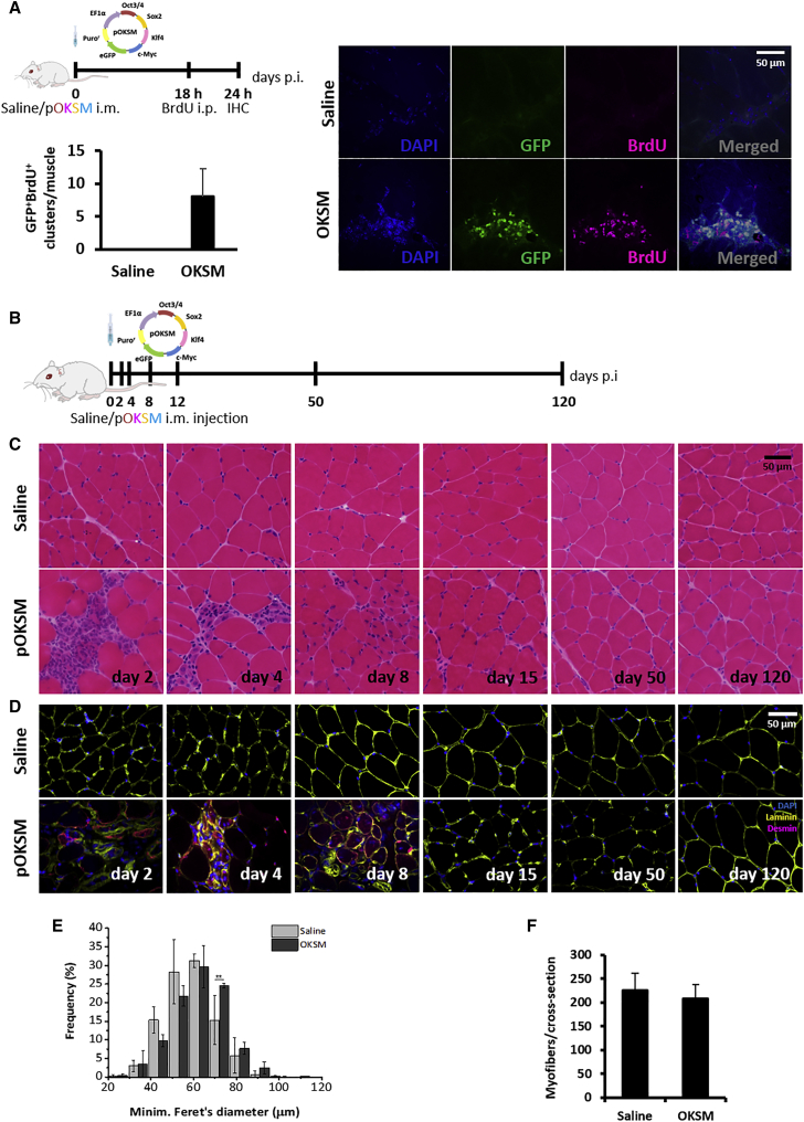
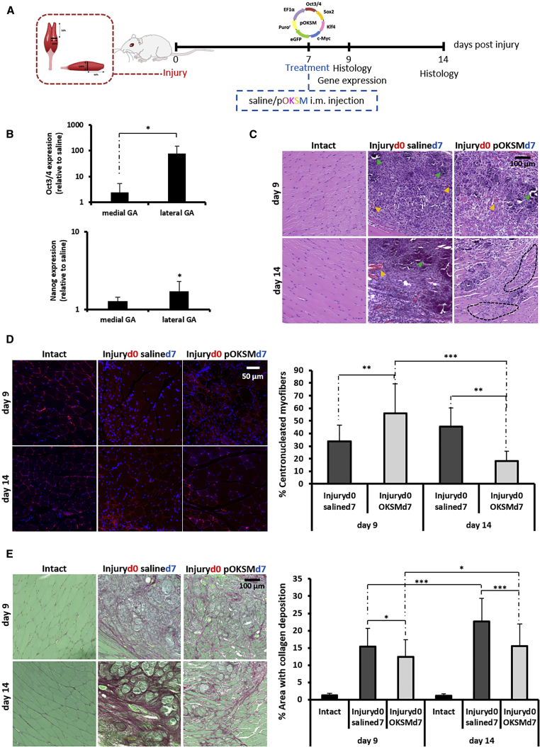
Overexpression of Oct3/4, Klf4, Sox2, and c-Myc (OKSM) transcription factors can de-differentiate adult cells in vivo. While sustained OKSM expression triggers tumorigenesis through uncontrolled proliferation of toti- and pluripotent cells, transient reprogramming induces pluripotency-like features and proliferation only temporarily, without teratomas. We sought to transiently reprogram cells within mouse skeletal muscle with a localized injection of plasmid DNA encoding OKSM (pOKSM), and we hypothesized that the generation of proliferative intermediates would enhance tissue regeneration after injury. Intramuscular pOKSM administration rapidly upregulated pluripotency (Nanog, Ecat1, and Rex1) and early myogenesis genes (Pax3) in the healthy gastrocnemius of various strains. Mononucleated cells expressing such markers appeared in clusters among myofibers, proliferated only transiently, and did not lead to dysplasia or tumorigenesis for at least 120 days. Nanog was also upregulated in the gastrocnemius when pOKSM was administered 7 days after surgically sectioning its medial head. Enhanced tissue regeneration after reprogramming was manifested by the accelerated appearance of centronucleated myofibers and reduced fibrosis. These results suggest that transient in vivo reprogramming could develop into a novel strategy toward the acceleration of tissue regeneration after injury, based on the induction of transiently proliferative, pluripotent-like cells in situ. Further research to achieve clinically meaningful functional regeneration is warranted.

Keywords: OKSM; in vivo reprogramming; muscle; plasmid DNA; pluripotency; regeneration.


Figures

None
Graphical abstract
Figure 1
Figure 1
Changes in Gene and Protein Expression after i.m. Administration of Reprogramming pDNA in Mouse GA (A) BALB/c mice were i.m. injected into the GA with 50 μg pOKSM or pGFP in 50 μL 0.9% saline or with 50 μL saline alone. (B–D) GAs were dissected 2, 4, and 8 days after injection with pOKSM or saline, and real-time qRT-PCR was performed to determine changes in gene expression of (B) reprogramming factors and GFP transgene encoded in pOKSM, (C) endogenous pluripotency markers, and (D) genes involved in myogenesis. Gene expression levels were normalized to day 2 for Oct3/4 and GFP mRNA. **p < 0.01 and *p < 0.05 indicate statistically significant differences between day 2 and day 4 post-injection (p.i.), assessed by one-way ANOVA; n.a. indicates no amplification of the target. Expression levels of other transcripts were normalized to saline-injected controls and *p < 0.05 indicates statistically significant differences in gene expression between pOKSM- and saline-injected groups, assessed by one-way ANOVA or Welch ANOVA. Data are presented as 2ˆ-ΔΔCt ± propagated error, n = 3. (E) 10-μm-thick GA sections, obtained 2 or 4 days after saline, pOKSM, or pGFP injection, were stained with anti-NANOG and anti-GFP antibodies. Images were taken with a slide scanner at 20× magnification; scale bars represent 50 μm. (F) Number of GFP+ cell clusters per GA. *p < 0.05 and **p < 0.01 indicate statistically significant differences in the number of GFP+ clusters compared to saline-injected controls, and ꞩ for p < 0.05 indicates statistically significant differences between 2 and 4 days after pOKSM injection, assessed by one-way ANOVA and Tukey's post-hoc test (n = 2 GAs, 3 whole sections/muscle). (G) Number of NANOG+GFP+ cell clusters per GA. ***p < 0.001 indicates statistically significant differences in the number of NANOG+GFP+ clusters between pOKSM-injected muscles (2 days p.i.) and the rest of the groups, assessed by one-way ANOVA and Tukey’s test (n = 2 GAs, 3 whole sections/muscle).
Figure 2
Figure 2
Changes in Gene Expression after i.m. Administration of Reprogramming pDNA in Nanog-GFP Mice (A) Nanog-GFP transgenics were i.m. administered, 50 μg pOKS and 50 μg pM in 50 μL 0.9% saline or 50 μL saline alone, into the GA. (B–F) At 2, 4, and 8 days p.i. GA tissues were dissected, and real-time qRT-PCR was performed to determine the relative gene expression of (B) reprogramming factors, (C) endogenous pluripotency markers, (D) genes involved in myogenesis, (E) satellite cell markers, and (F) pericyte markers. Relative expression was normalized to day 2 values (Oct3/4 and GFP mRNAs) or to saline-injected controls (other genes). *p < 0.05 and **p < 0.01 indicate statistically significant differences in gene expression between pDNA and saline-injected groups, assessed by one-way ANOVA or Welch ANOVA. Data are presented as 2ˆ-ΔΔCt ± propagated error, n = 4.
Figure 3
Figure 3
Characterization of In Vivo-Reprogrammed Cell Clusters in the GAs of Nanog-GFP and Pax3-GFP Mice (A) Nanog-GFP and Pax3-GFP transgenics were administered, 50 μg pOKS and 50 μg pM in 50 μL 0.9% saline, into the GA and dissected 2 days p.i. for histological analysis. (B) Clusters of reprogrammed cells were identified by H&E and the green fluorescence resulting from either Nanog or Pax3 upregulation (100×; scale bars represent 50 μm). Bright-field and fluorescence images show the same region within the tissue. *p < 0.05 and ***p < 0.001 indicate statistically significant differences in the number of GFP+ clusters found in reprogrammed tissues compared to saline-injected controls, assessed by one-way ANOVA, n = 4 (Nanog-GFP mice) and n = 2 (PAX3-GFP mice). (C) IHC for the expression of pluripotency markers in Nanog-GFP GA administered with reprogramming pDNA (100×; scale bar represents 50 μm). (D) IHC for NANOG expression in Pax3-GFP GA administered with reprogramming pDNA (100×; scale bar represents 50 μm). *p < 0.05 indicates statistically significant differences in the number of NANOG+PAX3+ clusters found in reprogrammed tissues compared to saline-injected controls, one-way ANOVA; n = 2, 5 whole sections/muscle.
Figure 4
Figure 4
The Effect of In Vivo Reprogramming toward Pluripotency on Healthy Skeletal Muscle (A) BALB/c mice were administered 50 μg pOKSM in 50 μL 0.9% saline or 50 μL saline alone in the GA. 18 hr later, BALB/c mice were i.p. administered 500 mg/kg BrdU. GAs were dissected 24 hr after pDNA injection. 10-μm-thick tissue sections were stained with anti-BrdU antibody. Green fluorescence corresponds to the GFP reporter encoded in the pDNA. Images were captured with a confocal microscope (100×). Scale bar represents 50 μm. (B) BALB/c mice were administered 50 μg pOKSM in 50 μL 0.9% saline or 50 μL saline alone in the GA. (C and D) Muscles were dissected 2, 4, 8, 12, 50, and 120 days p.i., sectioned, and stained with (C) H&E (100×; scale bar represents 50 μm) and (D) anti-desmin and anti-laminin antibodies (40×; scale bar represents 50 μm). (E) Fiber size distribution. *p < 0.05 indicates statistically significant differences in the frequency of 70-μm fibers between pOKSM and saline-injected groups, analyzed by one-way ANOVA; n = 3; data are presented as mean ± SD. (F) Number of myofibers per cross section. No statistically significant differences were found between pOKSM- and saline-injected animals, analyzed by one-way ANOVA; n = 3; data are presented as mean ± SD.
Figure 5
Figure 5
The Effect of In Vivo Reprogramming toward Pluripotency after Laceration of the Medial Head of the GA (A) The medial head of the GA of BALB/c mice was surgically transected, and 100 μg pOKSM in 40 μL 0.9% saline or 40 μL saline alone were i.m. administered 7 days after surgery. GA muscles were dissected 9 and 14 days after injury. (B) Oct3/4 and Nanog gene expression was normalized to saline-injected controls. *p < 0.05 indicates statistically significant differences between Oct3/4 expression in the medial and lateral heads of the GA of pDNA-injected mice and in the Nanog expression between the lateral head of the GA of pDNA- and saline-injected animals, assessed by one-way ANOVA and Tukey’s test (n = 4). (C) H&E staining (40×; scale bar represents 100 μm). Orange arrowheads point to sites of likely neovascularization. Green arrowheads point to areas of likely mineralization. Black dashed lines delineate areas of randomly organized regenerating myofibers. (D) Laminin/DAPI staining (63×; scale bar represents 50 μm) and quantification of the percentage of centronucleated myofibers. **p < 0.01 and ***p < 0.001 indicate statistically significant differences between pDNA- and saline-injected groups and different time points, assessed by Welch ANOVA and Games Howell’s test (n = 4 GAs per group, 2 sections per muscle, 3 random fields per section). All data are presented as mean ± SD and numerical values are provided in Table S2. (E) Picrosirius red-fast green staining (40×; scale bar represents 100 μm) and measurement of areas with collagen deposition. *p < 0.05 and ***p < 0.001 indicate statistically significant differences between pDNA- and saline-injected groups and different time points, assessed by one-way ANOVA and Tukey’s test (n = 4 GAs per group, 2 sections per muscle, 5 random fields per section). All data are presented as mean ± SD and numerical values are provided in Table S3.

References

    1. Takahashi K., Yamanaka S. Induction of pluripotent stem cells from mouse embryonic and adult fibroblast cultures by defined factors. Cell. 2006;126:663–676. - PubMed
    1. Aoi T., Yae K., Nakagawa M., Ichisaka T., Okita K., Takahashi K., Chiba T., Yamanaka S. Generation of pluripotent stem cells from adult mouse liver and stomach cells. Science. 2008;321:699–702. - PubMed
    1. Okita K., Yamakawa T., Matsumura Y., Sato Y., Amano N., Watanabe A., Goshima N., Yamanaka S. An efficient nonviral method to generate integration-free human-induced pluripotent stem cells from cord blood and peripheral blood cells. Stem Cells. 2013;31:458–466. - PubMed
    1. Vivien C., Scerbo P., Girardot F., Le Blay K., Demeneix B.A., Coen L. Non-viral expression of mouse Oct4, Sox2, and Klf4 transcription factors efficiently reprograms tadpole muscle fibers in vivo. J. Biol. Chem. 2012;287:7427–7435. - PMC - PubMed
    1. Yilmazer A., de Lázaro I., Bussy C., Kostarelos K. In vivo cell reprogramming towards pluripotency by virus-free overexpression of defined factors. PLoS ONE. 2013;8:e54754. - PMC - PubMed

Publication types

Substances