

Review
doi: 10.1085/jgp.201812186.
Epub 2018 Nov 23.
Visualization of expanding fusion pores in secretory cells
Affiliations
- PMID: 30470717
- PMCID: PMC6279363
- DOI: 10.1085/jgp.201812186
Item in Clipboard
Review
Visualization of expanding fusion pores in secretory cells
J Gen Physiol.
.
Abstract
Abbineni et al. examine recent imaging work on fusion pores and discuss the dynamics of PI-4,5-P2 accumulation on granule membranes.
Figures
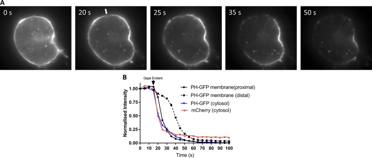
Digitonin permeabilization reveals reversible binding of PH-GFP to the plasma membrane of chromaffin cells. Bovine chromaffin cells were cotransfected with plasmids encoding PH-GFP and mCherry and imaged 5 d later in epifluorescence. Cells were bathed in Na glutamate solution (139 mM Na glutamate, 20 mM PIPES, 0.5 mM EGTA, 0.5 mM MgCl2, and 2 mM ATP) at 27°C and individually perfused with bath solution containing 10 µM digitonin through a 100-µm-inner-diameter glass pipette. (A) Within 30 s of digitonin application, gaps appeared in the PH-GFP–labeled plasma membrane (arrow) and a wave of loss of membrane PH-GFP fluorescence occurred starting at the plasma membrane proximal to the gaps. (B) PH-GFP intensities of segments of the plasma membrane proximal and distal to the gaps and in the cytosol were measured. Cytosolic mCherry fluorescence is rapidly lost coincident with the appearance of a gap in the plasma membrane, and PH-GFP fluorescence on the plasma membrane proximal to the gap decreases almost as rapidly as cytosolic mCherry and cytosolic PH-GFP. These results are similar to those from three other cells in which an initial gap in the PH-GFP fluorescence was detected.
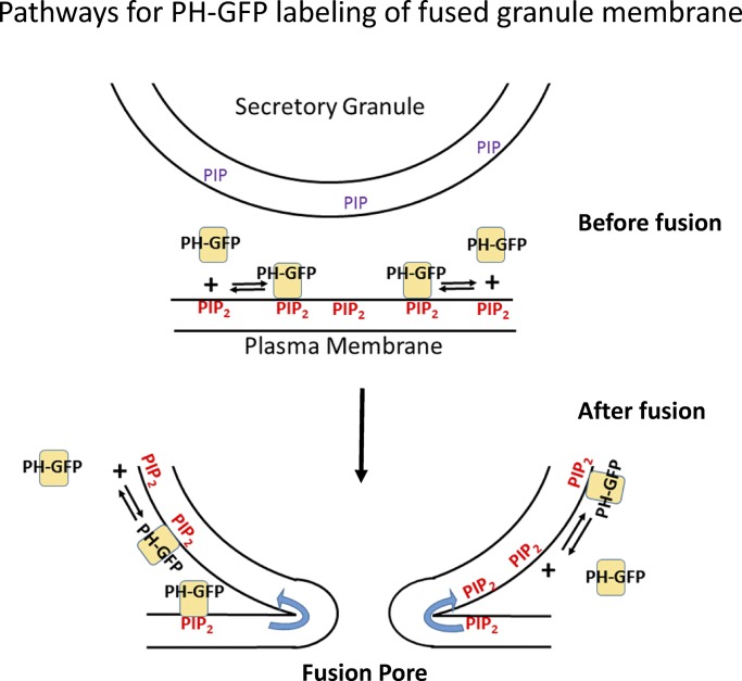
Dynamic binding of PH-GFP and PI-4,5-P2 and postfusion labeling of the secretory granule membrane. PH-GFP reversibly binds to PI-4,5-P2. The stable labeling of the fused granule membrane by PH-GFP probably reflects diffusion of PI-4,5-P2, bound or unbound to PH-GFP, with simultaneous association and dissociation of PH-GFP and PI-4,5-P2. The left and right side of the fusion pore depict diffusion of PH-GFP/PI-4,5-P2 or unbound PI-4,5-P2, respectively. De novo synthesis of PI-4,5-P2 on the fused granule membrane (see text) would also enhance PH-GFP binding.
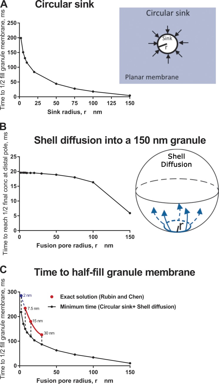
Diffusion time for a lipid probe in the plasma membrane to accumulate in a fused secretory granule with a fusion pore. The following calculations were performed with a diffusion constant of 1 × 10⁻⁸ cm2/s for PH-GFP (Hammond et al., 2009). (A) Circular sink. Minimum time for diffusional flow from a planar plasma membrane to supply a fused granule with enough molecules to attain a concentration of one half its final concentration. The calculation from Eq. 5.79 in Crank (1967) assumes that the (fusion pore) ring is a “perfect sink” that instantly and permanently transfers every molecule that hits it into the granule. (B) Shell diffusion. Time to attain a concentration of one half the final concentration at the distal pole of a fused granule (300-nm diameter) if all of the protein that is to enter the granule is distributed at zero time along a ring (fusion pore) of specific radius. The calculation is based on Velez and Axelrod (1988). (C) Time to half fill the granule membrane with plasma membrane probe through the fusion pore. The exact solution to this complex diffusion pathway has been solved (Rubin and Chen, 1990). It was applied to the diffusion of PH-GFP/PI-4,5-P2 from the plasma membrane of a 10.5-µm-radius cell into a fused granule membrane with a radius of 150 nm for fusion pores of radii of 7.5, 15, and 30 nm (red symbols). The three points were fitted with a parabola (red line), which was extrapolated to 2-nm pore radius (blue symbol and blue dashed line).The exact solution requires considerable computational power to extend to other fusion pore dimensions. Instead, the minimum time was estimated for a protein to reach one half of the final concentration by summing (black circles) the analyses in A and B. The minimum estimates of time and the exact solutions are remarkably consistent.
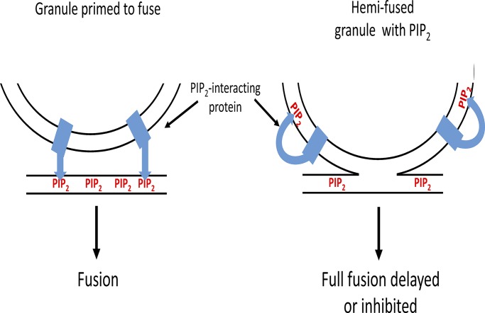
Appearance of PI-4,5-P2 on the granule membrane upon hemifusion may delay or inhibit full fusion by misdirecting PI-4,5-P2–dependent reactions. The functions of several proteins in the fusion pathway (e.g., synaptotagmin, CAPS, and Munc13) require binding to PI-4,5-P2. It is generally thought that these interactions occur with plasma membrane PI-4,5-P2. The functions of these proteins may be thwarted if the proteins interact with PI-4,5-P2 on the granule membrane before full fusion. The cartoon illustrates how the membrane interaction of a granule protein such as synaptotagmin would be altered by the appearance of PI-4,5-P2 in the granule membrane.
References
-
- Artalejo C.R., Elhamdani A., and Palfrey H.C.. 2002. Sustained stimulation shifts the mechanism of endocytosis from dynamin-1-dependent rapid endocytosis to clathrin- and dynamin-2-mediated slow endocytosis in chromaffin cells. Proc. Natl. Acad. Sci. USA. 99:6358–6363. 10.1073/pnas.082658499 - DOI - PMC - PubMed
Publication types
MeSH terms
Substances
Grants and funding
LinkOut - more resources
Full Text Sources
Research Materials
Miscellaneous
