. 2018 Nov 23;9(1):4964.
doi: 10.1038/s41467-018-07384-7.

Inflammasome activation negatively regulates MyD88-IRF7 type I IFN signaling and anti-malaria immunity

Affiliations

Inflammasome activation negatively regulates MyD88-IRF7 type I IFN signaling and anti-malaria immunity

Xiao Yu et al. Nat Commun. .

Abstract

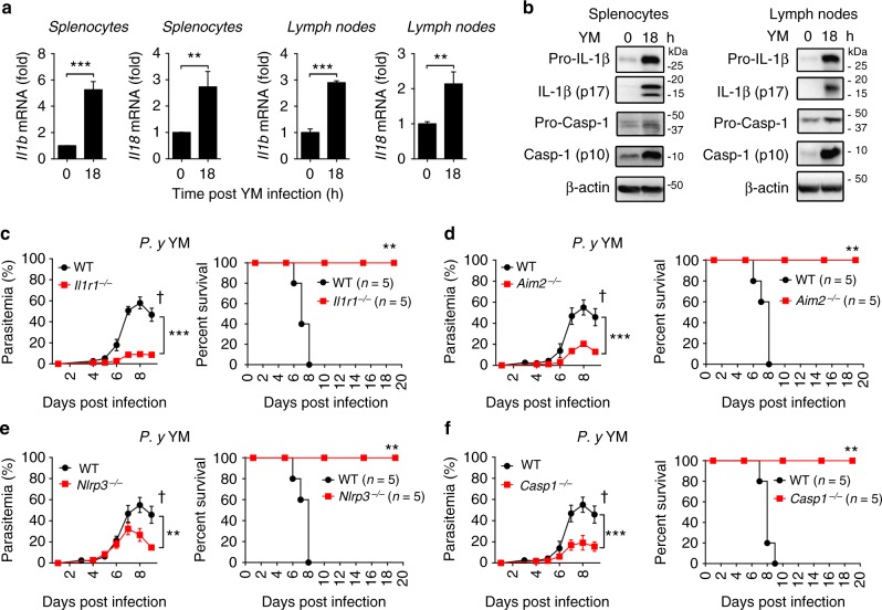
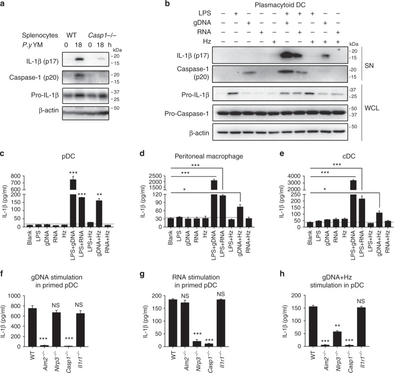
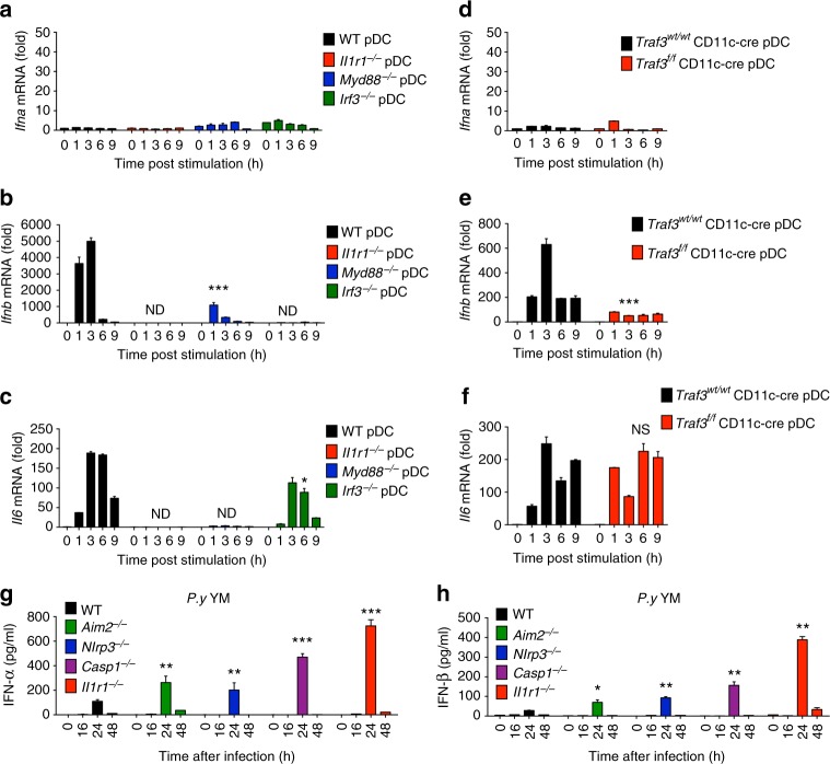
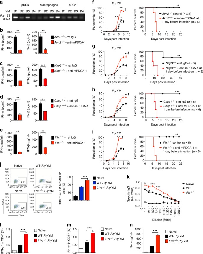
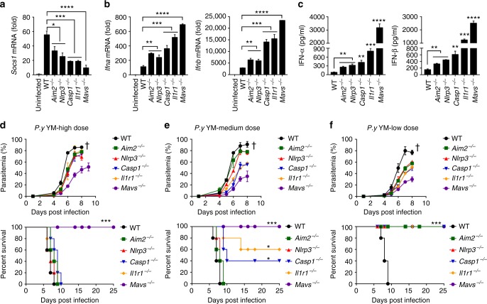
The inflammasome plays a critical role in inflammation and immune responses against pathogens. However, whether or how inflammasome activation regulates type I interferon (IFN-I) signaling in the context of malaria infection remain unknown. Here we show mice deficient in inflammasome sensors AIM2, NLRP3 or adaptor Caspase-1 produce high levels of IFN-I cytokines and are resistant to lethal Plasmodium yoelii YM infection. Inactivation of inflammasome signaling reduces interleukin (IL)-1β production, but increases IFN-I production. Mechanistically, we show inflammsome activation enhances IL-1β-mediated MyD88-TRAF3-IRF3 signaling and SOCS1 upregulation. However, SOCS1 inhibits MyD88-IRF7-mediated-IFN-I signaling and cytokine production in plasmacytoid dendritic cells. By contrast, ablation of inflammsome components reduces SOCS1 induction, and relieves its inhibition on MyD88-IRF7-dependent-IFN-I signaling, leading to high levels of IFN-α/β production and host survival. Our study identifies a previously unrecognized role of inflammasome activation in the negative regulation of IFN-I signaling pathways and provides potential targets for developing effective malaria vaccines.


Conflict of interest statement

The authors declare no competing interests.

Figures

Fig. 1
Fig. 1
Inflammasome deficiency augments the resistance to lethal P. yoelii YM infection in vivo. a, b WT (n = 5) mice were intraperitoneally infected with P. yoelii YM (0.5 × 106 iRBCs); spleen and lymph nodes were collected at indicated times post infection and subjected to qPCR (a) and immunoblotting analyses (b). cf WT and Il1r1−/− (c), Aim2−/− (d), Nlrp3−/− (e), Casp1−/− (f) mice (n = 5) were intraperitoneally infected with P. yoelii YM (0.5 × 106 iRBCs). Daily parasitemias and mortality rates are monitored. Data are representative of three independent experiments and plotted as mean ± SD. **p < 0.01, ***p < 0.001 vs. corresponding control. Dagger denotes mouse death
Fig. 2
Fig. 2
Plasmodium gDNA activates innate immune AIM2 inflammasome, while RNA and hemozoin trigger NLRP3 inflammasome activation. a WT and Casp1−/− mice (n = 5) were infected with P. yoelii YM, and splenocytes were harvested at 18 h after infection. Uninfected samples served as control group. Cell lysates were analyzed by immunoblotting with the indicated antibodies. b, c WT pDCs were stimulated as indicated for 24 h, cell lysate and supernatants were collected for immunoblotting analysis (b), and supernatants were collected for quantization of IL-1β cytokine by ELISA analysis (c). d, e WT peritoneal macrophages (d) and cDCs (e) were stimulated as indicated for 24 h, and supernatants were collected for quantization of IL-1β cytokine by ELISA analysis. f, g LPS-primed WT, Il1r1−/−, Aim2−/−, Nlrp3−/− and Casp1−/− pDCs were stimulated with gDNA (f) or RNA (g) for 24 h, and supernatants were collected for quantization of IL-1β cytokine by ELISA analysis. h WT, Il1r1−/−, Aim2−/−, Nlrp3−/−, and Casp1−/− pDCs were stimulated with gDNA/hemozoin complex for 24 h, supernatants were collected for quantization of IL-1β cytokine by ELISA analysis. Data are representatives of three independent experiments with similar results and plotted as mean ± SD. *p < 0.05, **p < 0.01 vs. corresponding control. NS, not significant
Fig. 3
Fig. 3
IL-1 signaling activates MyD88-TRAF3-IRF3-dependent type I IFN signaling in vitro and negatively regulates type I IFN cytokine production in vivo. ac WT, Il1r1−/−, Myd88−/−, and Irf3−/− pDCs were stimulated with recombinant mouse IL-1β for indicated times, and RNA from pDCs was isolated and used for expression analysis of Ifna (a), Ifnb (b), and Il6 (c) by using qPCR. df Traf3wt/wt CD11c-cre and Traf3f/f CD11c-cre pDCs were stimulated with recombinant mouse IL-1β for indicated times, and RNA from cells was isolated and used for expression analysis of Ifna (d), Ifnb (e), and Il6 (f) by using qPCR. g, h WT, Aim2−/−, Nlrp3−/−, Casp1−/−, and Il1r1−/− mice were intraperitoneally infected with P. yoelii YM. Serum was collected at indicated times and subjected to ELISA analysis of IFN-α (g) and IFN-β (h). Data are representatives of three independent experiments with similar results and plotted as mean ± SD. *p < 0.05, **p < 0.01, ***p < 0.001 vs. corresponding control. NS, not significant, ND, not detected
Fig. 4
Fig. 4
Activation of inflammasome negatively regulates type I IFN cytokine production in a MyD88-dependent manner. WT, Myd88−/−, Il1r1−/−, Casp1−/−, Myd88−/−Il1r1−/−, and Myd88−/−Casp1−/− mice (n = 5) were intraperitoneally infected with P. yoelii YM. Serum was collected at 24 h post infection and subjected to ELISA analysis of IFN-α (a) and IFN-β (b). Parasitemias and survivals (c and d) were monitored daily. Data are representatives of three independent experiments with similar results and plotted as mean ± SD. **p < 0.01, ***p < 0.001 vs. corresponding control. ND, not detected. Dagger denotes mouse death
Fig. 5
Fig. 5
pDCs contribute to the production of type I IFN at the early time post P. yoelii YM infection and ameliorate pathogenesis in inflammasome-deficient mice. a The cell populations of pDCs, cDCs, and macrophages were isolated from WT mice splenocytes at indicated times post YM infection using cell isolation kits, and then analyzed for cell-specific expression of P. yoelii 18 S rRNA by PCR. be Aim2−/− (b), Nlrp3−/− (c), Casp1−/− (d), and Il1r1−/− (e) mice (n = 5) were injected with anti-mPDCA-1 antibody at 12 h before and after infection, followed by P. yoelii YM infection. Serum was collected at 24 h post infection and subjected to ELISA analysis for IFN-α and IFN-β. fi Aim2−/− (f), Nlrp3−/− (g), Casp1−/− (h), and Il1r1−/− (i) mice (n = 5) were injected with anti-mPDCA-1 antibody at 12 h before and after infection, followed by P. yoelii YM infection. Parasitemias and survivals were monitored daily. j WT and Il1r1−/− mice (n = 5) were infected with YM. Splenocytes were collected at day 4 post infection, and CD86+ cells in CD11c+MHC-II+ cells were measured by flow cytometry. Representative FACS and statistical analysis of CD86-positive cells are shown. k Malaria specific IgG in serum from WT and Il1r1−/− mice (n = 5) at day 14 post YM infection, evaluated using ELISA.
 ln Intracellular staining of IFN-γ were measured by flow cytometry in splenocytes of WT and Il1r1−/− mice (n = 5) at day 10 post YM infection, followed by stimulation with crude antigens (YM iRBCs). Statistical analysis is shown in l and m. Splenocytes from YM-infected WT and Il1r1−/− mice were cultured with crude antigens (iRBCs) overnight, and cell supernatants were collected for ELISA analysis (n).
 Data are representative of three independent experiments and plotted as mean ± SD. *p < 0.05, **p < 0.01, ***p < 0.001 vs. corresponding control. Dagger denotes mouse death
Fig. 6
Fig. 6
Negative regulator SOCS1 is directly induced by IL-1β signaling in a MyD88-TRAF3-IRF3-dependent type I IFN signaling. a, b WT and Il1r1−/− mice (n = 5) were infected with P. yoelii YM for indicated times, and RNA from splenocytes was isolated and used for expression analysis of Socs1 (a), Ifna and Ifnb (b) by using qPCR. c WT pDCs were stimulated with recombinant mouse IL-1β (2 μg/ml) for indicated times, RNA from the pDCs was isolated and used for expression analysis of Socs1 by using qPCR. d WT, Il1r1−/−, Myd88−/−, and Irf3−/− pDCs were stimulated with recombinant IL-1β (2 μg/ml) for indicated times, RNA from pDCs was isolated and used for expression analysis of Socs1 by using qPCR. e WT and Traf3f/f CD11c-cre pDCs were stimulated with recombinant IL-1β (2 μg/ml) for indicated times, RNA from pDCs was isolated and used for expression analysis of Socs1 by using qPCR. fh WT, Traf3f/f CD11c-cre, and Traf3f/wt CD11c-cre mice (n = 5) were infected with P. yoelii YM. Serum was collected at 24 h post infection and subjected to ELISA analysis for IFN-α, IFN-β (f). Parasitemias (g) and survivals (h) were monitored daily. i A model to show whether SOCS1 is directly induced by IRF3-dependent signaling or the downstream IFNAR-mediated signaling. j WT and Irf3−/− cDCs were pretreated with or without anti-IFNAR antibody for 24 h and stimulated with YM gDNA plus RNA for indicated times. RNA from cDCs was isolated and used for expression analysis of Socs1 by using qPCR. kl WT and Stat1−/− cDCs were stimulated with YM gDNA plus RNA (k) or mouse IFN-α/β (l) for indicated times. RNA from splenocytes was isolated and used for expression analysis of Socs1 by using qPCR. Data are representative of three independent experiments and plotted as mean ± SD. *p < 0.05, **p < 0.01, ***p < 0.001 vs. corresponding control. NS, not significant. Dagger denotes mouse death
Fig. 7
Fig. 7
Fine-tuned regulation of SOCS1 induction by inflammasome and type I IFN signaling dictates host resistance to YM infection. ac WT and deficient (Aim2−/−, Nlrp3−/−, Casp1−/−, Il1r1−/−, and Mavs−/−) mice (n = 5) were infected with YM, RNA from pDCs was isolated at 18 h post infection and used for expression analysis of Socs1 (a), Ifna and Ifnb (b) by using qPCR, and serum levels of IFN-α and IFN-β in WT and deficient mice at 24 h after P. yoelii YM infection are shown in c. df WT and deficient (Aim2−/−, Nlrp3−/−, Casp1−/−, Il1r1−/−, and Mavs−/−) mice (n = 5) were infected with high dose (1 × 106 iRBCs, d), medium dose (0.75 × 106 iRBCs, e) or low dose (0.5 × 106 iRBCs, f) of P. yoelii YM. Daily parasitemias and mortality rates of WT and deficient mice after P. yoelii YM infection are shown. Data are plotted as mean ± SD and representative of three independent experiments with similar results. *p < 0.05, **p < 0.01, ***p < 0.001 vs. corresponding control. NS, not significant. Dagger denotes mouse death

References

    1. Crompton PD, et al. Malaria immunity in man and mosquito: insights into unsolved mysteries of a deadly infectious disease. Annu. Rev. Immunol. 2014;32:157–187. doi: 10.1146/annurev-immunol-032713-120220. - DOI - PMC - PubMed
    1. Stevenson MM, Riley EM. Innate immunity to malaria. Nat. Rev. Immunol. 2004;4:169–180. doi: 10.1038/nri1311. - DOI - PubMed
    1. Langhorne J, Ndungu FM, Sponaas AM, Marsh K. Immunity to malaria: more questions than answers. Nat. Immunol. 2008;9:725–732. doi: 10.1038/ni.f.205. - DOI - PubMed
    1. Gazzinelli RT, Kalantari P, Fitzgerald KA, Golenbock DT. Innate sensing of malaria parasites. Nat. Rev. Immunol. 2014;14:744–757. doi: 10.1038/nri3742. - DOI - PubMed
    1. Guo H, Callaway JB, Ting JP. Inflammasomes: mechanism of action, role in disease, and therapeutics. Nat. Med. 2015;21:677–687. doi: 10.1038/nm.3893. - DOI - PMC - PubMed

Publication types

MeSH terms