. 2018 Nov 23;8(1):17312.
doi: 10.1038/s41598-018-35531-z.

The presence of extracellular microRNAs in the media of cultured Drosophila cells

Affiliations

The presence of extracellular microRNAs in the media of cultured Drosophila cells

Stijn Van den Brande et al. Sci Rep. .

Abstract

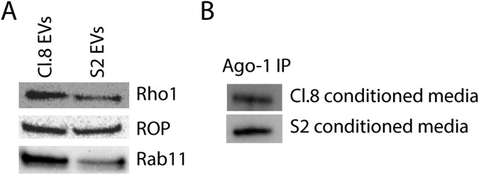
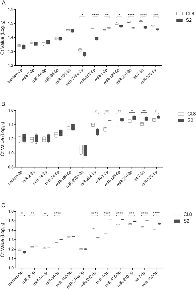
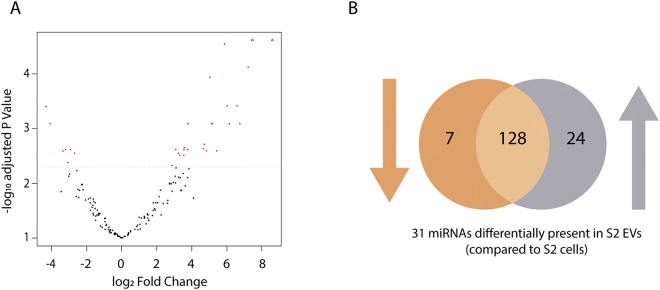
While regulatory RNA pathways, such as RNAi, have commonly been described at an intracellular level, studies investigating extracellular RNA species in insects are lacking. In the present study, we demonstrate the presence of extracellular microRNAs (miRNAs) in the cell-free conditioned media of two Drosophila cell lines. More specifically, by means of quantitative real-time PCR (qRT-PCR), we analysed the presence of twelve miRNAs in extracellular vesicles (EVs) and in extracellular Argonaute-1 containing immunoprecipitates, obtained from the cell-free conditioned media of S2 and Cl.8 cell cultures. Next-generation RNA-sequencing data confirmed our qRT-PCR results and provided evidence for selective miRNA secretion in EVs. To our knowledge, this is the first time that miRNAs have been identified in the extracellular medium of cultured cells derived from insects, the most speciose group of animals.


Conflict of interest statement

The authors declare no competing interests.

Figures

Figure 1
Figure 1
Immunoblot analysis on EVs and EAgo-1 immunoprecipitates. (A) EVs of both cell lines were subjected to immunoblot analysis for three common EV markers, which were clearly detected in the EV fractions purified from the extracellular media of both cell lines. (B) EAgo-1 IP was performed on the conditioned S2 and Cl.8 media as described in Methods section, but eluted in lithium dodecyl sulfate (LDS) instead of Qiazol. After elution, S2 and Cl.8 IP products were subjected to immunoblot analysis for EAgo-1, which was clearly detected in both IP products. Full-length blots are presented in Supplementary Fig. S1.
Figure 2
Figure 2
qRT-PCR data for microRNAs detected in extra- and intracellular fractions of Cl.8 and S2 cell lines. (A) Extracellular vesicles (EVs) and (B) extracellular Ago-1 containing immunoprecipitates (EAgo-1) were isolated from the cell-free conditioned media of Cl.8 and S2 cell cultures and different miRNAs were detected using qRT-PCR. (C) The intracellular expression levels of the selected miRNAs were measured as well. The data (log10 Ct values) are shown as box plots (min to max with the rectangle spanning the first to the third quartile including median) of four biological replicates, run in duplicate. Statistical analyses were performed with GraphPad Prism 6 (Graphpad Software Inc.). Significant differences (p < 0.05, p < 0.01, p < 0.001 and p < 0.0001) between the data obtained for specific miRNAs in the extracellular media of the two cell lines were found, after a log transformation, via a t-test (with or without two-sided Welch’s correction) and are indicated by (an) asterisk(s) (*, **, *** and **** respectively).
Figure 3
Figure 3
Differentially expressed miRNAs as measured in small RNA-seq data of S2 cells and derived EVs. Differential expression analysis was performed using edgeR on three biological replicates of both groups. MiRNAs were retained with a CPM above 1 in at least 3 samples. (A) Volcano plot indicating 31 significantly different miRNAs in S2 cells and EVs (red dots) and (B) the corresponding Venn diagram presenting the amount of miRNAs with increased (24) or decreased (7) abundance in S2 EVs compared to S2 cells. Abbreviations: EV: extracellular vesicle.

References

    1. Lee RC, Feinbaum RL, Ambros V. The C. elegans heterochronic gene lin-4 encodes small RNAs with antisense complementarity to lin-14. Cell. 1993;75:843–854. doi: 10.1016/0092-8674(93)90529-Y. - DOI - PubMed
    1. Berezikov E. Evolution of microRNA diversity and regulation in animals. Nat. Rev. Genet. 2011;12:846–860. doi: 10.1038/nrg3079. - DOI - PubMed
    1. Gomez-Orte E, Belles X. MicroRNA-dependent metamorphosis in hemimetabolan insects. Proc. Natl. Acad. Sci. USA. 2009;106:21678–82. doi: 10.1073/pnas.0907391106. - DOI - PMC - PubMed
    1. Caygill EE, Johnston LA. Temporal Regulation of Metamorphic Processes in Drosophila by the let-7 and miR-125 Heterochronic MicroRNAs. Curr. Biol. 2008;18:943–950. doi: 10.1016/j.cub.2008.06.020. - DOI - PMC - PubMed
    1. Jayachandran B, Hussain M, Asgari S. An insect trypsin-like serine protease as a target of microRNA: Utilization of microRNA mimics and inhibitors by oral feeding. Insect Biochem. Mol. Biol. 2013;43:398–406. doi: 10.1016/j.ibmb.2012.10.004. - DOI - PubMed

Publication types

MeSH terms

LinkOut - more resources