Review
. 2019 Mar;76(5):921-940.
doi: 10.1007/s00018-018-2972-z. Epub 2018 Nov 23.

Transcription factor Ptf1a in development, diseases and reprogramming

Affiliations
Review

Transcription factor Ptf1a in development, diseases and reprogramming

Kangxin Jin et al. Cell Mol Life Sci. 2019 Mar.

Abstract

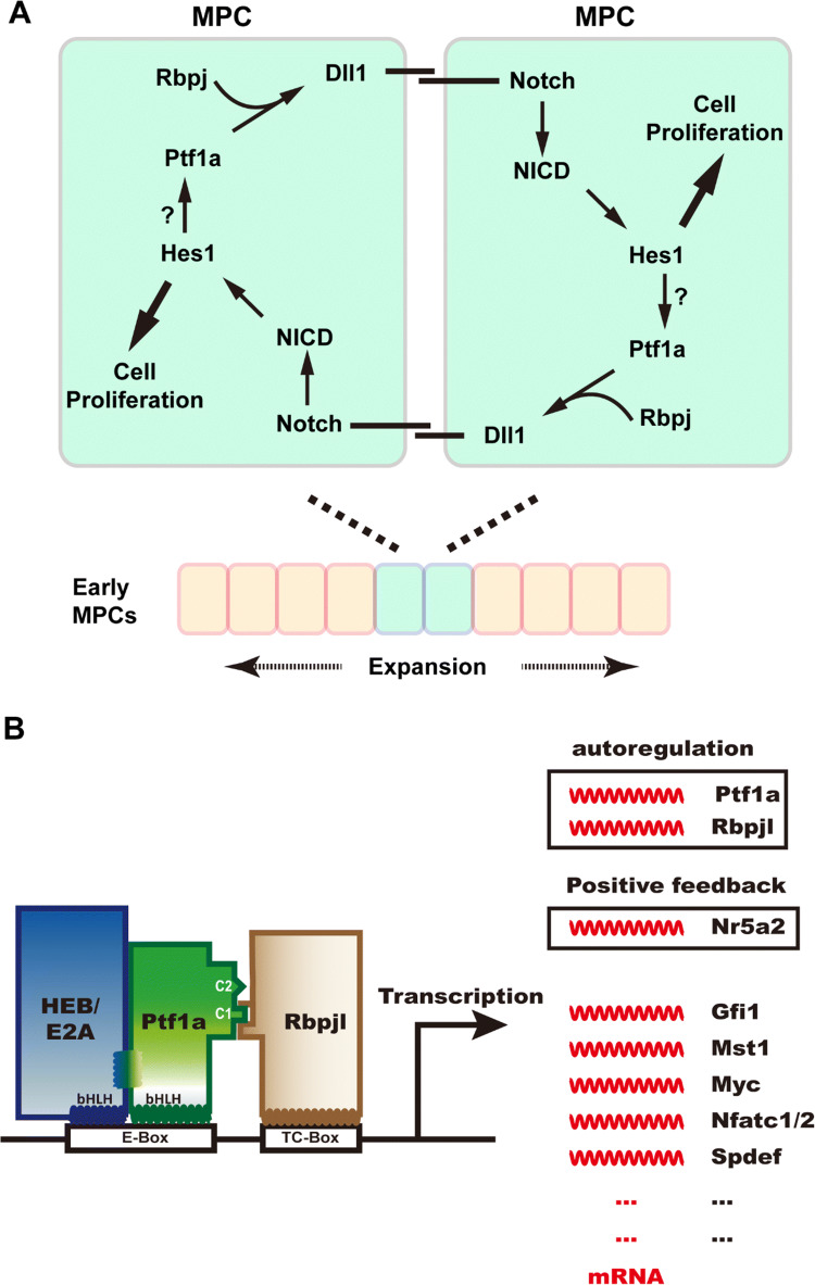
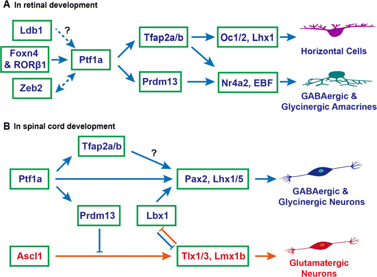
The transcription factor Ptf1a is a crucial helix-loop-helix (bHLH) protein selectively expressed in the pancreas, retina, spinal cord, brain, and enteric nervous system. Ptf1a is preferably assembled into a transcription trimeric complex PTF1 with an E protein and Rbpj (or Rbpjl). In pancreatic development, Ptf1a is indispensable in controlling the expansion of multipotent progenitor cells as well as the specification and maintenance of the acinar cells. In neural tissues, Ptf1a is transiently expressed in the post-mitotic cells and specifies the inhibitory neuronal cell fates, mostly mediated by downstream genes such as Tfap2a/b and Prdm13. Mutations in the coding and non-coding regulatory sequences resulting in Ptf1a gain- or loss-of-function are associated with genetic diseases such as pancreatic and cerebellar agenesis in the rodent and human. Surprisingly, Ptf1a alone is sufficient to reprogram mouse or human fibroblasts into tripotential neural stem cells. Its pleiotropic functions in many biological processes remain to be deciphered in the future.

Keywords: Acinar cells; Cell fate specification; Diabetes; GABAergic; Glutamatergic; Glycinergic; Inheritable; Inhibitory neurotransmitter; Pancreatic development; Retinal development; Somatic cell reprogramming; Transcriptional regulation.


Figures

Fig. 1
Fig. 1
Genomic structure and protein sequences of Ptf1a. a The upper portion of the image shows the genomic structure of the mouse Ptf1a gene. The blue bars represent two exons; the heightened parts are coding regions for Ptf1a. TSS, transcription start site. The lower portion of the image shows the corresponding multiz alignment and conservation of Ptf1a DNA sequences in 60 vertebrate species by UCSC genome browser. b The mouse Ptf1a protein is composed of 324 amino acid residues. The bHLH domain and several important residues are shown. c Multiple alignment of amino acid residues of the bHLH domains (upper image) and C termini (lower image) of Ptf1a proteins from seven vertebrate species. All sequences were downloaded from NCBI. The gray boxes in the C-terminal region (C1 and C2) outline two conserved regions important for interaction with Rbpj or Rbpjl protein, and one conserved lysine residue required for ubiquitination and proteasome degradation. d The phylogenetic tree for seven vertebrate Ptf1a proteins was constructed by the neighbor-joining method. The distance was calculated with Poisson correction. Gaps were distributed proportionally. The scale bar represents a distance of 0.05
Fig. 2
Fig. 2
DNA binding with Ptf1a and PTF1 complex. a The cartoon illustrates that two bHLH domains (from SCL-E47, PDB bank) bind to the major groove of DNA with their larger helices. The smaller helices fold and allow dimerization of the domains. b The common DNA binding consensus E-box and TC-box for the PTF1 complex. c Ptf1a and HEB (or E47, E12, isoforms of E2A) bind to the E-box, and Rbpj binds to the TC-box. Transcription activation requires that the PTF1 complex binds to E-box and TC-box simultaneously, which depends on Ptf1a interfacing with Rbpj via the conserved C1 and C2 regions at the C terminus. d The Notch intracellular domain (NICD) can compete with Ptf1a and E protein heterodimers for the binding sites on Rbpj protein
Fig. 3
Fig. 3
Ptf1a is required for early pancreatic genesis and adult acinar cell identity. a Ptf1a regulates early pancreatic MPC expansion in a positive feed-forward loop (adapted from Ahnfelt-Ronne et al. [38]). Ptf1a and Rbpj activate the expression of Notch ligand Dll1, which in turn binds to Notch receptors on the neighbor MPC, and triggers the downstream pathway of Notch. The activated Notch intracellular domain (NICD) enters the nucleus and activates transcription of target genes such as Hes1, which promotes cell proliferation. Hes1 may also maintain the Ptf1a level by stabilizing the Ptf1a structure. This positive feed-forward mechanism guarantees the proliferation and expansion of early MPCs in pancreatic primordia. b Ptf1a participates not only in the fate specification of acinar cells during development, but also in the maintenance of their physiological function and identity in the adult pancreas. The PTF1 triplex is capable of autoactivation by transactivating Ptf1a and Rbpjl expression. PTF1 activates the expression of Nr5a2 which can positively regulate the expression of Ptf1a and Rbpjl in return. PTF1 also activates the expression of many transcription factors crucial for acinar cell development and function, such as Gfi1, Mst1, Myc, Nfatc1/2, and Spdef
Fig. 4
Fig. 4
Ptf1a signaling pathways to specify inhibitory neuronal fates in retinal and spinal cord development. a During retinal development, Foxn4 and RORβ1 jointly turn on the expression of Ptf1a, which in turn activates the expression of Tfap2a/b and Prdm13, determining the cell fates of horizontal and amacrine cells. Both GABAergic and glycinergic amacrine cells are inhibitory neurons. Zeb2 directly activates Ptf1a expression and Ptf1a may enhance Zeb2 expression during amacrine and horizontal cell development. Ldb1 complexes may also be involved in activating Ptf1a expression and vice versa. b During spinal cord development, the upstream regulator of Ptf1a is unknown. Ptf1a activates expression of downstream targets Pax2 and Lhx1/5 directly or indirectly through Tfap2a/b, specifying the progenitors towards inhibitory GABAergic and glycinergic neuronal fates. Meanwhile, Ptf1a inhibits the excitatory glutamatergic neuronal fates indirectly through the downstream gene Prdm13. The coordinate interaction between Ptf1a and Lbx1 is unclear. In cerebellar and brainstem development, the same mechanism may be also at play as in the spinal cord
Fig. 5
Fig. 5
Ptf1a participates in brain development and cell fate reprogramming. a The image shows the expression pattern of GFP/Ptf1a (brownish signal) in the E15.5 brain (sagittal section) of a BAC transgenic mouse line harboring Ptf1a regulatory sequences and GFP as a reporter gene (image adapted from http://GENSAT.org). Ptf1a is modestly or strongly expressed in many regions in the forebrain, midbrain and hindbrain. b Around perinatal stage is the critical period when male brain is exposed to testosterone signal and female exposed to non-testosterone signals to establish sexual differentiation of the brain. Forebrain Ptf1a confers the sex differentiation competence during brain development. Ptf1a-deficient mice display sexually dimorphic behavior abnormalities. Image adapted from Fujiyama et al. [101]
Fig. 6
Fig. 6
Known disease-causing mutations in human PTF1A. The human genomic (DNA) structure of PTF1A gene is illustrated (nonproportionally). Exons 1 and 2 with the corresponding numbers of coded amino acid residues, and the 3′ downstream pancreatic progenitor-specific enhancer are highlighted. The disease-causing mutations at protein level (p.xxx) or genomic level (g.xxx) are listed on top of the DNA structure. Red arrows point to the mutation sites. The critical peptide motifs, bHLH, C1 and C2, and their positions in PTF1A protein, are placed below the corresponding DNA structure
Fig. 7
Fig. 7
Ptf1a alone is sufficient to reprogram human and mouse fibroblasts into induced neural stem cells (iNSCs). a After infected with lentiviruses expressing Ptf1a, human or mouse fibroblasts were transformed into neurospheres. Individual neurosphere can be further expanded and form typical single layered iNSCs in culture dishes. The iNSCs have the tripotence to differentiate into various types of neurons, astrocytes and oligodendrocytes both in vitro and in vivo. The iNSCs constitute an ideal cell source for preclinical transplantation experiments and clinical studies. b To determine the therapeutic effect of the Ptf1a-derived iNSCs, they were transplanted into the hippocampus of the Alzheimer disease mouse models. After training for 5 days, the iNSCs-transplanted mice spent much less time (bottom left) to find the target platform in the Morris water maze test. An example of travel pathways for a control mouse and an iNSC-transplanted mouse (top left) was given. Target annulus crossovers revealed that iNSC-transplanted mice displayed a preference for the target platform location (top right). Moreover, they also spent more time in the target quadrant (bottom right). These results demonstrate that iNSC transplantation improves the memory and cognitive functions of the AD mouse models

Similar articles

Cited by

References

    1. Murre C, et al. Interactions between heterologous helix–loop–helix proteins generate complexes that bind specifically to a common DNA sequence. Cell. 1989;58(3):537–544. doi: 10.1016/0092-8674(89)90434-0. - DOI - PubMed
    1. Chaudhary J, Skinner MK. Basic helix–loop–helix proteins can act at the E-box within the serum response element of the c-fos promoter to influence hormone-induced promoter activation in Sertoli cells. Mol Endocrinol. 1999;13(5):774–786. doi: 10.1210/mend.13.5.0271. - DOI - PubMed
    1. Grove CA, et al. A multiparameter network reveals extensive divergence between C. elegans bHLH transcription factors. Cell. 2009;138(2):314–327. doi: 10.1016/j.cell.2009.04.058. - DOI - PMC - PubMed
    1. Murre C, et al. Structure and function of helix–loop–helix proteins. Biochim Biophys Acta. 1994;1218(2):129–135. doi: 10.1016/0167-4781(94)90001-9. - DOI - PubMed
    1. Massari ME, Murre C. Helix–loop–helix proteins: regulators of transcription in eucaryotic organisms. Mol Cell Biol. 2000;20(2):429–440. doi: 10.1128/MCB.20.2.429-440.2000. - DOI - PMC - PubMed

MeSH terms

Substances

LinkOut - more resources