Review
. 2018 Dec;10(6):1537-1551.
doi: 10.1007/s12551-018-0469-5. Epub 2018 Nov 23.

From solution to surface to filament: actin flux into branched networks

Affiliations
Review

From solution to surface to filament: actin flux into branched networks

R Dyche Mullins et al. Biophys Rev. 2018 Dec.

Abstract

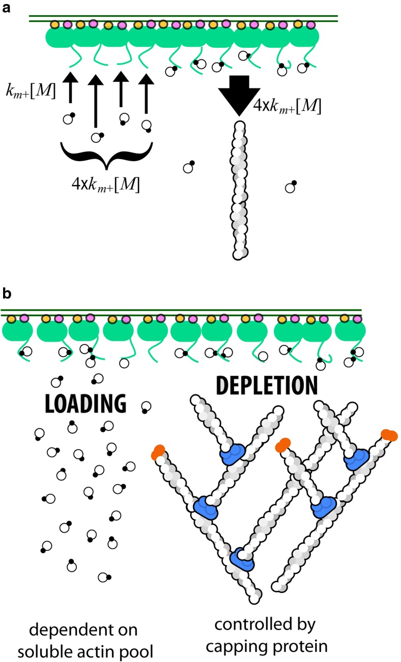
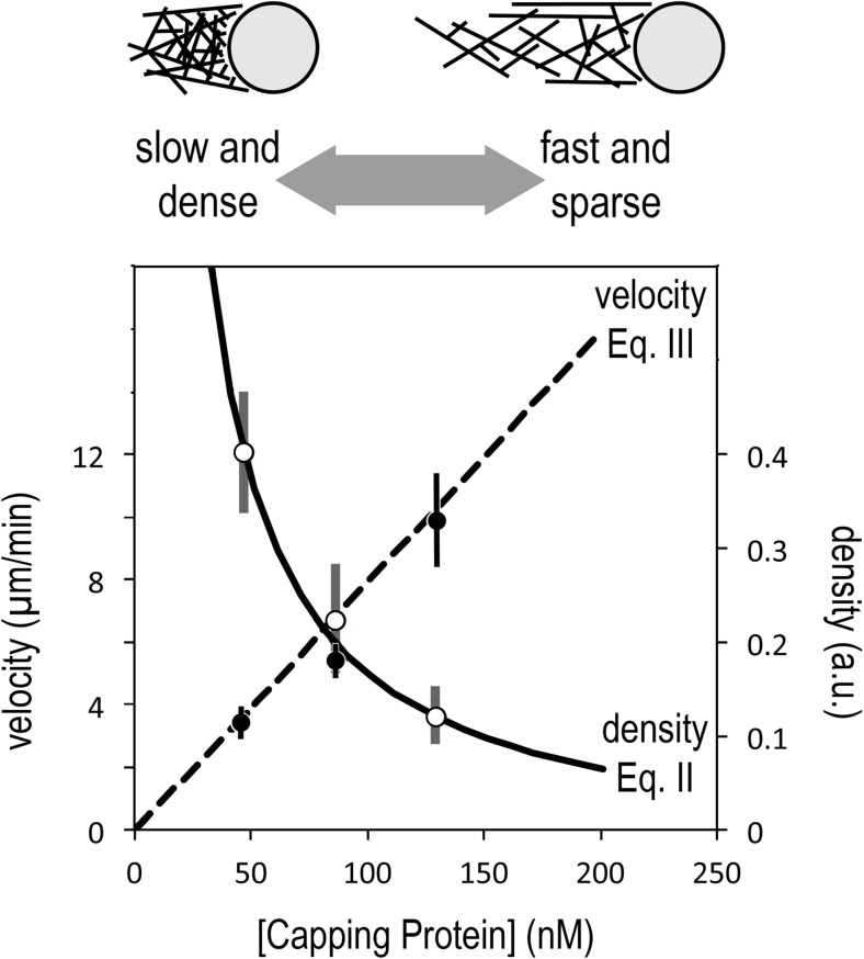
The actin cytoskeleton comprises a set of filament networks that perform essential functions in eukaryotic cells. The idea that actin filaments incorporate monomers directly from solution forms both the "textbook picture" of filament elongation and a conventional starting point for quantitative modeling of cellular actin dynamics. Recent work, however, reveals that filaments created by two major regulators, the formins and the Arp2/3 complex, incorporate monomers delivered by nearby proteins. Specifically, actin enters Arp2/3-generated networks via binding sites on nucleation-promoting factors clustered on membrane surfaces. Here, we describe three functions of this surface-associated actin monomer pool: (1) regulating network density via product inhibition of the Arp2/3 complex, (2) accelerating filament elongation as a distributive polymerase, and (3) converting profilin-actin into a substrate for the Arp2/3 complex. These linked functions control the architecture of branched networks and explain how capping protein enhances their growth.

Keywords: Actin cytoskeleton; Actin filament; Arp2/3 complex; Branched actin network; Capping protein; WASP family protein.


Conflict of interest statement

Conflict of interest

R. Dyche Mullins declares that he has no conflict of interest. Peter Bieling declares that he has no conflict of interest. Daniel Fletcher declares that he has no conflict of interest.

Ethical approval

This article does not contain any studies with human participants or animals performed by any of the authors.

Figures

Fig. 1
Fig. 1
Actin filament assembly and the surface-associated monomer pool. a In vitro, an actin filament (helical structure on right) can elongate by incorporation of monomers and/or profilin-actin complexes (linked filled and empty circles) directly from solution. b The filaments in a branched actin network grow by incorporating monomers from membrane-associated nucleation-promoting factors such as the WAVE Regulatory Complex (green). c Recruitment and activation of the WAVE Regulatory Complex. Two small, membrane-associated G proteins (Rac and/or Arf) bind a WAVE Regulatory complex and induce a conformational change that releases a natively unstructured region, called a PWCA domain. PWCA sequences, which are found in all WASP-family nucleation-promoting factors, comprise a proline-rich sequence (P) that binds profilin and profilin-actin complexes, a WH2 domain (W) that binds monomeric actin, and a central/acidic region (CA) that interacts with the Arp2/3 complex
Fig. 2
Fig. 2
Autocatalytic filament formation with and without negative feedback. Nucleation of new actin filaments by the Arp2/3 complex is autocatalytic, because the product of the reaction—a “daughter” filament—is also a substrate—an additional “mother” filament. a If autocatalysis were unregulated, filaments would beget filaments and the network density would grow exponentially. b In reality, however, the activity of the Arp2/3 complex is product inhibited, because growing filaments leech actin monomers away from WASP family proteins on the membrane surface, thus slowing the rate of nucleation. c Time evolution of filament density predicted by simple autocatalysis (Eq. 1, top curve) and autocatalysis with negative feedback (bottom curve)
Fig. 3
Fig. 3
Layout of the WAVE1 PWCA domain and flux of actin and profilin-actin through the proline-rich and WH2 domains. a Linear model of the PWCA domain from WAVE1, consisting of amino acids 277–559. Profilin-binding sites in the proline-rich domain are marked in yellow, the actin-binding WH2 domain is red, and the central/acidic region is blue. Actin monomers and profilin-actin complexes are shown on the same scale as the linear extent of the PWCA polypeptide (note scale bar). The affinity (dissociation equilibrium constant) of each profilin or actin-binding site is marked above in micromolar. b Pathways by which actin flows through surface-associated WASP family proteins and into growing filament networks. Left: poly-proline sequences bind profilin-actin complexes and can transfer monomeric actin onto the WH2 domain. From there, the actin can be used to activate the Arp2/3 complex and create a new filament, branching from the side of a preexisting filament. Middle: Actin can be transferred from the WH2 domain and poly-proline sequences onto the ends of nearby filaments. Decreased transfer from the poly-proline sequences and increased depletion by nearby filaments decreases the occupancy of the WH2 domain and slows the rate of nucleation. Right: capping protein decreases the number of growing filaments and increases the steady-state occupancy of both the WH2 domain and the poly-proline sequences
Fig. 4
Fig. 4
Sequence of steps in the transfer of actin from a proline-rich (a) or WH2 (b) domain of a membrane-associated nucleation-promoting factor onto the end of an actin filament. a A profilin-actin complex (linked circles) binds to the proline-rich (yellow) region (step 1), the bound profilin-actin complex attaches to the barbed end of an actin filament (step 2), the terminal actin monomer undergoes a rapid conformational change (step 3), and the profilin-poly-proline complex dissociates from the barbed end of the filament (step 4). In the absence of a conformational change in the terminal actin subunit, dissociation of the profilin is slow. b Monomeric actin (open circle) binds to the WH2 (red) sequence (step 1), the WH2-bound actin monomer attaches to the barbed end of a nearby filament (step 2), the terminal actin monomer undergoes a rapid conformational change (step 3), and the WH2 domain rapidly dissociates from the filament barbed end (step 4). Effective polymerase activity requires that the local density of PWCA domains be high enough to support frequent monomer transfer events
Fig. 5
Fig. 5
The steady-state occupancy of actin-binding sites on the membrane surface depends on the rates at which they are loaded and depleted. The WH2 and proline-rich domains are loaded by the binding of soluble actin monomers or profilin-actin complexes (left). These sites are depleted primarily by interactions between the bound actin and growing filament ends close to the membrane (right). Loading depends primarily on the concentration of soluble actin and profilin-actin, while depletion is proportional to the number of growing filament ends in proximity to the membrane. By capping fast-growing barbed ends, capping protein lowers the rate of depletion and increases the steady-state occupancy of surface actin pool
Fig. 6
Fig. 6
The antagonistic relationship between elongation and nucleation of filaments in a branched actin network means that the velocity and density have reciprocal responses to capping protein. Low rates of capping create networks that are denser but slower, while high rates of capping produce networks that are sparser but faster (top). This effect is described quantitatively by Eqs. 2 and 3 (bottom), which predict a linear increase in network velocity with capping protein concentration (Eq. 3, dashed line), and an inverse relationship between network density and capping (Eq. 2, solid line). Data points are taken from Fig. 2c, d of Akin (2008) and represent velocity and density of polarized, branched actin networks assembled from purified components by nucleation-promoting factors immobilized on polystyrene microspheres. Density (right axis) is expressed in arbitrary units based on intensity of fluorescently labeled actin (for more experimental details, see Akin, 2008). To account for mechanical effects associated with propulsion of spherical particles coated with immobile nucleation-promoting factors, we have adjusted the data points by subtracting the capping protein concentration required for the actin network to break symmetry and begin moving

References

    1. Achard V, Martiel JL, Michelot A, Guérin C, Reymann AC, Blanchoin L, Boujemaa-Paterski R. A "primer"-based mechanism underlies branched actin filament network formation and motility. Curr Biol. 2010;20(5):423–428. - PubMed
    1. Akin O, Mullins RD. Capping protein increases the rate of actin-based motility by promoting filament nucleation by the Arp2/3 complex. Cell. 2008;133(5):841–851. - PMC - PubMed
    1. Alberts B, Johnson A, Lewis J, Raff M, Roberts K, Walter P. Molecular biology of the cell. 6. New York: Garland Science; 2015.
    1. Arasada R, Pollard TD. Distinct roles for F-BAR proteins Cdc15p and Bzz1p in actin polymerization at sites of endocytosis in fission yeast. Curr Biol. 2011;21(17):1450–1459. - PMC - PubMed
    1. Bernheim-Groswasser A, Wiesner S, Golsteyn RM, Carlier MF, Sykes C. The dynamics of actin-based motility depend on surface parameters. Nature. 2002;417(6886):308–311. - PubMed