. 2019 Aug;29(8):669-682.
doi: 10.1002/hipo.23058. Epub 2018 Dec 11.

Remodeling of cholinergic input to the hippocampus after noise exposure and tinnitus induction in Guinea pigs

Affiliations

Remodeling of cholinergic input to the hippocampus after noise exposure and tinnitus induction in Guinea pigs

Liqin Zhang et al. Hippocampus. 2019 Aug.

Abstract

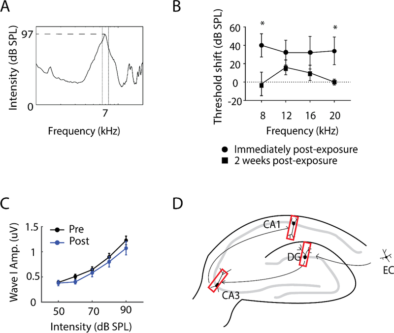
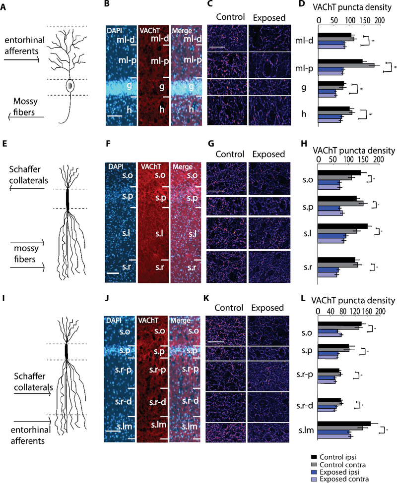
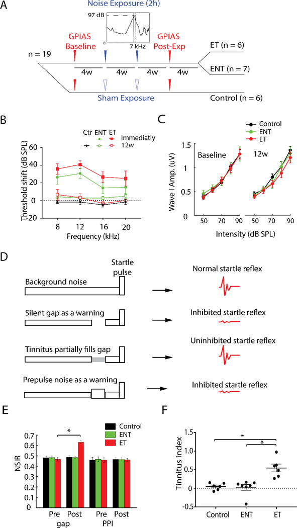
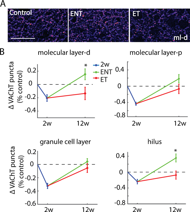
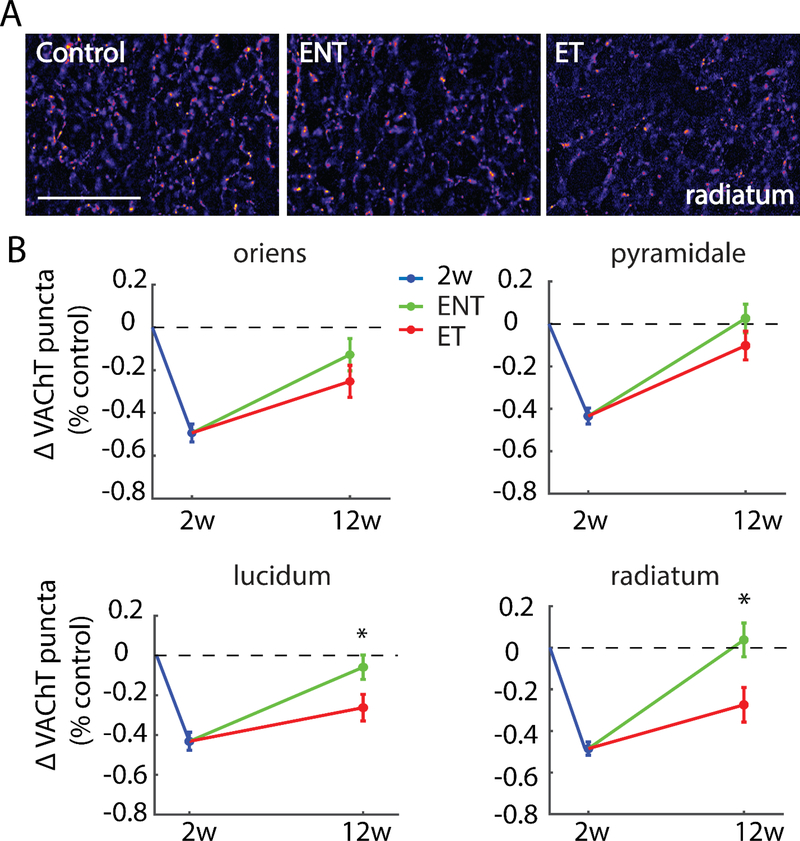
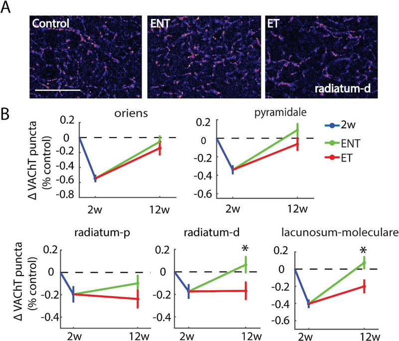
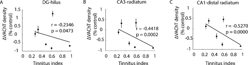
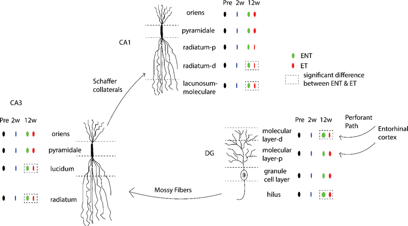
Here, we investigate remodeling of hippocampal cholinergic inputs after noise exposure and determine the relevance of these changes to tinnitus. To assess the effects of noise exposure on the hippocampus, guinea pigs were exposed to unilateral noise for 2 hr and 2 weeks later, immunohistochemistry was performed on hippocampal sections to examine vesicular acetylcholine transporter (VAChT) expression. To evaluate whether the changes in VAChT were relevant to tinnitus, another group of animals was exposed to the same noise band twice to induce tinnitus, which was assessed using gap-prepulse Inhibition of the acoustic startle (GPIAS) 12 weeks after the first noise exposure, followed by immunohistochemistry. Acoustic Brainstem Response (ABR) thresholds were elevated immediately after noise exposure for all experimental animals but returned to baseline levels several days after noise exposure. ABR wave I amplitude-intensity functions did not show any changes after 2 or 12 weeks of recovery compared to baseline levels. In animals assessed 2-weeks following noise-exposure, hippocampal VAChT puncta density decreased on both sides of the brain by 20-60% in exposed animals. By 12 weeks following the initial noise exposure, changes in VAChT puncta density largely recovered to baseline levels in exposed animals that did not develop tinnitus, but remained diminished in animals that developed tinnitus. These tinnitus-specific changes were particularly prominent in hippocampal synapse-rich layers of the dentate gyrus and areas CA3 and CA1, and VAChT density in these regions negatively correlated with tinnitus severity. The robust changes in VAChT labeling in the hippocampus 2 weeks after noise exposure suggest involvement of this circuitry in auditory processing. After chronic tinnitus induction, tinnitus-specific changes occurred in synapse-rich layers of the hippocampus, suggesting that synaptic processing in the hippocampus may play an important role in the pathophysiology of tinnitus.

Keywords: auditory; limbic system; memory; neuroplasticity; vesicular acetylcholine transporter (VAChT).


Figures

Figure 1.
Figure 1.. Experimental procedures of two-weeks post-noise-exposure animals.
(A) Features of the noise band to which experimental animals were unilaterally exposed for two hours. (B) Ipsilateral Acoustic Brainstem Response (ABR) thresholds of noise-exposed animals(n=5) at 8, 12, 16, 20 kHz immediately following noise exposure, and following a two-week recovery period. ABR thresholds recovered to baseline levels at 8 and 20 kHz and to near baseline levels at 12 and 16 kHz within two weeks. *p<0.05. (C) ABR wave I amplitude-intensity functions for noise-exposed animals prior to and two weeks following noise exposure. No differences were apparent two weeks post-exposure compared to baseline levels (pre-exposure). (D) Schematic diagram of hippocampal circuit with red rectangles depicting where images in the dentate gyrus (DG), area CA3 and CA1 were taken for immunohistochemistry. EC, entorhinal cortex.
Figure 2.
Figure 2.. Robust decreases in VAChT puncta density in dentate gyrus (DG), area CA3, and area CA1 two weeks following noise exposure.
(A) Schematic granule cell, depicting organization of inputs corresponding to the layers in (B), which are images of VAChT labeling in DG at 100X magnification. (C) Representative images at 400X magnification in DG with layers corresponding to (D), which depicts mean (±SEM) VAChT puncta density (per 104 μm2) in the indicated layers. (E) Schematic pyramidal neuron, depicting organization of inputs corresponding to the layers in (F), which are images of VAChT labeling in area CA3 at 100X magnification. (G) Representative images at 400X magnification in area CA3 with layers corresponding to (H), which depicts mean (±SEM) VAChT puncta density (per 104 μm2) in the indicated layers. (I) Schematic pyramidal neuron, depicting organization of inputs corresponding to the layers in (J), which are images of VAChT labeling in area CA1 at 100X magnification. (K) Representative images at 400X magnification in area CA1 with layers corresponding to (L), which depicts mean (±SEM) VAChT puncta density (per 104 μm2) in the indicated layers. In (B), (F), (J), scale bar is 100 μm. In (C), (G), (K), scale bar is 50 μm. Abbreviations: ml-d, distal region of molecular layer; ml-p, proximal region of molecular layer; g, granule cell layer; h, hilus; s.o, stratum oriens; s.p, stratum pyramidale; s.l, stratum lucidum; s.r, stratum radiatum; s.r-p, proximal region of stratum radiatum; s.r-d, distal region of stratum radiatum; s.lm, stratum lacunosum-moleculare. *p < 0.05
Figure 3.
Figure 3.. Repeated noise exposure induces tinnitus in a subset of experimental animals.
(A) Timeline of the experimental procedures of the chronically-exposed group. Nineteen animals were grouped into sham controls (n=6) and noise-exposed animals(n=13). GPIAS was used as tinnitus assessment and baseline thereof was acquired for four weeks pre-noise exposure. Animals were exposed to the same noise band/sham for two hours twice in sessions conducted four weeks apart, and then assessed for tinnitus eight weeks following the first noise exposure. ABR measurements were performed before and after each noise exposure and GPIAS. Noise-exposed animals were divided into two groups according to GPIAS assessment: noise exposed animals that exhibit no signs of tinnitus (ENT, n=7), and exposed animals that exhibit tinnitus (ET, n=6). (B) Mean (±SEM) ABR thresholds of animals with tinnitus (ET) and without tinnitus (ENT). ABR thresholds on the ipsilateral side were elevated immediately following noise exposure in both groups, but recovered to baseline levels at 8, 12, 16, 20 kHz 12 weeks after the first noise exposure. (C) Mean (±SEM) ABR wave I amplitude-intensity functions for ENT and ET animals pre- (baseline) and post-noise exposure (12w) were not significantly different, suggesting no underlying cochlear synaptopathy in both ENT and ET animals after the noise exposure. (D) Rationale of GPIAS (adapted from Turner et al., 2006). Row 1: Normal animals respond with a robust startle to the presentation of a startle pulse (20ms, 95 dB) embedded in a continuous background sound (65 dB). Row 2: When a silent gap (50ms) is introduced in the background sound, normal animals use the gap to predict the incoming startle pulse and respond with decreased startle amplitude. Row 3: Animals with tinnitus fail to detect the gap due to their tinnitus percept and respond with an uninhibited startle to the pulse presentation. Row 4: The gap is replaced with a prepulse noise (75dB). Both normal hearing and tinnitus animals respond with decreased startle amplitude due to alarm effects of the prepulse noise. Animals with hearing loss fail to detect the prepulse noise and thus respond with an uninhibited startle to the pulse presentation. This assessment tells whether animals’ inhibited responses to gap trials are due to hearing impairment. (E) Mean (±SEM) normalized startle inhibition ratio (NSIR) was the ratio of the startle amplitudes for the gap (or prepulse inhibition, PPI) trials and those for the no-gap trials. NSIR for gap trials was significantly higher post-exposure(Post) relative to baseline levels (Pre) for ET animals, but not for ENT or control animals. All animals exhibited stable responses to PPI trials both pre and post noise exposure. (F) Tinnitus indices of animals with tinnitus (ET) were significantly higher than those of controls and no-tinnitus animals (ENT).
Figure 4.
Figure 4.. Tinnitus animals exhibit diminished recovery of VAChT labeling relative to no-tinnitus animals in the dentate gyrus(DG).
(A) Representative images from distal molecular layer at 400X magnification; Scale bar = 50 μm. (B) Mean (±SEM) change of VAChT density (normalized to respective control) in the DG in two-weeks and 12-weeks post-noise-exposure animals. Distal molecular layer (ml-d) and hilus (h) showed significantly higher VAChT labeling in no-tinnitus animals (ENT) than in tinnitus animals (ET), whereas granule cell layer (g) and proximal molecular layer (ml-p) showed similar VAChT labeling in ENT and ET animals; *p < 0.05.
Figure 5.
Figure 5.. Tinnitus animals exhibit persistent decreases in VAChT labeling relative to no-tinnitus animals in synapse-rich areas of hippocampal area CA3.
(A) Representative images from stratum radiatum at 400x magnification; Scale bar = 50 μm. (B) Mean (±SEM) change of VAChT density (normalized to respective control) in area CA3 in two-weeks and 12-weeks post-noise-exposure animals. Strata lucidum (s.l) and radiatum (s.r) showed significantly lower VAChT labeling in tinnitus animasl (ET) than in no-tinnitus animals (ENT), whereas strata oriens (s.o) and pyramidale (s.p) showed similar VAChT labeling in ENT and ET animals; *p < 0.05.
Figure 6.
Figure 6.. Tinnitus animals exhibit persistent decreases in VAChT labeling relative to no-tinnitus animals in synapse-rich areas of hippocampal area CA1.
(A) Representative images from distal half of stratum radiatum at 400X magnification; Scale bar = 50 μm. (B) Mean (±SEM) change of VAChT density (normalized to respective control) in area CA1 in two-weeks and 12-weeks post-noise-exposure animals. Distal half of stratum radiatum(s.r-d) and stratum lacunosum-moleculare (s.lm) showed significantly lower VAChT labeling in tinnitus animals (ET) than in no-tinnitus animals (ENT), whereas strata oriens (s.o) and pyramidale (s.p) showed similar VAChT labeling in ENT and ET animals; *p < 0.05.
Figure 7.
Figure 7.. Tinnitus severity correlates with VAChT puncta density changes in tinnitus animals.
Scatterplots of VAChT density (normalized to control) vs tinnitus index in the hilus of DG (A), stratum radiatum of CA3 (B), and in distal stratum radiatum of CA1 (C). Severity of tinnitus is indicated by increasing values of tinnitus index; each data point represents an individual animal. Shown in inset are Pearson Correlation Coefficients (r) and accompanying p value. VAChT density in all 3 areas correlate with tinnitus severity, though the association is stronger in synapse-rich layers of CA3 and CA1.
Figure 8.
Figure 8.. Summary of changes in hippocampus after noise exposure and tinnitus.
VAChT labeling decreased in all three hippocampal sub-regions two weeks after noise exposure, suggesting short-term alterations in cholinergic neurotransmission after noise exposure. Twelve weeks after noise exposure, VAChT labeling remained low in animals that exhibited signs of tinnitus (ET), but recovered in exposed animals that exhibited no signs of tinnitus (ENT). Animals with tinnitus showed persistent disruption of VAChT in synapse-rich layers of hippocampus that receive inputs from upstream stages in the “trisynaptic circuit”, including distal region of molecular layer and hilus in the DG, strata lucidum and radiatum in area CA3, distal region of stratum radiatum and stratum lacunosum-moleculare in area CA1. This pattern of results raises the possibility that synaptic processing in the hippocampus plays an important role in the physiopathology of tinnitus.

References

    1. Al-Onaizi MA, Parfitt GM, Kolisnyk B, Law CS, Guzman MS, Barros DM, … Prado VF (2017). Regulation of Cognitive Processing by Hippocampal Cholinergic Tone. Cereb Cortex, 27(2), 1615–1628. doi: 10.1093/cercor/bhv349 - DOI - PubMed
    1. Altman NS (1992). An introduction to kernel and nearest-neighbor nonparametric regression. The American Statistician, 46(3), 10. doi: 10.1080/00031305.1992.10475879 - DOI
    1. Azman KF, Zakaria R, Abdul Aziz CB, & Othman Z (2016). Tualang Honey Attenuates Noise Stress-Induced Memory Deficits in Aged Rats. Oxid Med Cell Longev, 2016, 1549158. doi: 10.1155/2016/1549158 - DOI - PMC - PubMed
    1. Basura GJ, Koehler SD, & Shore SE (2015). Bimodal stimulus timing-dependent plasticity in primary auditory cortex is altered after noise exposure with and without tinnitus. J Neurophysiol, 114(6), 3064–3075. doi: 10.1152/jn.00319.2015 - DOI - PMC - PubMed
    1. Bauer CA, Turner JG, Caspary DM, Myers KS, & Brozoski TJ (2008). Tinnitus and inferior colliculus activity in chinchillas related to three distinct patterns of cochlear trauma. J Neurosci Res, 86(11), 2564–2578. doi: 10.1002/jnr.21699 - DOI - PMC - PubMed

Publication types

Substances