Review
. 2019 Apr;139(4):760-768.
doi: 10.1016/j.jid.2018.11.005. Epub 2018 Nov 22.

Mutations in Recessive Congenital Ichthyoses Illuminate the Origin and Functions of the Corneocyte Lipid Envelope

Affiliations
Review

Mutations in Recessive Congenital Ichthyoses Illuminate the Origin and Functions of the Corneocyte Lipid Envelope

Debra Crumrine et al. J Invest Dermatol. 2019 Apr.

Abstract

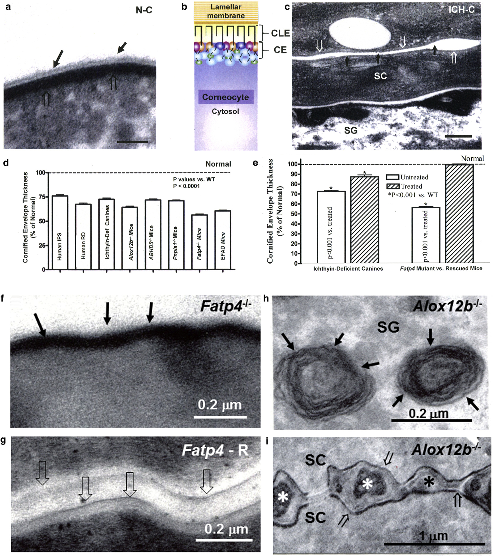
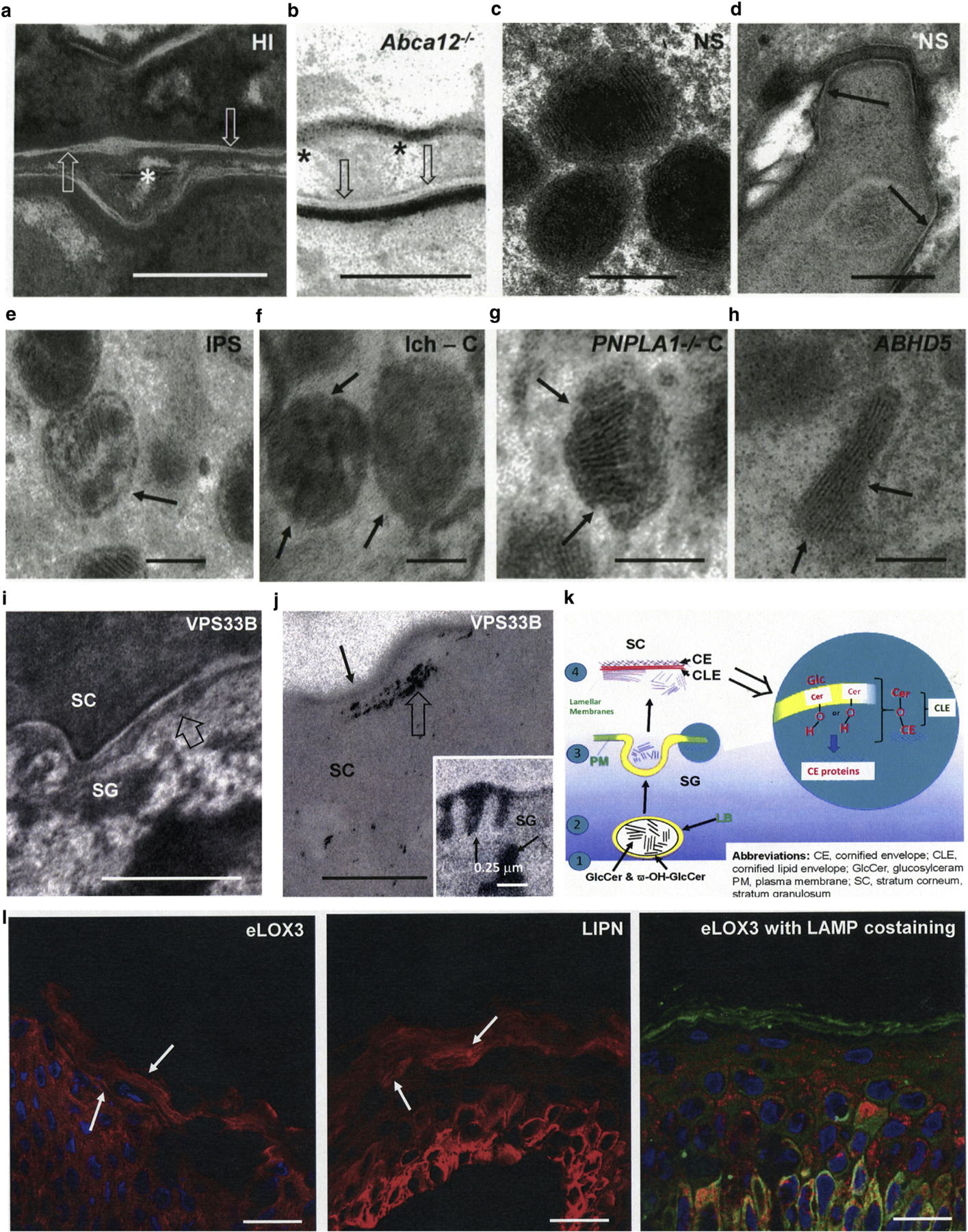
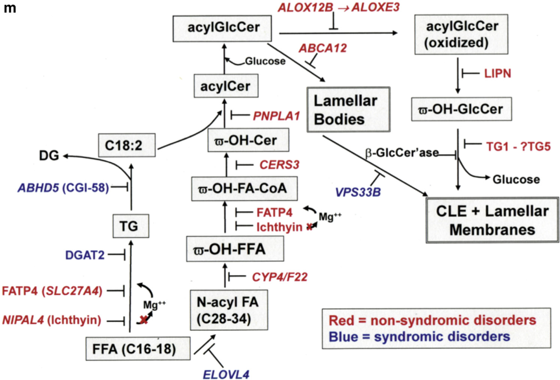
The corneocyte lipid envelope (CLE), a monolayer of ω-hydroxyceramides whose function(s) remain(s) uncertain, is absent in patients with autosomal recessive congenital ichthyoses with mutations in enzymes that regulate epidermal lipid synthesis. Secreted lipids fail to transform into lamellar membranes in certain autosomal recessive congenital ichthyosis epidermis, suggesting the CLE provides a scaffold for the extracellular lamellae. However, because cornified envelopes are attenuated in these autosomal recessive congenital ichthyoses, the CLE may also provide a scaffold for subjacent cornified envelope formation, evidenced by restoration of cornified envelopes after CLE rescue. We provide multiple lines of evidence that the CLE originates as lamellar body-limiting membranes fuse with the plasma membrane: (i) ABCA12 patients and Abca12-/- mice display normal CLEs; (ii) CLEs are normal in Netherton syndrome, despite destruction of secreted LB contents; (iii) CLEs are absent in VSP33B-negative patients; (iv) limiting membranes of lamellar bodies are defective in lipid-synthetic autosomal recessive congenital ichthyoses; and (v) lipoxygenases, lipase activity, and LIPN co-localize within putative lamellar bodies.


Conflict of interest statement

CONFLICT OF INTEREST

The authors state no conflict of interest.

Figures

Figure 1.
Figure 1.. Dual scaffold functions of CLEs in several ARCIs.
(a, c) Loss of both CLE (solid arrows) and attenuation of cornified envelopes (CE) (open arrows in ichthyin-deficient canines [ICH-C] vs. replete CLEs and CEs in normal littermates [N-C]). (b) Model of the relationship between CLE, CE, lamellar membranes, and corneocyte cytosol in normal stratum corneum. (d, e) Significant decline in CE thickness in several ARCI patients and animal models (mean ± standard error of the mean). (e) Normalization of CE in ichthyin-deficient canines treated with topical ω-O-acylceramide versus vehicle alone. (f) Loss of CLE in Fatp4 knockout (−/−) (solid arrows) and (g) reappearance of CLE (open arrows) after suprabasal transgenic rescue (Fatp4-R) (see Mauldin et al. [2018] for images of CLE in ichthyin-deficient canines]. (h) Despite replete lamellar body contents (solid arrows), (i) secreted contents fail to transform into lamellar membranes in Alox12b−/− mice (asterisk). Note also the loss of CLEs and attenuation of CEs in Alox12b−/− SC (i, open arrows). (c, g, i) osmium tetroxide postfixation. (a, f, m) Ruthenium tetroxide postfixation after pyridine pretreatment. Scale bars = 100 nm in a; 0.2 µm in c, f, g, and h; and 1 µm in i. ARCI, autosomal recessive congenital ichthyosis; CE, cornified envelope; CLE, corneocyte lipid envelope; Def, deficient; IPS, ichthyosis prematurity syndrome; RD, Refsum disease; SC, stratum corneum; SG, stratum granulosum; WT, wild type.
Figure 2.
Figure 2.. Evidence that CLEs form during LB exocytosis.
(a, b) Intact CLEs (open arrows), but paucity of extracellular lamellar membranes in harlequin ichthyosis patients and Abca12−/− mice (asterisks). (c, d) Normal LBs and CLEs in Netherton syndrome. (e–h) Abnormal lamellar body-limiting membranes in different ARCI and animal models. Solid arrows point to defects in LB-limiting membranes. (c.f. Figure 1h for another example). (i, j) Paucity of secreted LB contents at the SG-SC interface (i, open arrows) and absence of CLEs (j, solid arrows) in patients with VPS33B mutation (i, open arrow). Note also blockade in secretion results in entombment of lipase activity in the corneocyte cytosol (j, open arrow). (j) (insert) Lipase activity in lamellar bodies and at the SG-SC interface in normal human epidermis (solid arrows). (k) Proposed model of CLE formation with fusion of LB-limiting membranes to plasma membrane in outer SG. (l) Immunolocalization of eLOX3 (EI303 antibody) and LAMP1 in vesicular structures and lamellar membranes in frozen sections of normal human epidermis (solid arrows). (m) Epidermal lipid synthetic pathway leading to formation of the CLE and extracellular lamellar membranes, as well as sites of enzyme blockade in the lipid synthetic ARCI. Scale bars in a, b, d, e, and j = 0.2 µm; in c, f, g, and h = 0.1 µm; in i = 0.5 µm; in j = 0.25 µm; and in j, insert = l–10 µm. ARCI, autosomal recessive congenital ichthyosis; C, canine; CE, cornified envelop; CLE, corneocyte lipid envelope; HI, harlequin ichthyosis; ICH, ichthyin; IPS, ichthyosis prematurity syndrome; LB, lamellar body; M, transgenic mice; NS, Netherton syndrome; SC, stratum corneum; SG, stratum granulosum.

References

    1. Akiyama M Corneocyte lipid envelope (CLE), the key structure for skin barrier function and ichthyosis pathogenesis. J Dermatol Sci 2017;88: 3–9. - PubMed
    1. Behne M, Uchida Y, Seki T, de Montellano PO, Elias PM, Holleran WM. Omega-hydroxyceramides are required for corneocyte lipid envelope (CLE) formation and normal epidermal permeability barrier function. J Invest Dermatol 2000;114:185–92. - PubMed
    1. Brash AR. Lipoxygenases: occurrence, functions, catalysis, and acquisition of substrate. J Biol Chem 1999;274:23679–82. - PubMed
    1. Brash AR, Yu Z, Boeglin WE, Schneider C. The hepoxilin connection in the epidermis. FEBS J 2007;274:3494–502. - PubMed
    1. Candi E, Schmidt R, Melino G. The cornified envelope: a model of cell death in the skin. Nat Rev Mol Cell Biol 2005;6:328–40. - PubMed

Publication types