. 2018 Dec 6;103(6):1009-1021.
doi: 10.1016/j.ajhg.2018.10.019. Epub 2018 Nov 21.

MACF1 Mutations Encoding Highly Conserved Zinc-Binding Residues of the GAR Domain Cause Defects in Neuronal Migration and Axon Guidance

Affiliations

MACF1 Mutations Encoding Highly Conserved Zinc-Binding Residues of the GAR Domain Cause Defects in Neuronal Migration and Axon Guidance

William B Dobyns et al. Am J Hum Genet. .

Abstract

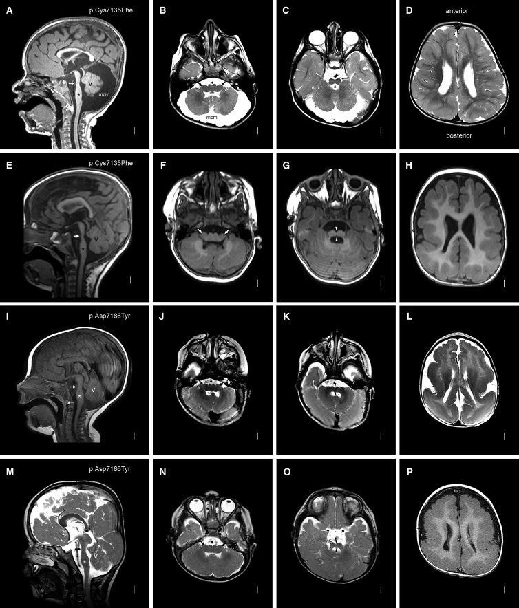
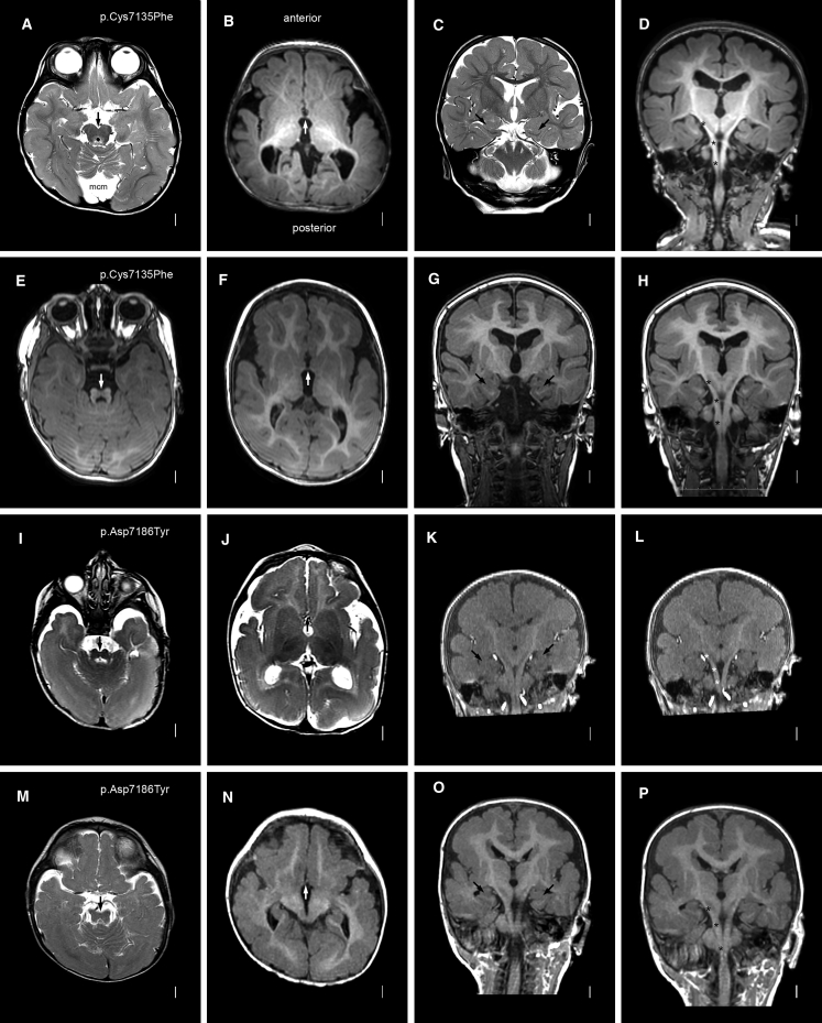
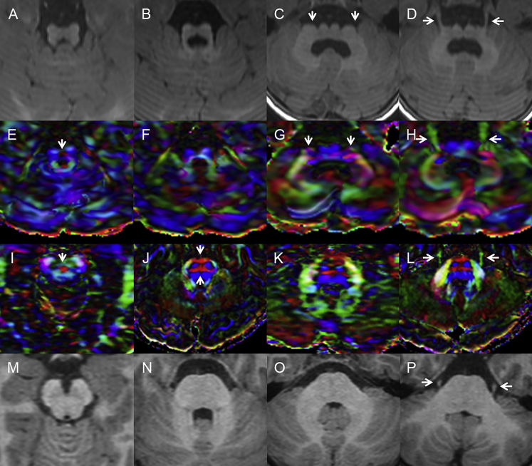
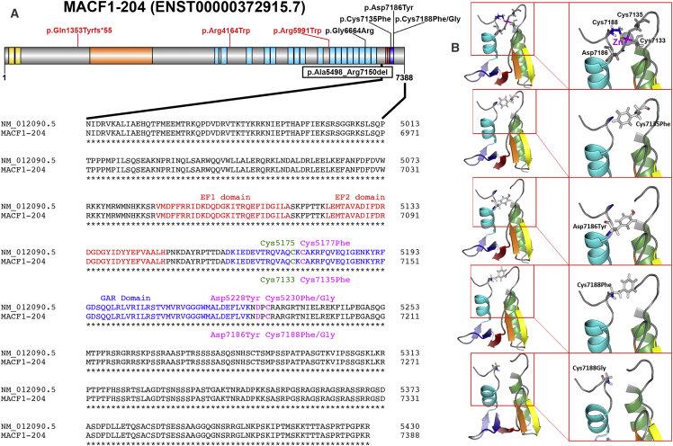
To date, mutations in 15 actin- or microtubule-associated genes have been associated with the cortical malformation lissencephaly and variable brainstem hypoplasia. During a multicenter review, we recognized a rare lissencephaly variant with a complex brainstem malformation in three unrelated children. We searched our large brain-malformation databases and found another five children with this malformation (as well as one with a less severe variant), analyzed available whole-exome or -genome sequencing data, and tested ciliogenesis in two affected individuals. The brain malformation comprised posterior predominant lissencephaly and midline crossing defects consisting of absent anterior commissure and a striking W-shaped brainstem malformation caused by small or absent pontine crossing fibers. We discovered heterozygous de novo missense variants or an in-frame deletion involving highly conserved zinc-binding residues within the GAR domain of MACF1 in the first eight subjects. We studied cilium formation and found a higher proportion of mutant cells with short cilia than of control cells with short cilia. A ninth child had similar lissencephaly but only subtle brainstem dysplasia associated with a heterozygous de novo missense variant in the spectrin repeat domain of MACF1. Thus, we report variants of the microtubule-binding GAR domain of MACF1 as the cause of a distinctive and most likely pathognomonic brain malformation. A gain-of-function or dominant-negative mechanism appears likely given that many heterozygous mutations leading to protein truncation are included in the ExAC Browser. However, three de novo variants in MACF1 have been observed in large schizophrenia cohorts.

Keywords: ACF7; MACF1; actin; axonal pathfinding; brainstem hypoplasia; cilia; cytoskeleton; lissencephaly; microtubules; midline crossing.


Figures

Figure 1
Figure 1
Brain MRI in Subjects with MACF1 Zinc-Binding-Pocket Mutations Images from mid-sagittal (far-left column) and three axial planes are shown from subjects LR14-088 (A–D), LR17-434 (E–H), LR16-306 (I–L), and LR17-450 (M–P). MACF1 mutations are shown in the far-left column for each subject. The mid-sagittal images all show striking brainstem hypoplasia and dysplasia with a mildly narrow midbrain, a dramatically narrow pons with tiny (white arrows in A and E) or no (white arrows in I and M) bumps on the ventral surface, and a mildly thick medulla. The midline images also show a mildly thin and variably short corpus callosum and mild cerebellar vermis hypoplasia with mega-cisterna magna (A) or borderline vermis hypoplasia (E, I, and M). The medulla is very wide and flat—almost double the typical width—and has small pyramids visible on the ventral surface (arrows in B, F, J, and N). The pons is also very wide and flat with a deep ventral cleft in the midline (arrowheads in C, G, K, and O). Images of the cortex show diffuse mild (D and H) or severe (L and P) pachygyria with a posterior gradient more severe than the anterior gradient.
Figure 2
Figure 2
Brain MRI in Subjects with MACF1 Zinc-Binding-Pocket Mutations: Additional Features Axial (left two columns), coronal (third column), and reconstructed coronal (far-right column) images are shown for (the same as in Figure 1) subjects LR14-088 (A–D), LR17-434 (E–H), LR16-306 (I–L), and LR17-450 (M–P). MACF1 mutations are again shown in the far-left column. The low midbrain or isthmus appears small and narrow with variably prominent superior cerebellar peduncles that are smaller than seen in the molar-tooth malformation associated with Joubert syndrome (especially E, I, and M). The anterior commissures are thin (arrows in B, F, J, and N). The hippocampi are small and dysplastic (C, G, K, and O). The pyramidal tracts are easy to follow given the paucity of transverse pontine crossing fibers; a few are seen in the top two images (asterisks over the right pyramidal tracts in D and H), but none are seen in the lower images (asterisks in L and P).
Figure 3
Figure 3
Diffusion Tensor Imaging Confirms Defects in Midline Crossing Tracts Axial T1-weighted magnetic resonance images and matched color-coded fractional-anisotropy maps from subject LR17-434 (A–H) and from a healthy control individual (I–P). Images at the level of the decussation of the superior cerebellar peduncles (first column) show a small midbrain (A) with the decussation represented as small (arrow in E) or medium-sized (arrow in I) red dots. Axial images through the upper pons (second column) demonstrate a ventral cleft in the midline of the small and dysplastic pons (B) and absence of the anterior and posterior transverse pontine fibers (F) in comparison with the healthy control individual (arrows in J). Axial images through the middle and low pons (third and fourth columns) demonstrate the narrow dorsal-ventral diameter and broad left-right axis of the brainstem (arrows in C and D) with descending and ventro-dorsal fibers (blue and green in G) seen in the far lateral pons (arrows pointing to blue and green tracts in G). The trigeminal nerves originate from the far lateral pons (arrows in D and H), a sharp contrast from the location of the trigeminal nerves in the control (arrows in L and P).
Figure 4
Figure 4
MACF1 Mutations Affect GAR Domain Amino Acids and Spectrin Rod Repeats (A) Schematic representation of MACF1 annotated according to transcript MACF1-204 shows the actin binding Calponin homology domains in yellow, plakin domain in orange, spectrin repeat domains in light blue, EF-hand domains in red, and GAR domain in blue. The missense variants associated with LIS and brainstem hypoplasia (black text) and schizophrenia (red text) are shown above the gene, and the intragenic deletion is shown below the gene. Alignment of the translated protein (GenBank: NM_012090.5) and MACF1-204 (UniProt: Q9UPN3) is shown below the gene. The EF-hand domains are shown in red text, and the zinc-binding GAR domain is shown in blue text. Mutations affecting the residues that coordinate zinc binding (Cys7135, Asp7186, and Cys7188) are shown in magenta above (GenBank: NM_012090.5) or below (UniProt: Q9UPN3) the normal sequence, and the fourth zinc binding residue is shown in green. (B) 3D models of the four GAR domain missense variants in the PyMOL Molecular Graphics System show significant changes in molecular structure. The top panel shows configuration of the normal GAR domain zinc-binding pocket. The divalent zinc ion is depicted as a gray sphere with a magenta label, and the four residues that coordinate zinc binding are depicted as gray sticks. In the simulated structures with pathogenic variants, the moderate negative charge of cysteine or aspartate is replaced by a bulky hydrophobic residue (phenylalanine or tyrosine; second to fourth panels) or by a small non-polar residue (glycine; bottom panel).

References

    1. Goryunov D., Liem R.K. Microtubule-actin cross-linking factor 1: domains, interaction partners, and tissue-specific functions. Methods Enzymol. 2016;569:331–353. - PubMed
    1. Moffat J.J., Ka M., Jung E.M., Smith A.L., Kim W.Y. The role of MACF1 in nervous system development and maintenance. Semin. Cell Dev. Biol. 2017;69:9–17. - PMC - PubMed
    1. Lane T.R., Fuchs E., Slep K.C. Structure of the ACF7 EF-hand-GAR module and delineation of microtubule binding determinants. Structure. 2017;25:1130–1138.e6. - PMC - PubMed
    1. Irahara K., Saito Y., Sugai K., Nakagawa E., Saito T., Komaki H., Nakata Y., Sato N., Baba K., Yamamoto T. Pontine malformation, undecussated pyramidal tracts, and regional polymicrogyria: a new syndrome. Pediatr. Neurol. 2014;50:384–388. - PMC - PubMed
    1. Di Donato N., Chiari S., Mirzaa G.M., Aldinger K., Parrini E., Olds C., Barkovich A.J., Guerrini R., Dobyns W.B. Lissencephaly: expanded imaging and clinical classification. Am. J. Med. Genet. A. 2017;173:1473–1488. - PMC - PubMed

Publication types