

Endoscopic evaluation of middle ear anatomic variations in autopsy series: analyses of 204 ears
- PMID: 30472004
- PMCID: PMC9422588
- DOI: 10.1016/j.bjorl.2018.10.002
Endoscopic evaluation of middle ear anatomic variations in autopsy series: analyses of 204 ears
Abstract
Introduction: Microsurgery of the ear requires complete evaluation of middle ear surgical anatomy, especially the posterior tympanic cavity anatomy. Preoperative assessment of the middle ear cavity is limited by the permeability of eardrum and temporal bone density. Therefore, middle ear exploration is an extremely useful method to identify structural abnormalities and anatomical variations.
Objective: The aim of this study is to determine anatomic variations of the middle ear in an autopsy series.
Methods: All evaluations were performed in the Forensic Medicine Institute Morgue Department. The cases over 18 years of age, with no temporal bone trauma and history of otologic surgery included in this study.
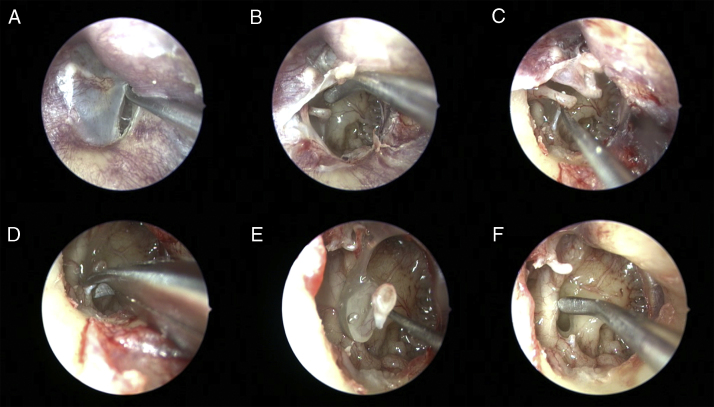
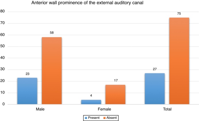
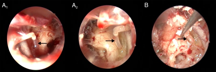
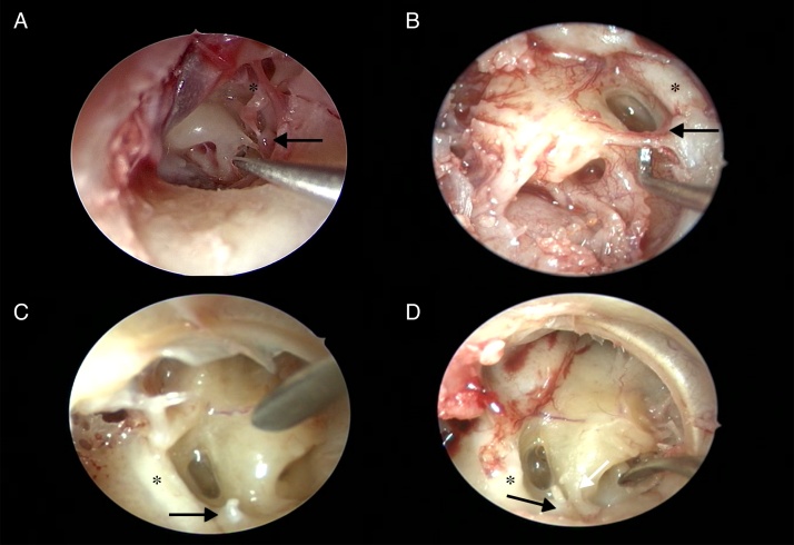
Results: One hundred and two cadavers were included in the study. The mean age was 49.08±17.76 years. Anterior wall prominence of the external auditory canal was present in 27 of all cadavers (26.4%). The tympanic membrane was normal in 192 ears (94%) while several eardrum pathologies were detected in 12 ears (6%). Agenesis of the pyramidal eminence and stapedial tendon was found in 3 ears. While the ponticulus was bony ridge-shaped in 156 of 204 ears (76.4%), it was bridge-shaped in 25 ears (12.3%). The ponticulus was absent in 23 ears (11.3%). While complete subiculum was present in 136 of all ears (66.7%), incomplete subiculum was present in 21 ears (10.3%). Subiculum was absent in 47 ears (23%). Facial dehiscence was found in 32 ears and the round window niche was covered by a pseudomembrane in 85 ears (41.6%). A fixed footplate was present in 7.4% of all ears, and no persistent stapedial artery was seen in any cases.
Conclusion: The pseudomembrane frequency covering the round window niche was found different from reports in the literature. In addition, the frequency of the external auditory canal wall prominence has been reported for the first time.
Introdução: A otomicrocirurgia requer avaliação completa da anatomia cirúrgica da orelha média, especialmente da anatomia da cavidade timpânica posterior. A avaliação pré-operatória da cavidade timpânica é limitada pela permeabilidade do tímpano e densidade do osso temporal. Portanto, a exploração da orelha média é um método extremamente útil para identificar anormalidades estruturais e variações anatômicas.
Objetivo: Determinar as variações anatômicas da orelha média em uma série de autópsias.
Método: Todas as avaliações foram realizadas no necrotério do Instituto Médico-Legal. Os casos com mais de 18 anos, sem trauma do osso temporal e história de cirurgia otológica foram incluídos neste estudo.
Resultados: Cento e dois cadáveres foram incluídos no estudo. A média de idade foi de 49,08 ± 17,76 anos. A proeminência da parede anterior do conduto auditivo externo estava presente em 27 de todos os cadáveres (26,4%). A membrana timpânica era normal em 192 orelhas (94%), enquanto várias alterações do tímpano foram detectadas em 12 orelhas (6%). Agenesia da eminência piramidal e do tendão do estapédio foi encontrada em 3 orelhas. Enquanto o pontículo tinha formato de crista óssea em 156 das 204 orelhas (76,4%), tinha o formato de ponte em 25 orelhas (12,3%). O pontículo estava ausente em 23 orelhas (11,3%). Enquanto o subículo completo estava presente em 136 de todas as orelhas (66,7%), encontrava-se incompleto em 21 orelhas (10,3%). O subículo estava ausente em 47 orelhas (23%). Deiscência facial foi encontrada em 32 orelhas e o nicho da janela redonda estava coberto por uma pseudomembrana em 85 orelhas (41,6%). A platina fixa foi observada em 7,4% de todas as orelhas e a artéria estapediana persistente não foi vista.
Conclusão: A frequência da pseudomembrana que cobre o nicho da janela redonda foi diferente daquela encontrada na literatura. Além disso, a frequência da proeminência da parede do canal auditivo externo foi relatada pela primeira vez.
Keywords: Anatomia da orelha média; Cirurgia endoscópica da orelha; Endoscopic ear surgery; Middle ear anatomy; Ponticulus; Pontículo; Retrotympanum; Retrotímpano; Subiculum; Subículo.
Copyright © 2018 Associação Brasileira de Otorrinolaringologia e Cirurgia Cérvico-Facial. Published by Elsevier Editora Ltda. All rights reserved.
Figures
Similar articles
-
The variants of the retro- and hypotympanum: an endoscopic anatomical study.Eur Arch Otorhinolaryngol. 2017 May;274(5):2141-2148. doi: 10.1007/s00405-017-4492-0. Epub 2017 Feb 27. Eur Arch Otorhinolaryngol. 2017. PMID: 28243781
-
Otoendoscopy in cholesteatoma surgery of the middle ear: what benefits can be expected?Otol Neurotol. 2008 Dec;29(8):1085-90. doi: 10.1097/MAO.0b013e318188e8d7. Otol Neurotol. 2008. PMID: 18836388
-
Pyramidal eminence and subpyramidal space: an endoscopic anatomical study.Laryngoscope. 2010 Mar;120(3):557-64. doi: 10.1002/lary.20748. Laryngoscope. 2010. PMID: 20013839
-
Endoscopic transcanal ear anatomy and dissection.Otolaryngol Clin North Am. 2013 Apr;46(2):131-54. doi: 10.1016/j.otc.2013.02.001. Otolaryngol Clin North Am. 2013. PMID: 23566901 Review.
-
Imaging anatomy of the retrotympanum: variants and their surgical implications.Br J Radiol. 2020 Jan;93(1105):20190677. doi: 10.1259/bjr.20190677. Epub 2019 Oct 8. Br J Radiol. 2020. PMID: 31593485 Free PMC article. Review.
Cited by
-
Videoautopsy-A Minimally Invasive Autopsy Method Using Endoscopic Techniques in Forensic Medicine: Clinical Features.Diagnostics (Basel). 2024 Apr 24;14(9):884. doi: 10.3390/diagnostics14090884. Diagnostics (Basel). 2024. PMID: 38732299 Free PMC article.
-
Optical Coherence Tomography-Based Atlas of the Human Cochlear Hook Region.J Clin Med. 2022 Dec 28;12(1):238. doi: 10.3390/jcm12010238. J Clin Med. 2022. PMID: 36615042 Free PMC article.
-
Landmarks for Proper Round Window Electrode Insertion in Cochlear Implantation.J Int Adv Otol. 2022 May;18(3):210-213. doi: 10.5152/iao.2022.21435. J Int Adv Otol. 2022. PMID: 35608488 Free PMC article.
-
Application of Otoform to Study Variations of Sinus Tympani: A Novel Technique.Indian J Otolaryngol Head Neck Surg. 2024 Feb;76(1):245-249. doi: 10.1007/s12070-023-04135-z. Epub 2023 Aug 20. Indian J Otolaryngol Head Neck Surg. 2024. PMID: 38440593 Free PMC article.
-
Minimally invasive autopsy - endoscopic approach to post-mortem diagnostics.Wideochir Inne Tech Maloinwazyjne. 2024 Mar;19(1):122-128. doi: 10.5114/wiitm.2023.134122. Epub 2023 Dec 29. Wideochir Inne Tech Maloinwazyjne. 2024. PMID: 38974768 Free PMC article. Review.
References
-
- Saito R., Igarashi M., Alford B.R., Guilford F.R. Anatomical measurement of the sinus tympani. Arch Otolaryngol Head Neck Surg. 1971;94:918–925. - PubMed
-
- Abdel Baki F., El Dine M.B., El Saiid I., Bakry M. Sinus tympani endoscopic anatomy. Otolaryngol Head Neck Surg. 2002;127:158–162. - PubMed
-
- Pickett B., Cail W., Lamburt P. Sinus tympani: anatomic consideration, computed tomography and a discussion of retro facial approach for removal of disease. Am J Otol. 1995;16:741–750. - PubMed
-
- Donaldson J.A., Anson B.J., Warpeha R.L., Rensink M.J. The perils of the sinus tympani. Trans Pacific Coast Otolaryngol Ophthalmol Soc. 1968;49:99–106. - PubMed
-
- Thomassin J.M., Korchia D., Doris J.M. Endoscopic guided otosurgery in the prevention of residual cholesteatomas. Laryngoscope. 1993;103:939–943. - PubMed
MeSH terms
LinkOut - more resources
Full Text Sources
Medical
