. 2019 Feb;23(2):750-760.
doi: 10.1111/jcmm.13897. Epub 2018 Nov 24.

Therapeutic effects of lentinan on inflammatory bowel disease and colitis-associated cancer

Affiliations

Therapeutic effects of lentinan on inflammatory bowel disease and colitis-associated cancer

Yanrong Liu et al. J Cell Mol Med. 2019 Feb.

Erratum in

  • Corrigendum.
    [No authors listed] [No authors listed] J Cell Mol Med. 2023 Jan;27(1):163. doi: 10.1111/jcmm.17588. J Cell Mol Med. 2023. PMID: 36588279 Free PMC article. No abstract available.

Abstract

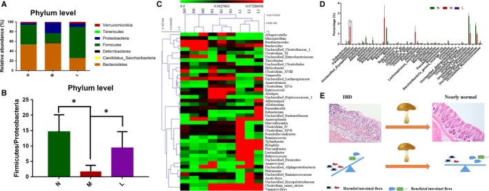
In this study, we investigated the therapeutic potential of lentinan in mouse models of inflammatory bowel disease (IBD) and colitis-associated cancer (CAC). Lentinan decreased the disease activity index and macroscopic and microscopic colon tissue damage in dextran sulphate sodium (DSS)-induced or TNBS-induced models of colitis. High-dose lentinan was more effective than salicylazosulfapyridine in the mouse models of colitis. Lentinan decreased the number of tumours, inflammatory cell infiltration, atypical hyperplasia and nuclear atypia in azoxymethane/DSS-induced CAC model. It also decreased the expression of pro-inflammatory cytokines, such as IL-13 and CD30L, in IBD and CAC model mice possibly by inhibiting Toll-like receptor 4 (TLR4)/NF-κB signalling and the expression of colon cancer markers, such as carcinoembryonic antigen, cytokeratin 8, CK18 and p53, in CAC model mice. In addition, lentinan restored the intestinal bacterial microbiotal community structure in IBD model mice. Thus, it shows therapeutic potential in IBD and CAC model mice possibly by inhibiting TLR4/NF-κB signalling-mediated inflammatory responses and disruption of the intestinal microbiotal structure.

Keywords: TLR4; colitis-associated cancer; inflammatory bowel disease; lentinan.


Figures

Figure 1
Figure 1
Effects of lentinan treatment on body weight, disease activity index (DAI), and colon length in DSS‐induced and TNBS‐induced mouse models of colitis. A, Schematic representation showing the establishment of mouse models of DSS‐induced acute or chronic colitis and TNBS‐induced colitis and lentinan administration in (i) acute colitis, (ii) chronic colitis and (iii) TNBS‐induced colitis model mice. B, Body weight changes in low‐, medium‐, and high‐dose lentinan‐treated (i) acute colitis, (ii) chronic colitis and (iii) TNBS‐induced colitis model mice. C, DAI in low‐, medium‐, and high‐dose lentinan‐treated (i) acute colitis, (ii) chronic colitis and (iii) TNBS‐induced colitis model mice. D, Colon lengths in low‐, medium‐, and high‐dose lentinan‐treated (i) acute colitis, (ii) chronic colitis and (iii) TNBS‐induced colitis model mice. *denotes < 0.05; **denotes < 0.01
Figure 2
Figure 2
Histopathological analysis of mouse models of colitis after treatment with lentinan. A, Left panel shows representative photographs of the colons from normal, DSS‐induced acute, and chronic colitis and TNBS‐induced colitis mice treated with or without SASP and low‐, medium‐, and high‐dose lentinan. The right panel shows the macroscopic damage scores based on visual inspection of the histopathology. As shown, lentinan decreases colon damage scores in a concentration‐dependent manner. B, Left panel shows representative photographs of H&E‐stained colon pathological sections from normal, acute and chronic colitis and TNBS‐induced colitis mice treated with or without SASP and low‐, medium‐, and high‐dose lentinan. The inflammatory cell infiltration and crypt erosion are marked with blue or yellow arrows respectively. The foci of inflammatory cells in the model group are enlarged in the upper left corner. The right panel shows the microscopic damage scores based on the analysis of H&E‐stained pathological sections. Lentinan ameliorates colitis‐associated microscopic damage in the mouse model. *denotes < 0.05; **denotes < 0.01
Figure 3
Figure 3
Lentinan pre‐treatment prevents DSS‐induced colitis. A, DAIs of normal, untreated and lentinan pre‐treated colitis model mice. B, Body weights of normal, untreated and lentinan pre‐treated colitis model mice. C, Colon lengths of normal, untreated and lentinan pre‐treated colitis model mice. D, Representative photographs of colons from normal, untreated and lentinan pre‐treated model mice and their macroscopic damage scores. E, Representative photographs of H&E‐stained colon sections from normal, untreated and lentinan pre‐treated model mice and their colon histological scores
Figure 4
Figure 4
Lentinan treatment prevents CAC. A, Body weights of normal, untreated and lentinan pre‐treated CAC model mice. B, Representative photographs and data of colons from normal, untreated and lentinan pre‐treated CAC model mice showing tumor counts and volume. C, Representative photographs of H&E‐stained pathological sections of colons from normal, untreated and lentinan pre‐treated CAC model mice. 15× and 200× represent magnification of photographs; *denotes < 0.05; ** denotes < 0.01
Figure 5
Figure 5
Lentinan inhibits TLR4 signalling pathway. A, Expression of TLR4 signalling pathway proteins including CaMKII, IRAK4, MyD88, IKBKB and TRAF6 in LPS‐stimulated RAW264.7 cells treated with or without 0.5, 1 and 2 mg/mL lentinan. B, FACS analysis of calcium influx in LPS‐stimulated RAW264.7 cells treated with or without 0.5, 1 and 2 mg/mL lentinan. The calcium influx signal decreases in a dose‐dependent manner upon lentinan treatment. C, Representative Western blot showing expression of NF‐κB p65 subunit in the nuclear extract from LPS‐stimulated RAW264.7 cells treated with or without 0.5, 1 and 2 mg/mL lentinan. D, Immunofluorescence micrographs showing the status of nuclear translocation of NF‐κB‐p65 in LPS‐stimulated RAW264.7 cells treated with or without 0.5, 1 and 2 mg/mL lentinan. E, Immunohistochemical staining images showing NF‐κB‐p65 levels in the colon from acute colitis model mice with or without lentinan treatment. F, Dual luciferase reporter assay analysis showing changes in NF‐κB activity in the presence and absence of lentinan. All the above data are represented as mean ± SD. *denotes < 0.05; **denotes < 0.01
Figure 6
Figure 6
Lentinan inhibits the expression of inflammatory cytokines and colon cancer markers. A, Cytokine antibody chip assay analysis of the concentrations of cytokines from RAW264.7 cells treated with 1 mg/mL LPS in the presence or absence of 1 μg/mL lentinan, detected by cytokine antibody chip assay. B, Grouping analysis of the cytokine antibody chip assay results. C, Representative images of IHC staining of the cancer markers CEA, CK8, CK18 and p53 (all in brown) and the cytokines IL‐13 and CD30L (both in red) in colon tissues from CAC model mice treated with or without lentinan. The nuclear staining is coloured blue. D, Quantitative analysis of cancer markers and inflammatory cytokines based on the IHC staining in 5C. Data are represented as mean ± SD. *denotes < 0.05; **denotes < 0.01
Figure 7
Figure 7
Lentinan restores intestinal bacteria microbiotal community structure in acute colitis model mice. A, Bacteroidetes, Firmicutes and Proteobacteria were the dominant bacteria at the phylum level in the mouse faecal samples. Lentinan‐treated mice were more similar to normal mice. B, Lentinan treatment recovered the Firmicutes/Proteobacteria ratio to nearly normal levels. C, Cluster analysis showed that the gut microbiota at the genus level in the lentinan‐treated mice was more similar to normal mice without acute colitis. D, Gut microbiota such as Eubacterium and Parabacteroides reduced to nearly normal levels after lentinan treatment. E, Schematic showing the role of lentinan in protecting the intestinal bacteria microbiota and mucosa

Similar articles

Cited by

References

    1. Crohn BB, Rosenberg H. The sigmoidoscopic picture of chronic ulcerative colitis (non‐specific). Am J Med Sci. 1925;170:220‐227.
    1. Eaden JA, Abrams KR, Mayberry JF. The risk of colorectal cancer in ulcerative colitis: a meta‐analysis. Gut. 2001;48:526‐535. - PMC - PubMed
    1. Gyde S, Prior P, Dew MJ, et al. Mortality in ulcerative colitis. Gastroenterology. 1982;83:36‐43. - PubMed
    1. Baumgart DC, Carding SR. Inflammatory bowel disease: cause and immunobiology. Lancet. 2007;369:1627‐1640. - PubMed
    1. Brubaker SW, Bonham KS, Zanoni I, Kagan JC. Innate immune pattern recognition: a cell biological perspective. Annu Rev Immunol. 2015;33:257‐290. - PMC - PubMed

Publication types

MeSH terms