Review
. 2018 Dec;43(12):970-983.
doi: 10.1016/j.tibs.2018.09.011. Epub 2018 Oct 26.

Greasing the Wheels of Lipid Biology with Chemical Tools

Affiliations
Review

Greasing the Wheels of Lipid Biology with Chemical Tools

Timothy W Bumpus et al. Trends Biochem Sci. 2018 Dec.

Abstract

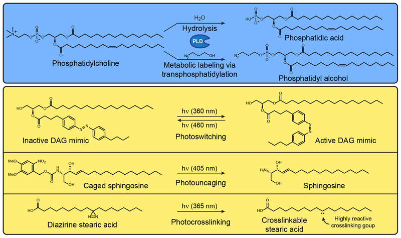
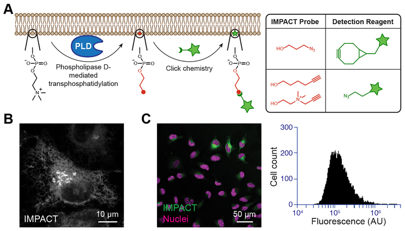
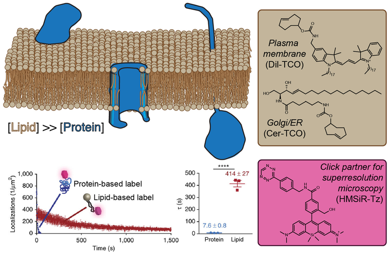
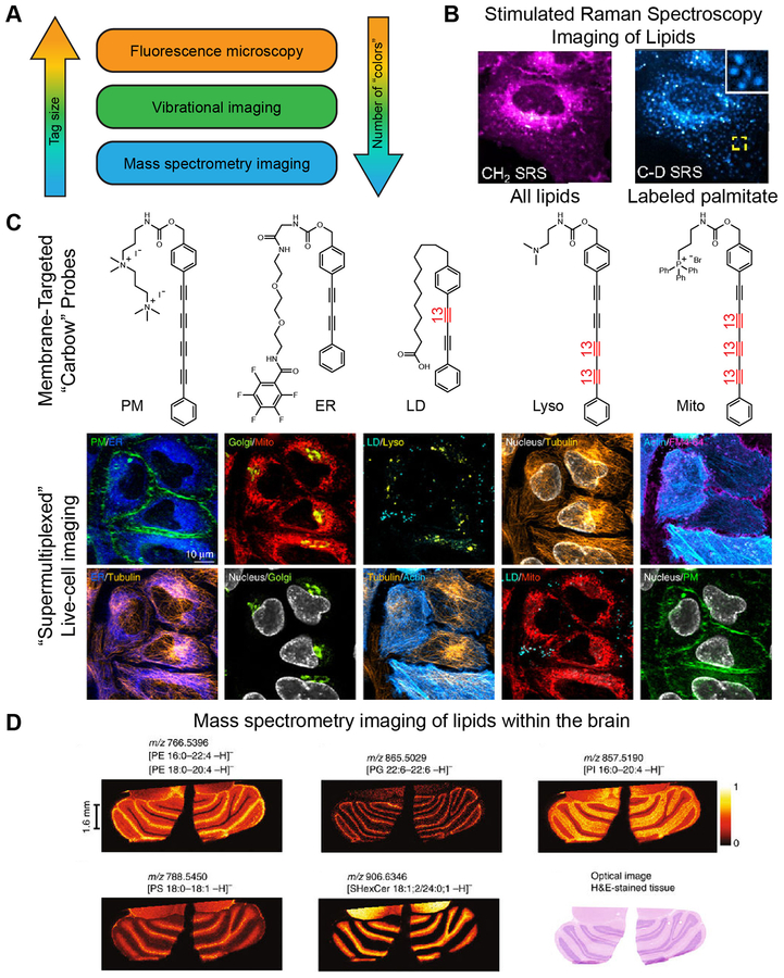
Biological lipids are a structurally diverse and historically vexing group of hydrophobic metabolites. Here, we review recent advances in chemical imaging techniques that reveal changes in lipid biosynthesis, metabolism, dynamics, and interactions. We highlight tools for tagging many lipid classes via metabolic incorporation of bioorthogonally functionalized precursors, detectable via click chemistry, and photocaged, photoswitchable, and photocrosslinkable variants of different lipids. Certain lipid probes can supplant traditional protein-based markers of organelle membranes in super-resolution microscopy, and emerging vibrational imaging methods, such as stimulated Raman spectroscopy (SRS), enable simultaneous imaging of more than a dozen different types of target molecule, including lipids. Collectively, these chemical imaging techniques will illuminate, in living color, previously hidden aspects of lipid biology.

Keywords: bioorthogonal,; click chemistry; imaging; lipids; metabolic labeling.


Figures

Figure 1.
Figure 1.. Biochemical and Photochemical Reactions Used in Conjunction with Lipid Probes.
(A) Metabolic labeling of phospholipase D (PLD) activity. PLD functions naturally as a hydrolase that produces phosphatidic acid from phosphatidylcholine, but the addition of low concentrations of exogenous primary alcohols, such as 3-azido-1-propanol, causes PLDs to catalyze a transphosphatidylation reaction to form a functionalized phosphatidyl alcohol. (B) Photochemical reactions that can be used to turn on inactive precursors, including those that can activate photoswitchable (i), photocaged (ii), and photocrosslinkable (iii) lipids. Abbreviation: DAG, diaglycerol.
Figure 2.
Figure 2.. Imaging Phospholipase D (PLD) Activity with Clickable Alcohols via Transphosphatidylation (IMPACT).
(A) (i) Schematic of IMPACT labeling. Functionalized alcohols are installed into reporter lipids by PLD transphosphatidylation. These unnatural reporter lipids can then be visualized with fluorescent click reagents. (ii) Examples of fluorescent click reagents. (B) IMPACT can be used to visualize intracellular organelle membranes bearing PLD activity. (C) IMPACT can also be used to visualize PLD activity at the single-cell level within a population by fluorescence microscopy (i) or flow cytometry (ii).
Figure 3.
Figure 3.. Targeting Lipids Enables Time-Lapse Super-Resolution Microscopy Visualization of Organelles.
(A) Lipids (tan) have a higher molar density than proteins (blue) within a given surface area of a membrane bilayer and, therefore, intracellular membranes can be densely tagged with lipid probes bearing reactive handles capable of rapid ligation with a flA) Lipids bearing the opposing click partner. (B) Clickable lipid-based probes for targeting specific organelles. (C) An appropriately functionalized click partner bearing a hydroxymethylene silicon rhodamine dye. This probe switches between nonfluorescent and fluorescent forms within a hydrophobic environment, enabling the extended imaging times relative to identically labeled proteins. Graphs show photobleaching time course (D) and fluorescence half-life (E) of lipid and protein probes targeted to the same organelle and bearing the same silicon rhodamine dye.
Figure 4.
Figure 4.. Less Is More: Non-Fluorescence Methods for ‘Supermultiplexed’ Imaging with Smaller Tags.
(A) Comparison of fluorescence imaging with vibrational imaging and mass spectrometry imaging in terms of tag size and number of species that can be detected simultaneously (‘colors’). (B) Stimulated Raman spectroscopy (SRS) imaging can be used to visualize lipids by exciting the vibrational modes of the CH2 groups within lipid tails (left) without any exogenous labels. Deuterated palmitate allows for pulse-chase imaging of the subset of lipids containing this particular lipid tail (right). (C) SRS imaging can be combined with fluorescence microscopy to enable ten-color imaging in live cells. Multiple vibrational ‘colors’ are created by extending the length of the dyes in polyyne chain and by introducing 13C–12C pairs into the chain, indicated in red. Such probes are dubbed ‘carbow’ (for carbon rainbow) and can target different organelles, including the plasma membrane (PM), endoplasmic reticulum (ER), lipid droplets (LD), lysosomes (lyso), and mitochondria (mito). (D) Mass spectrometry enables label-free imaging of lipids within tissues. Shown are images of phosphatidylethanolamine (PE), phosphatidylglycerol (PG), phosphatidylinositol (PI), phosphatidylserine (PS), and sulfatide (SHexCer) within a brain section taken from a mouse.

References

    1. Wymann MP and Schneiter R (2008) Lipid signalling in disease. Nat. Rev. Mol. Cell Biol 9, 162–176 - PubMed
    1. Brown HA et al. (2017) Targeting phospholipase D in cancer, infection and neurodegenerative disorders. Nat. Rev. Drug Discov 16, 351–367 - PMC - PubMed
    1. Afzelius BA and Maunsbach AB (2004) Biological ultrastructure research; the first 50 years. Tissue Cell 36, 83–94 - PubMed
    1. Cubitt AB et al. (1995) Understanding, improving and using green fluorescent proteins. Trends Biochem. Sci 20, 448–455 - PubMed
    1. Platre MP and Jaillais Y (2016) Guidelines for the use of protein domains in acidic phospholipid imaging. Methods Mol. Biol 1376, 175–194 - PMC - PubMed

Publication types

MeSH terms

LinkOut - more resources