Review
. 2018:143:1-30.
doi: 10.1016/bs.irn.2018.09.007. Epub 2018 Oct 23.

Neuroimaging Applications in Dystonia

Affiliations
Review

Neuroimaging Applications in Dystonia

Kristina Simonyan. Int Rev Neurobiol. 2018.

Abstract

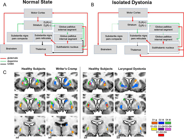
Dystonia is a neurological disorder characterized by involuntary, repetitive movements. Although the precise mechanisms of dystonia development remain unknown, the diversity of its clinical phenotypes is thought to be associated with multifactorial pathophysiology, which is linked not only to alterations of brain organization, but also environmental stressors and gene mutations. This chapter will present an overview of the pathophysiology of isolated dystonia through the lens of applications of major neuroimaging methodologies, with links to genetics and environmental factors that play a prominent role in symptom manifestation.

Keywords: Diffusion imaging; Functional magnetic resonance imaging; Isolated dystonia; Neuroreceptor mapping; Pathophysiology; Positron emission tomography; Voxel-based morphometry.


Figures

Fig. 1
Fig. 1
Schematic representation of multifactorial pathophysiology of isolated dystonia with major factors including abnormal brain structural, functional and neurochemical organization, underlying gene mutations, and environmental triggers, a combination of which leads to the manifestation of dystonic symptoms.
Fig. 2
Fig. 2
Microstructural gray matter abnormalities in isolated dystonia. (A) Anatomic likelihood estimation meta-analysis of common gray matter volumetric abnormalities across focal dystonias. (B) Gray matter volumetric increases in bilateral putamen in non-manifesting DYT1 mutation carriers and non-DYT1 dystonia patients compared to manifesting DYT1 mutation carriers and healthy subjects. The plot shows crossover interaction between genotype and phenotype factors. DYT M+, mutation-positive dystonia; DYT M−, mutation negative dystonia. (C) Cortical thickness changes across different phenotypes and putative genotypes (without confirmed gene mutation but based on a family history of dystonia) in patients with laryngeal dystonia. MTG, middle temporal gyrus; STG, superior temporal gyrus; SMA, supplementary motor area; R, right; L, left. Panel (A): Adapted from Zheng, Z., Pan, P., Wang, W., & Shang, H. (2012). Neural network of primary focal dystonia by an anatomic likelihood estimation meta-analysis of gray matter abnormalities. Journal of the Neurological Sciences, 316(1–2), 51–55. https://doi.org/10.1016/j.jns.2012.01.032. Panel (B): Adapted from Draganski, B., Schneider, S. A., Fiorio, M., Kloppel, S., Gambarin, M., Tinazzi, M., et al. (2009). Genotype-phenotype interactions in primary dystonias revealed by differential changes in brain structure. NeuroImage, 47(4), 1141–1147. doi:10.1016/j.neuro-image.2009.03.057. Panel (C): Adapted from Bianchi, S., Battistella, G., Huddlestone, H., Scharf, R., Fleysher, L., Rumbach, A. F., et al. (2017). Phenotype- and genotype-specific structural alterations in spasmodic dysphonia. Movement Disorders, 32(4), 560–568. https://doi.org/10.1002/mds.26920.
Fig. 3
Fig. 3
(A) Decreased gray matter volume in the facial portion of the precentral gyrus and reduced volume of the corticobulbar tract in healthy subjects (left) and patients with blepharospasm (right). (B) Specificity of white matter changes depending on the phenotype of dystonia: reduced white matter integrity along the corticobulbar tract in laryngeal dystonia, with subsequent postmortem neuropathology confirming axonal degeneration and demyelination within the corticobulbar tract in laryngeal dystonia. Put, putamen; ic, internal capsule. Panel (A): Adapted from Horovitz, S. G., Ford, A., Najee-Ullah, M. A., Ostuni, J. L., & Hallett, M. (2012). Anatomical correlates of blepharospasm. Translational Neurodegeneration, 1(1), 12. https://doi.org/10.1186/2047–9158-1–12. Panel (B): Adapted from Simonyan, K., Tovar-Moll, F., Ostuni, J., Hallett, M., Kalasinsky, V. F., Lewin-Smith, M. R., et al. (2008). Focal white matter changes in spasmodic dysphonia: A combined diffusion tensor imaging and neuropathological study. Brain, 131(Pt. 2), 447–459. https://doi.org/10.1093/brain/awm303.
Fig. 4
Fig. 4
Schematic representation of common abnormalities of brain metabolism and activation across different forms of dystonia and their relevance to the pathophysiology of this disorder. MGF, middle frontal gyrus; PreM, premotor cortex; M1/S1, primary sensorimotor cortex; IPC, inferior parietal cortex; SMA, supplementary motor area; BG, basal ganglia; Th, thalamus; Cbl, cerebellum.
Fig. 5
Fig. 5
(A) Large-scale network organization based on resting-state functional MRI group-averaged networks in healthy subjects and patients with different forms of focal isolated dystonia. This panel shows the regional distribution of neural communities based on the inter-regional coupling. Patients show disintegration of neural communities compared to healthy subjects. (B) Functional architecture of the neural network is influenced by the phenotype of dystonia; an example in abductor vs adductor forms of laryngeal dystonia. The panel depicts phenotype-specific abnormal distribution of connector and provincial hubs (regions of highest information transfer), which, as a result, establish a characteristic pattern of connectivity with non-hub regions across the entire brain. 6/17, area 6/17; 7A/7P, subdivisions of area 7; CbI-I/IV/Cbl-V/Cbl-VI, cerebellar lobules I/IV/V/VI; Cu/PCu, cuneus/precuneus; FG, fusiform gyrus; Ig1, part Ig1 of the insula; LG, lingual gyrus; MCC/PCC, middle/posterior cingulate cortex; SOG, superior orbital gyrus; Tp/Tpf/Ts/Tt, parietal/prefrontal/somatosensory/temporal subdivisions of the thalamus; hOC4v, ventral part of area hOC4; L, left; R, right. Panel (A): Adapted from Battistella, G., Termsarasab, P., Ramdhani, R. A., Fuertinger, S., & Simonyan, K. (2017). Isolated focal dystonia as a disorder of large-scale functional networks. Cerebral Cortex, 27(2), 1203–1215. doi:10.1093/cercor/bhv313. Panel (B): Adapted from Fuertinger, S., & Simonyan, K. (2017). Connectome-wide phenotypical and genotypical associations in focal dystonia. The Journal of Neuroscience, 37(31), 7438–7449. https://doi.org/10.1523/JNEUROSCI.0384-17.2017.
Fig. 6
Fig. 6
Schematic representation of neurotransmission during (A) normal state and (B) in isolated dystonia. Striatal dopaminergic input from the substantia nigra, pars compacta, is weakened; dopaminergic neurotransmission is enhanced via the direct pathway and diminished via the indirect pathway. (C) Topological distribution of striatal dopaminergic function in healthy subjects and patients with writer’s cramp and laryngeal dystonia. Within each patient and control group, a conjunction analysis was used to examine the overlap and distinct distribution between the significant clusters derived from three measures of dopaminergic function: D1 receptor binding; D2 receptor binding, and striatal phasic dopamine release during finger tapping (for the comparison with writer’s cramp) and sentence production (for the comparison with laryngeal dystonia). Healthy subjects have a great degree of overlap between all three measures as well as smaller regions of distinct receptor distribution. This topology is reversed in patients with dystonia, where the overlap is largely diminished. The legend provides the color scheme for overlapping as well as distinct regions of receptor activation and dopamine release. DA, dopamine.

References

    1. Albanese A, Bhatia K, Bressman SB, Delong MR, Fahn S, Fung VS, et al. (2013). Phenomenology and classification of dystonia: A consensus update. Movement Disorders, 28(7), 863–873. 10.1002/mds.25475. - DOI - PMC - PubMed
    1. Ali SO, Thomassen M, Schulz GM, Hosey LA, Varga M, Ludlow CL, et al. (2006). Alterations in CNS activity induced by botulinum toxin treatment in spasmodic dysphonia: An H215O PET study. Journal of Speech, Language, and Hearing Research: JSLHR, 49(5), 1127–1146. 10.1044/1092-4388(2006/081). - DOI - PubMed
    1. Argyelan M, Carbon M, Niethammer M, Ulug AM, Voss HU, Bressman SB, et al. (2009). Cerebellothalamocortical connectivity regulates penetrance in dystonia. The Journal of Neuroscience, 29(31), 9740–9747. 10.1523/JNEUROSCI.2300-09. - DOI - PMC - PubMed
    1. Asanuma K, Ma Y, Okulski J, Dhawan V, Chaly T, Carbon M, et al. (2005). Decreased striatal D2 receptor binding in non-manifesting carriers of the DYT1 dystonia mutation. Neurology, 64(2), 347–349. 10.1212/01.WNL.0000149764.34953.BF. - DOI - PubMed
    1. Asgeirsson H, Jakobsson F, Hjaltason H, Jonsdottir H, & Sveinbjornsdottir S (2006). Prevalence study of primary dystonia in Iceland. Movement Disorders, 21(3), 293–298. 10.1002/mds.20674. - DOI - PubMed

MeSH terms