

Abnormal Biomechanics at 6 Months Are Associated With Cartilage Degeneration at 3 Years After Anterior Cruciate Ligament Reconstruction
- PMID: 30473456
- PMCID: PMC6361700
- DOI: 10.1016/j.arthro.2018.07.033
Abnormal Biomechanics at 6 Months Are Associated With Cartilage Degeneration at 3 Years After Anterior Cruciate Ligament Reconstruction
Abstract
Purpose: To investigate the changes in landing biomechanics over a 3-year period and their correlation with cartilage degenerative changes in the medial tibiofemoral joint of the knee after anterior cruciate ligament reconstruction (ACLR) using magnetic resonance T1ρ mapping.
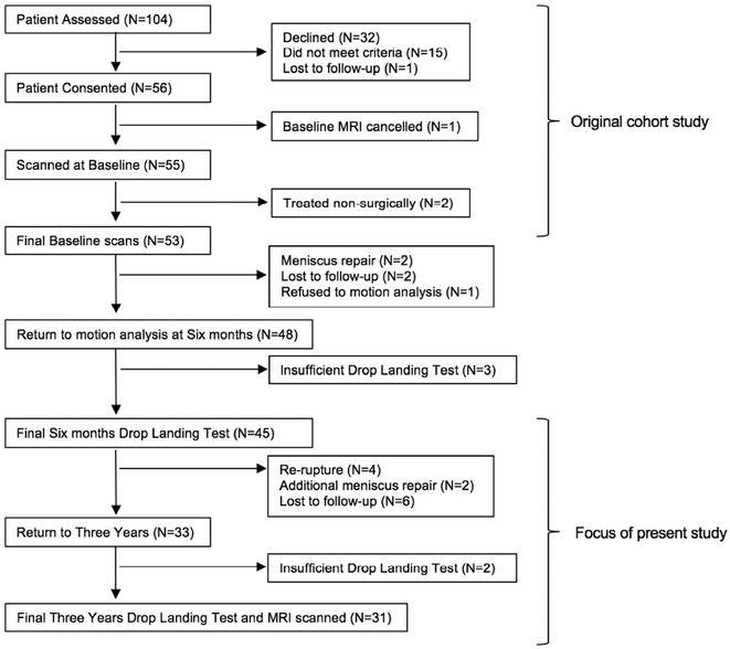
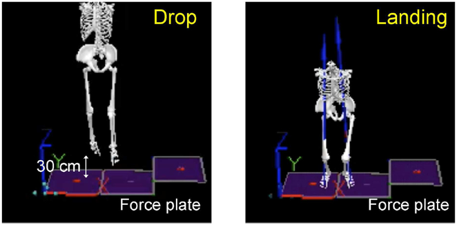
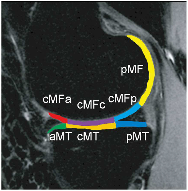
Methods: Thirty-one anterior cruciate ligament-injured patients underwent magnetic resonance imaging of the injured knee before ACLR and 3 years after ACLR, as well as biomechanical analysis of a drop-landing task at 6 months and 3 years after ACLR. Sixteen healthy individuals were recruited and underwent knee magnetic resonance imaging and biomechanical assessment during a drop-landing task. T1ρ cartilage relaxation times were calculated for the medial femur and tibia.
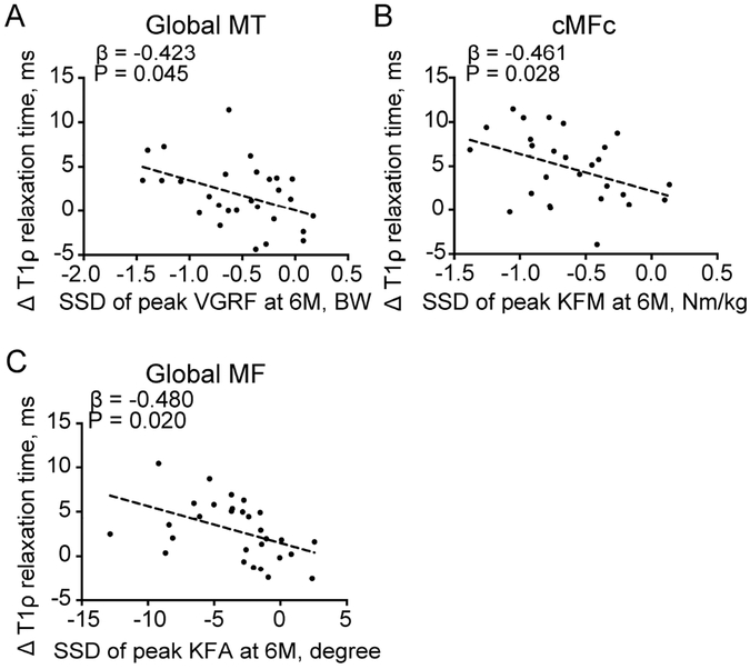
Results: ACLR patients exhibited increased peak vertical ground reaction force (VGRF), VGRF impulse, peak knee flexion moment (KFM), and KFM impulse from 6 months to 3 years (P < .001 for each). Although the ACLR knees showed significantly lower peak VGRF and KFM at 6 months (P < .001 for both) when compared with the controls, there were no significant differences at 3 years. At 3 years, ACLR patients showed higher T1ρ values over the medial femur (P < .001) and tibia (P = .012) when compared with their preoperative values and with healthy control values. Within the ACLR group, side-to-side differences in peak VGRF and sagittal knee biomechanics at 6 months were associated with increased T1ρ values from baseline to 3 years.
Conclusions: The results of this longitudinal study show that landing biomechanics are altered after ACLR but biomechanical abnormalities tend to recover at 3 years after ACLR. Differences in lower-extremity mechanics during a landing task at 6 months may be associated with cartilage degeneration at 3 years after anterior cruciate ligament injury and reconstruction.
Level of evidence: Level II, prospective trial.
Copyright © 2019 Arthroscopy Association of North America. Published by Elsevier Inc. All rights reserved.
Figures

Comment in
-
Early Abnormal Biomechanics May Lead to Increased Risk of Osteoarthritis and Poorer Outcomes After Anterior Cruciate Ligament Reconstruction.Arthroscopy. 2019 Apr;35(4):1012-1013. doi: 10.1016/j.arthro.2019.01.053. Arthroscopy. 2019. PMID: 30954092 No abstract available.
-
Author Reply to "Early Abnormal Biomechanics May Lead to Increased Risk of Osteoarthritis and Poorer Outcomes After Anterior Cruciate Ligament Reconstruction".Arthroscopy. 2019 Apr;35(4):1013. doi: 10.1016/j.arthro.2019.02.014. Arthroscopy. 2019. PMID: 30954093 No abstract available.
Similar articles
-
Gait Characteristics Associated With a Greater Increase in Medial Knee Cartilage T1ρ and T2 Relaxation Times in Patients Undergoing Anterior Cruciate Ligament Reconstruction.Am J Sports Med. 2017 Dec;45(14):3262-3271. doi: 10.1177/0363546517723007. Epub 2017 Sep 12. Am J Sports Med. 2017. PMID: 28898105 Clinical Trial.
-
Gait Mechanics and T1ρ MRI of Tibiofemoral Cartilage 6 Months after ACL Reconstruction.Med Sci Sports Exerc. 2019 Apr;51(4):630-639. doi: 10.1249/MSS.0000000000001834. Med Sci Sports Exerc. 2019. PMID: 30444797
-
Variations in Knee Kinematics After ACL Injury and After Reconstruction Are Correlated With Bone Shape Differences.Clin Orthop Relat Res. 2017 Oct;475(10):2427-2435. doi: 10.1007/s11999-017-5368-8. Clin Orthop Relat Res. 2017. PMID: 28451863 Free PMC article.
-
Progressive Changes in Walking Kinematics and Kinetics After Anterior Cruciate Ligament Injury and Reconstruction: A Review and Meta-Analysis.J Athl Train. 2017 Sep;52(9):847-860. doi: 10.4085/1062-6050-52.6.06. J Athl Train. 2017. PMID: 28985125 Free PMC article. Review.
-
Hip and Knee Kinematics and Kinetics During Landing Tasks After Anterior Cruciate Ligament Reconstruction: A Systematic Review and Meta-Analysis.J Athl Train. 2018 Feb;53(2):144-159. doi: 10.4085/1062-6050-334-16. Epub 2018 Jan 19. J Athl Train. 2018. PMID: 29350551 Free PMC article.
Cited by
-
Kinetic Asymmetry During a Repetitive Tuck Jump Task in Athletes with a History of Anterior Cruciate Ligament Reconstruction.Int J Sports Phys Ther. 2021 Oct 1;16(5):1278-1285. doi: 10.26603/001c.28088. eCollection 2021. Int J Sports Phys Ther. 2021. PMID: 34631248 Free PMC article.
-
Longitudinal Changes in Medial Meniscal Extrusion After ACL Injury and Reconstruction and Its Relationship With Cartilage Degeneration Assessed Using MRI-Based T1ρ and T2 Analysis.Am J Sports Med. 2025 Feb;53(2):350-359. doi: 10.1177/03635465241305734. Epub 2025 Jan 2. Am J Sports Med. 2025. PMID: 39743985 Free PMC article.
-
Gait Mechanics in Women of the ACL-SPORTS Randomized Control Trial: Interlimb Symmetry Improves Over Time Regardless of Treatment Group.J Orthop Res. 2019 Aug;37(8):1743-1753. doi: 10.1002/jor.24314. Epub 2019 May 20. J Orthop Res. 2019. PMID: 31042301 Free PMC article. Clinical Trial.
-
Association of Jump-Landing Biomechanics With Tibiofemoral Articular Cartilage Composition 12 Months After ACL Reconstruction.Orthop J Sports Med. 2021 Jul 21;9(7):23259671211016424. doi: 10.1177/23259671211016424. eCollection 2021 Jul. Orthop J Sports Med. 2021. PMID: 34368382 Free PMC article.
-
Short-Term Clinical Outcomes After Anterior Cruciate Ligament Reconstruction In Adolescents During The COVID-19 Pandemic.Int J Sports Phys Ther. 2022 Jun 1;17(4):585-592. doi: 10.26603/001c.35668. eCollection 2022. Int J Sports Phys Ther. 2022. PMID: 35693856 Free PMC article.
References
-
- Lohmander LS, Ostenberg A, Englund M, Roos H. High prevalence of knee osteoarthritis, pain, and functional limitations in female soccer players twelve years after anterior cruciate ligament injury. Arthritis Rheum. 2004;50:3145–3152. - PubMed
-
- Lohmander LS, Englund PM, Dahl LL, Roos EM. The long-term consequence of anterior cruciate ligament and meniscus injuries: osteoarthritis. Am J Sports Med. 2007;35:1756–1769. - PubMed
-
- Li RT, Lorenz S, Xu Y, Harner CD, Fu FH, Irrgang JJ. Predictors of radiographic knee osteoarthritis after anterior cruciate ligament reconstruction. Am J Sports Med. 2011;39:2595–2603. - PubMed
-
- Oiestad BE, Engebretsen L, Storheim K, Risberg MA. Knee osteoarthritis after anterior cruciate ligament injury: a systematic review. Am J Sports Med. 2009;37:1434–1443. - PubMed
Publication types
MeSH terms
Grants and funding
LinkOut - more resources
Full Text Sources
Medical
Research Materials
