

Endoscopic Goniosynechialysis for Acute Angle Closure Glaucoma Following Descemet's Stripping Automated Endothelial Keratoplasty
- PMID: 30473604
- PMCID: PMC6236120
- DOI: 10.5005/jp-journals-10008-1250
Endoscopic Goniosynechialysis for Acute Angle Closure Glaucoma Following Descemet's Stripping Automated Endothelial Keratoplasty
Abstract
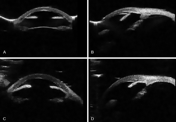
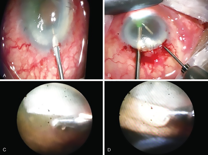
We describe a new modified technique to release the peripheral iridocorneal adhesions that formed after Descemet stripping automated endothelial keratoplasty. The usual technique of goniosynechialysis was modified and performed using endoscopic fiber-optic light and camera probe to aid visualization of the adherent iris tissue and carry out uneventful 270 degrees release of adhesions. The iris tissue was gently pulled away using micro forceps. The modified technique was conceptualized, as the view from the cornea was very poor due to recent lamellar surgery and corneal oedema secondary to poorly controlled intraocular pressure. The blocked trabecular meshwork system was successfully recanalized, which allowed an adequate control of intraocular pressure. The graft survived the insult and cornea gained complete clarity giving the patient the desired vision and improved quality of life. How to cite this article: Rana M, Shah S, Pandey P, Masood I. Endoscopic Goniosynechialysis for Acute Angle Closure Glaucoma Following Descemet's Stripping Automated Endothelial Keratoplasty. J Curr Glaucoma Pract 2018;12(2):90-93.
Keywords: Educational training; Glaucoma; Resident versus attending; Tube shunt surgery; Cohort study.
Conflict of interest statement
Source of support: Nil Conflict of interest: None NOTE: Video File: Surgical technique showing the endoscopic view of the occluded trabecular meshwork and use of micro-forceps to release the adhesions (available online only).
Figures
References
-
- Campbell DG, Vela A. Modern goniosynechialysis for the treatment of synechial angle-closure glaucoma. Ophthalmology. 1984 Sep;91(9):1052–1060. - PubMed
-
- Shingleton BJ, Chang MA, Bellows AR, Thomas JV. Surgical goniosynechialysisfor angle closure glaucoma. Ophthalmology. 1990 May; 97(5):551–556. - PubMed
-
- Tillet CW. Posterior lamellar keratoplasty. Am J Ophthalmol. 1956 Mar;41:530–533. - PubMed
-
- Lee JS, Desai NR, Schmidt GW, Jun AS, Schein OD, Stark WJ, Eghrari AO, Gottsch JD. Secondary angle closure caused by air migrating behind the pupil in Descemet stripping endothelial keratoplasty. Cornea. 2009 Jul; 28(6):652–656. - PubMed
-
- Ayyala RS. Penetrating keratoplasty and glaucoma. Surv Ophthalmol. 2000 Sep-Oct; 45(2):91–105. - PubMed
Publication types
LinkOut - more resources
Full Text Sources