Case Reports
. 2018 Oct 26:8:22.
doi: 10.1186/s13569-018-0109-7. eCollection 2018.

Descending thoracic aortic aneurysm revealing metastasis of a soft tissue fibrosarcoma: a case report and review of the literature

Affiliations
Case Reports

Descending thoracic aortic aneurysm revealing metastasis of a soft tissue fibrosarcoma: a case report and review of the literature

Clotilde Delerce et al. Clin Sarcoma Res. .

Abstract

Background: Review of the first documented case of aortic wall metastasis from a limb sarcoma.

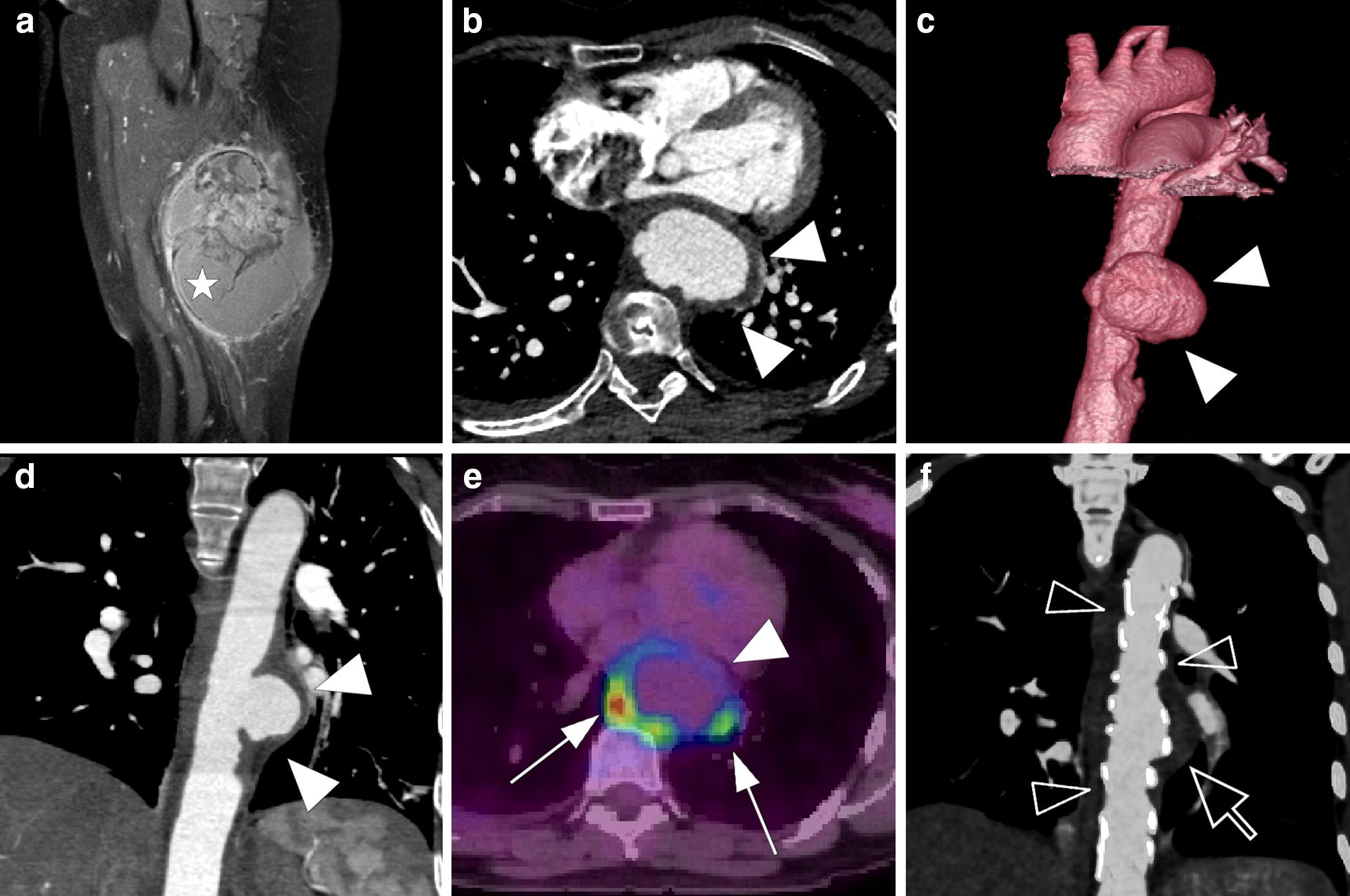
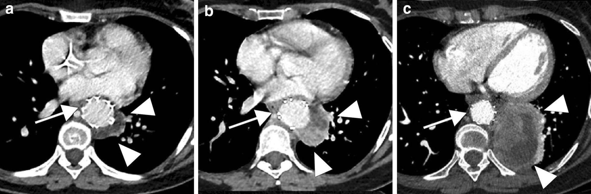
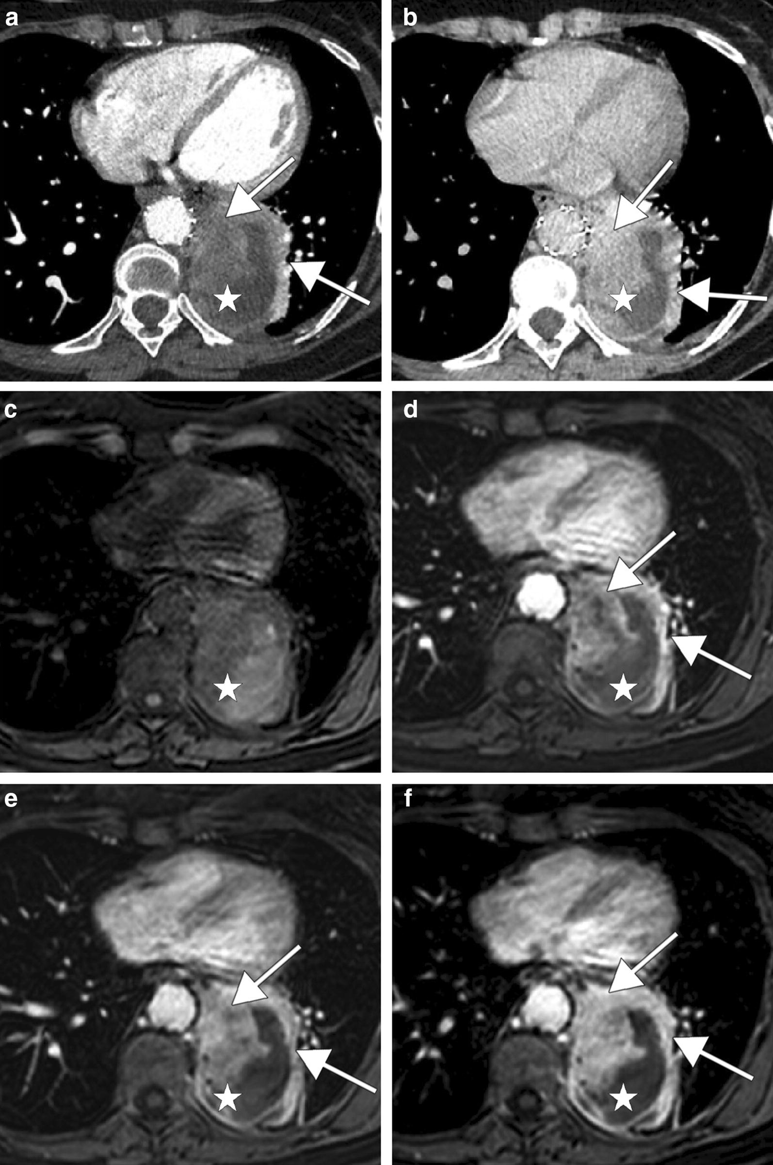
Case presentation: In a 56-year-old woman with a diagnosis of a high-grade limb fibrosarcoma, an aortic metastasis was revealed by a fast growing aneurysm of the descending thoracic aorta. This was managed with an endoprosthesis.

Conclusion: The presence of an aneurysm in a patient with a sarcoma with a high potential for metastasis and poor cardiovascular risk factors should alert physicians.

Keywords: Aortic aneurysm; Aortic metastasis; Soft tissue sarcoma.


Figures

Fig. 1
Fig. 1
a Coronal gadolinium-enhanced T1-weighted image with fat saturation shows a posterolateral muscular mass of the left thigh with both necrotic and enhanced portions (white star); Saccular aneurysm of the descendant thoracic aorta (white head arrows): arterial phase CT scan of the aneurysm with axial view (b), coronal reconstruction (c) and 3D reconstruction (d). e PET-CT image fusion shows FDG uptake (white arrows) around the aneurysm. f Coronal maximum intensity projection (MIP) reconstruction of postoperative CT shows the endoprosthesis (transparent head arrows) and the excluded aneurysmal cavity (transparent arrow)
Fig. 2
Fig. 2
Follow-up CT scans shows rapid progression of the lateral aortic metastasis (white head arrows) near the endoprosthesis (white arrows). a Initial postoperative CT scan shows a non-enhancing lateral aortic lesion which is difficult to differentiate from an excluded aneurysmal cavity. b One month later CT scan shows an important tumor’s size increase with a minimal peripheral enhancement (b). c Six months later, CT scan shows an important tumor progression
Fig. 3
Fig. 3
CT scan and MRI performed 6 months after endoprosthesis placement show a large lateral aortic tumor mass (white stars) with a progressive contrast enhancement (white arrows). CT scan shows a minimal contrast enhancement on portal phase (a) and an important one on delayed phase (b). Axial non-contrast (c) and gadolinium-enhanced T1-weighted images with fat saturation on arterial phase (d), portal phase (e) and delayed phase (f) show also a progressive tumor enhancement

References

    1. Hallahan DE, Vogelzang NJ, Borow KM, Bostwick DG, Simon MA. Cardiac metastases from soft-tissue sarcomas. J Clin Oncol. 1986;11:1662–1669. doi: 10.1200/JCO.1986.4.11.1662. - DOI - PubMed
    1. Fletcher CDM, Unni KK, Mertens F. Pathology and genetics of tumours of soft tissue and bone. Lyon: IARC press; 2002.
    1. Gerrand CH, Billingham LJ, Woll PJ, Grimer RJ. Follow up after primary treatment of soft tissue sarcoma: a survey of current practice in the United Kingdom. Sarcoma. 2007;2007:602–612. doi: 10.1155/2007/34128. - DOI - PMC - PubMed
    1. Dimitrakopoulou-Strauss A, Strauss LG, Schwarzbach M, Burger C, Heichel T, Willeke F, Mechtersheimer G, Lehnert T. Dynamic PET 18F-FDG studies in patients with primary and recurrent soft-tissue sarcomas: impact on diagnosis and correlation. J Nucl Med. 2001;42:713–720. - PubMed
    1. ESMO (European Sarcoma Network Working Group) Soft tissue and visceral sarcomas: ESMO-EURACAN clinical practice guidelines for diagnosis, treatment and follow-up. Ann Oncol. 2018;29:51–67. doi: 10.1093/annonc/mdy096. - DOI - PubMed

Publication types

LinkOut - more resources