Review
. 2018 Nov 9:7:F1000 Faculty Rev-1771.
doi: 10.12688/f1000research.16096.1. eCollection 2018.

Recent advances in imaging and understanding interstitial cystitis

Affiliations
Review

Recent advances in imaging and understanding interstitial cystitis

Pradeep Tyagi et al. F1000Res. .

Abstract

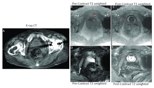
Interstitial cystitis/bladder pain syndrome (IC/BPS) is a debilitating condition associated with intense pelvic pain and bladder storage symptoms. Since diagnosis is difficult, prevalence estimates vary with the methodology used. There is also a lack of proven imaging tools and biomarkers to assist in differentiation of IC/BPS from other urinary disorders (overactive bladder, vulvodynia, endometriosis, and prostatitis). Current uncertainty regarding the etiology and pathology of IC/BPS ultimately impacts its timely and successful treatment, as well as hampers future drug development. This review will cover recent developments in imaging methods, such as magnetic resonance imaging, that advance the understanding of IC/BPS and guide drug development.

Keywords: Interstitial cystitis; MRI; bladder wall; fibrosis; imaging; radiation.


Conflict of interest statement

Competing interests: Joseph Janicki is an employee of Lipella Pharmaceuticals, whereas Jonathan Kaufman and Michael Chancellor are inventors of novel contrast mixture. Pradeep Tyagi, Chan-Hong Moon, Naoki Yoshimura and Christopher Chermansky declare that they have no competing interests.No competing interests were disclosed.No competing interests were disclosed.

Figures

Figure 1.
Figure 1.. Magnetic resonance imaging (MRI) is superior to computed tomography (CT) in bladder wall segmentation.
Contrast-enhanced T 1-weighted MRI (Panel C) is superior to CT (Panel A) and unenhanced T 1-weighted MRI (Panel B) for resolving the thickened bladder wall of the same female ulcerative interstitial cystitis/bladder pain syndrome (IC/BPS) patient as indicated by a red line drawn across the bladder wall. Representative T 1-weighted fast low angle shot (FLASH) images acquired at the flip angle of 14° in orthogonal orientation before (Panel B) and after novel contrast mixture (NCM) instillation (Panel C) demonstrate that NCM-enhanced MRI can non-invasively segment the bladder wall into a middle layer of bright signal intensity sandwiched between inner and outer layers of lower signal intensity. Fibrotic changes reported in the pathological results of the biopsy from the IC/BPS patient are visible as the bright middle layer from the diffusion of gadobutrol from NCM instilled in the lumen (hypointense circular region in Panel C). Fast acquisition of a single slice (5 mm thickness) over a single breath hold of approximately 17 seconds with imaging parameters of repetition time (TR)/echo time (TE) of 5.5/2 milliseconds, field of view of 180×180 mm 2, matrix of 256×256 and 10 averaging achieved in-plane resolution of 0.35 mm (after 2x interpolation) with minimal influence of motion and chemical shift artifacts (Panel B and C). Akin to CT (Panel A), T 1-weighted MRI (Panel B) with similar imaging parameters as in Panel C could not resolve the bladder wall from the lumen in the absence of NCM owing to the relatively long intrinsic T 1 relaxation time of >1 second for the bladder wall and of urine in the lumen. Although the bladder wall is demarcated clearly in pre-contrast T 2-weighted image (Panel D and E) acquired at the TR/TE of 6.9/3.1 milliseconds, there is underestimation of bladder wall thickness, especially of the inner layer from 3.62 mm in panel C to 1.21 mm in Panel E. Instillation of gadobutrol in the absence of ferumoxytol enhances the T 1 contrast of urine as well as the bladder wall, which precludes it from affording improved image contrast. This figure is an original image taken in our clinic for this publication.
Figure 2.
Figure 2.. Near infra-red imaging.
Representative images for the rat pelvic area in visible light and in near infra-red (NIR) light after instillation of liposomes containing a trace amount of NIR dye. Deep tissue imaging of the bladder wall is possible, as light emitted from the bladder lumen in the NIR band encounters low background auto-fluorescence and minimal absorption by tissue components. This figure is an original image taken in our clinic for this publication.
Figure 3.
Figure 3.. Contrast-enhanced magnetic resonance imaging (MRI) relies on the diffusion of gadolinium into the bladder wall.
Novel contrast mixture-enhanced MRI relies on the differences in the contrast mechanisms and molecular weight of two US Food and Drug Administration-approved agents (gadobutrol diluted 1:250 and ferumoxytol diluted 1:104) for increasing the contrast resolution of the bladder wall (Panel A). Gadobutrol, a transparent liquid, is a gadolinium-based contrast agent (GBCA) with a molecular weight of 604.71 Da that reaches the extracellular space in the lamina propria to shorten T 1 (positive contrast) or produce higher signal intensity (Panel A and B). Meanwhile, the large molecular weight of 750 kDa for ferumoxytol (vial with brown-colored liquid) restricts its bladder wall diffusion, and it produces a localized increase in proton dephasing, which decreases the signal intensity (negative contrast) in T 1-weighted images. Instillation of novel contrast mixture 50 mL can non-invasively segment the inner layer (urothelium) and the outer layer of adventitia (dark signal) from the bright signal in the middle layer composed of the lamina propria and detrusor smooth muscle (Panel C). Bladder wall histology in Panel C is shown for illustration and does not represent the pathological characterization of the interstitial cystitis/bladder pain syndrome (IC/BPS) patient shown in Panel B. This figure is an original image taken in our clinic for this publication.
Figure 4.
Figure 4.. Quantitative measurement of gadolinium diffusion.
T 1-weighted fast low angle shot (FLASH) images with constant repetition time (TR) of 5.5 milliseconds (ms) at flip angle (FA) of 6° (Panel A, C, and E) and 14° (Panel B, D, and E) demonstrate that gadobutrol-mediated signal enhancement (visible in the right tube containing gadobutrol 4 mM alone) is suppressed by the presence of ferumoxytol (5 mM) in the novel contrast mixture (NCM) tube, as the gadobutrol concentration of 4 mM is the same in both tubes (Panel A and B). T 1-weighted FLASH images demonstrates that greater separation of gadobutrol into the bladder wall away from the NCM instilled in the bladder lumen occurs in ulcerative IC/BPS patients (Panel E and F) than in non-ulcerative interstitial cystitis/bladder pain syndrome (IC/BPS) patient (Panel C and D), which is evident from the dramatic increase in signal intensity in Panel F relative to Panel D at FA of 14°. Catheter used for instillation is shown by C in panel CH. Constant TR of 5.5 ms at different FAs achieves the stable steady-state conditions necessary for the differences in signal intensity of the same slice to become a function of T 1 relaxation time as indicated by the color panel in Panel H. Greater shortening of T 1 relaxation time (blue color) in ulcerative IC/BPS patients is consistent with higher diffusion of gadobutrol into the expanded extracellular matrix of the thickened bladder wall of IC/BPS patients. This figure is an original image taken in our clinic for this publication.

References

    1. Wennevik GE, Meijlink JM, Hanno P, et al. : The Role of Glomerulations in Bladder Pain Syndrome: A Review. J Urol. 2016;195(1):19–25. 10.1016/j.juro.2015.06.112 - DOI - PubMed
    1. Doggweiler R, Whitmore KE, Meijlink JM, et al. : A standard for terminology in chronic pelvic pain syndromes: A report from the chronic pelvic pain working group of the international continence society. Neurourol Urodyn. 2017;36(4):984–1008. 10.1002/nau.23072 - DOI - PubMed
    1. Mullins C, Bavendam T, Kirkali Z, et al. : Novel research approaches for interstitial cystitis/bladder pain syndrome: thinking beyond the bladder. Transl Androl Urol. 2015;4(5):524–33. 10.3978/j.issn.2223-4683.2015.08.01 - DOI - PMC - PubMed
    1. Suskind AM, Berry SH, Ewing BA, et al. : The prevalence and overlap of interstitial cystitis/bladder pain syndrome and chronic prostatitis/chronic pelvic pain syndrome in men: results of the RAND Interstitial Cystitis Epidemiology male study. J Urol. 2013;189(1):141–5. 10.1016/j.juro.2012.08.088 - DOI - PMC - PubMed
    1. Tung A, Hepp Z, Bansal A, et al. : Characterizing Health Care Utilization, Direct Costs, and Comorbidities Associated with Interstitial Cystitis: A Retrospective Claims Analysis. J Manag Care Spec Pharm. 2017;23(4):474–82. 10.18553/jmcp.2017.23.4.474 - DOI - PMC - PubMed
    2. F1000 Recommendation

Publication types