. 2018 Oct 8:7:1617.
doi: 10.12688/f1000research.16473.2. eCollection 2018.

Using zebrafish larval models to study brain injury, locomotor and neuroinflammatory outcomes following intracerebral haemorrhage

Affiliations

Using zebrafish larval models to study brain injury, locomotor and neuroinflammatory outcomes following intracerebral haemorrhage

Siobhan Crilly et al. F1000Res. .

Abstract

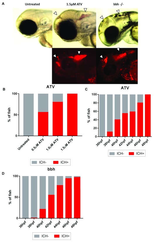
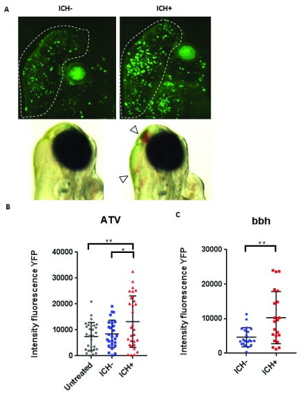
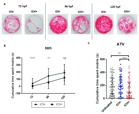
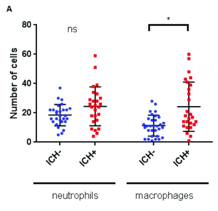
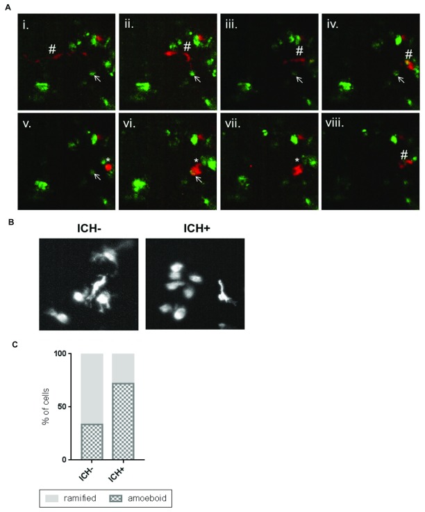
Intracerebral haemorrhage (ICH) is a devastating condition with limited treatment options, and current understanding of pathophysiology is incomplete. Spontaneous cerebral bleeding is a characteristic of the human condition that has proven difficult to recapitulate in existing pre-clinical rodent models. Zebrafish larvae are frequently used as vertebrate disease models and are associated with several advantages, including high fecundity, optical translucency and non-protected status prior to 5 days post-fertilisation. Furthermore, other groups have shown that zebrafish larvae can exhibit spontaneous ICH. The aim of this study was to investigate whether such models can be utilised to study the pathological consequences of bleeding in the brain, in the context of pre-clinical ICH research. Here, we compared existing genetic (bubblehead) and chemically inducible (atorvastatin) zebrafish larval models of spontaneous ICH and studied the subsequent disease processes. Through live, non-invasive imaging of transgenic fluorescent reporter lines and behavioural assessment we quantified brain injury, locomotor function and neuroinflammation following ICH. We show that ICH in both zebrafish larval models is comparable in timing, frequency and location. ICH results in increased brain cell death and a persistent locomotor deficit. Additionally, in haemorrhaged larvae we observed a significant increase in macrophage recruitment to the site of injury. Live in vivo imaging allowed us to track active macrophage-based phagocytosis of dying brain cells 24 hours after haemorrhage. Morphological analyses and quantification indicated that an increase in overall macrophage activation occurs in the haemorrhaged brain. Our study shows that in zebrafish larvae, bleeding in the brain induces quantifiable phenotypic outcomes that mimic key features of human ICH. We hope that this methodology will enable the pre-clinical ICH community to adopt the zebrafish larval model as an alternative to rodents, supporting future high throughput drug screening and as a complementary approach to elucidating crucial mechanisms associated with ICH pathophysiology.

Keywords: Intracerebral haemorrhage; animal models; neuroinflammation; pre-clinical; zebrafish.


Conflict of interest statement

No competing interests were disclosed.

Figures

Figure 1.
Figure 1.. Atorvastatin (ATV)-induced and bubblehead (bbh) mutant intracerebral haemorrhage (ICH) show comparable models of brain-specific bleeding.
( A) Brain-specific bleeds were observed in both ATV and bbh models maintained on the transgenic gata1:DsRed reporter background using both brightfield (top panels) and fluorescence (bottom panels) microscopy. Bleeds formed in both forebrain and mid-hindbrain regions, as described by others ( Eisa-Beygi et al., 2013) (arrows denotes haemorrhages). Bleeds in bbh mutants are frequently associated with severe cranial oedema making blood pooling more disperse. Original magnification, x20. ( B) ATV treatment causes ICH to occur in a dose-dependent manner. ( C) Timeline of ICH development in ATV-treated and untreated embryos and ( D) bbh homozygotes.
Figure 2.
Figure 2.. Intracerebral haemorrhage (ICH) in zebrafish larvae results in a quantifiable brain injury.
( A) Representative images of the brain injury phenotype in ICH+ larvae (right panels), in comparison to ICH- siblings (left panels), at 72 hpf. Bright-field images (bottom panels) demonstrate the presence of brain bleeds (arrows) in ICH+ larvae. Fluorescent microscopy was performed to visualise cell death in the ubiq:secAnnexinV-mVenus reporter line (top panels). Clusters of dying cells were observed in peri-haematomal regions. Images were cropped to brain only regions and analysed for total green fluorescence intensity in round particles bigger than 30 pixels in diameter (white line). ( B) Quantification of fluorescent signal in the brains of untreated, ICH- and ICH+ larvae obtained through the ATV model (n=12 per group; 3 independent replicates) at 72 hpf. Significant differences were observed when comparing ICH+ with untreated (**p=0.004) and with ICH- (*p=0.03) siblings. ( C) Quantification of fluorescent signal as a read out for annexinV binding in the brains of ICH- and ICH+ larvae obtained through the bubblehead (bbh) model (n=12 per group; 2 independent replicates) at 72 hpf. A significant difference in mVenus fluorescence was observed between ICH+ and ICH- age-matched siblings (**p=0.002). Original magnification, x20.
Figure 3.
Figure 3.. ICH-induced brain injury results in a quantifiable locomotor deficit in bubblehead (bbh) zebrafish larvae.
( A) Representative examples of the swimming tracks in ICH- and ICH+ larvae at 72, 96 and 120 hpf. ( B) ICH+ larvae exhibited a significant decrease in the cumulative time spent mobile during the 10 minute recording period at both 72 and 96 hpf. Significance was lost at the 120 hpf time point potentially alluding to recovery from brain injury (n=24 larvae per group; 3 independent replicates; ****p=0.00006; **p=0.003 ns p=0.08) ( C) Quantification of cumulative time spent moving in untreated and ATV-treated ICH- and ICH+ larvae at 120 hpf. ICH+ larvae exhibited a significant decrease in the cumulative time spent mobile during the 10 minute recording period. Three technical replicates (n=24 larvae per group) were used to calculate s.d from the mean (***p=0.00004, **p=0.0003).
Figure 4.
Figure 4.. Intracerebral haemorrhage (ICH) initiates an innate cellular immune response in the zebrafish larval brain.
Numbers of leukocytes quantified within the brains of mpo:GFP; mpeg1:dsRed double transgenic larvae (n=8 per group; 2 independent replicates) at 72 hpf reveals a significant increase in macrophages (*p=0.01), but not neutrophils (p=0.5), in response to ICH.
Figure 5.
Figure 5.. Activated macrophage cells show a phagocytic response to the brain lesion.
( A) Representative time-lapse stills (from Supplementary Video 1) showing a ramified patrolling macrophage migrating towards an annexinV positive cell (i - vi). The macrophage acquired an amoeboid morphology (v) before phagocytosing the annexinV-positive cell (vi, vii). After phagocytosis the macrophage resumes a ramified morphology and migrates away and the annexinV-positive cell can no longer be seen (viii). Ramified macrophage (#), annexinV positive cell (arrow), amoeboid macrophage (*). ( B) Representative images of mpeg1-positive cells in the intracerebral haemorrhage (ICH)- and ICH+ larval brain exhibiting amoeboid and ramified morphologies. ( C) An increased proportion of amoeboid (phagocytic) and decreased proportion of ramified (inactive) macrophages was observed in ICH+ brains in comparison to ICH- siblings.
Figure 6.
Figure 6.. Graphic of experimental timeline to characterise brain injury, locomotor and neuroinflammatory outcomes.
ICH, intracerebral haemorrhage; bbh, bubblehead.

References

    1. Abbott NJ: Inflammatory mediators and modulation of blood-brain barrier permeability. Cell Mol Neurobiol. 2000;20(2):131–147. 10.1023/A:1007074420772 - DOI - PMC - PubMed
    1. Alharbi BM, Tso MK, Macdonald RL: Animal models of spontaneous intracerebral hemorrhage. Neurol Res. 2016;38(5):448–455. 10.1080/01616412.2016.1144671 - DOI - PubMed
    1. An SJ, Kim TJ, Yoon BW: Epidemiology, Risk Factors, and Clinical Features of Intracerebral Hemorrhage: An Update. J Stroke. 2017;19(1):3–10. 10.5853/jos.2016.00864 - DOI - PMC - PubMed
    1. Andaluz N, Zuccarello M, Wagner KR: Experimental animal models of intracerebral hemorrhage. Neurosurg Clin N Am. 2002;13(3):385–393. 10.1016/S1042-3680(02)00006-2 - DOI - PubMed
    1. ASPA 1986 amendments 2012: Animals (Scientific Procedures) Act 1986. Reference Source

Publication types

LinkOut - more resources