. 2018 Nov 6;138(19):2130-2144.
doi: 10.1161/CIRCULATIONAHA.118.035231.

Rapid Self-Assembly of Bioengineered Cardiovascular Bypass Grafts From Scaffold-Stabilized, Tubular Bilevel Cell Sheets

Affiliations

Rapid Self-Assembly of Bioengineered Cardiovascular Bypass Grafts From Scaffold-Stabilized, Tubular Bilevel Cell Sheets

Daniel von Bornstädt et al. Circulation. .

Abstract

Background: Cardiovascular bypass grafting is an essential treatment for complex cases of atherosclerotic disease. Because the availability of autologous arterial and venous conduits is patient-limited, self-assembled cell-only grafts have been developed to serve as functional conduits with off-the-shelf availability. The unacceptably long production time required to generate these conduits, however, currently limits their clinical utility. Here, we introduce a novel technique to significantly accelerate the production process of self-assembled engineered vascular conduits.

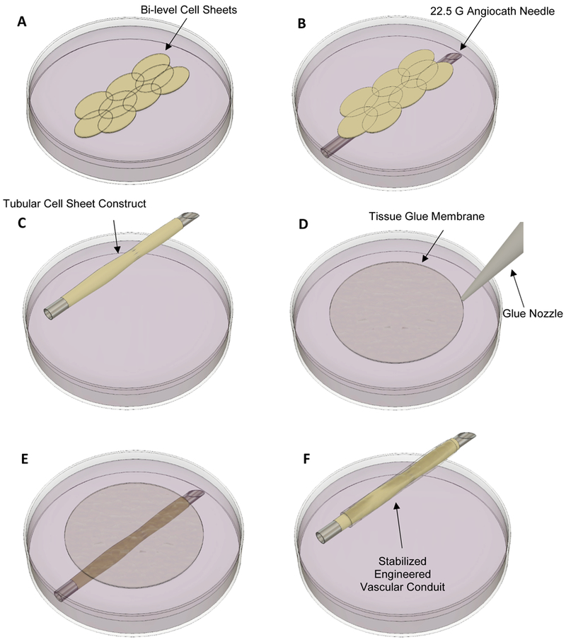
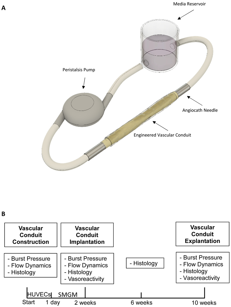
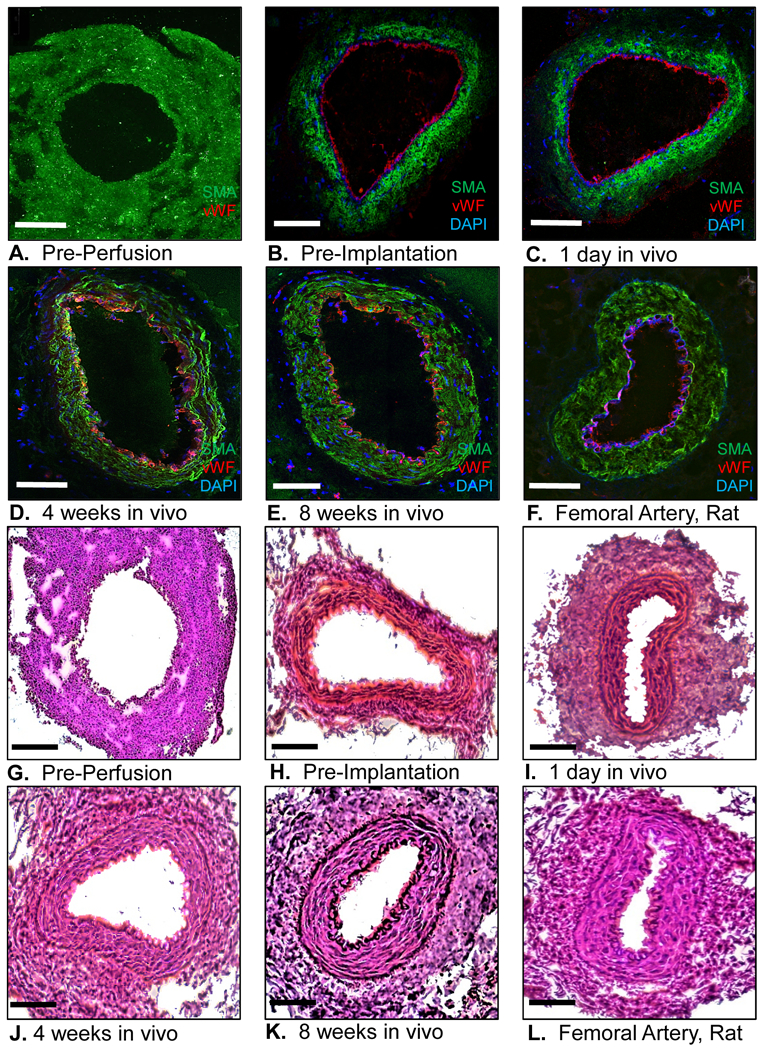
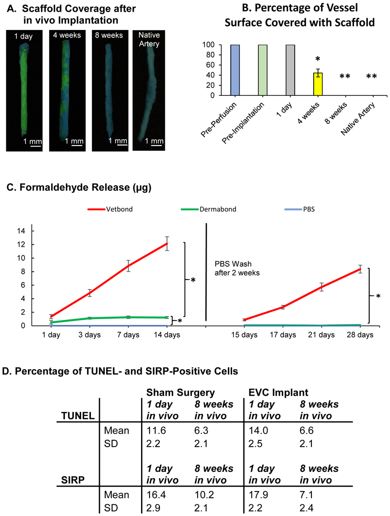
Methods: Human aortic smooth muscle cells and skin fibroblasts were used to construct bilevel cell sheets. Cell sheets were wrapped around a 22.5-gauge Angiocath needle to form tubular vessel constructs. A thin, flexible membrane of clinically approved biodegradable tissue glue (Dermabond Advanced) served as a temporary, external scaffold, allowing immediate perfusion and endothelialization of the vessel construct in a bioreactor. Subsequently, the matured vascular conduits were used as femoral artery interposition grafts in rats (n=20). Burst pressure, vasoreactivity, flow dynamics, perfusion, graft patency, and histological structure were assessed.

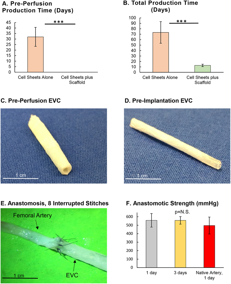
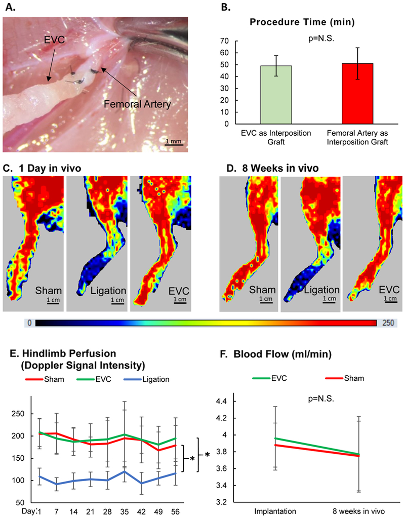
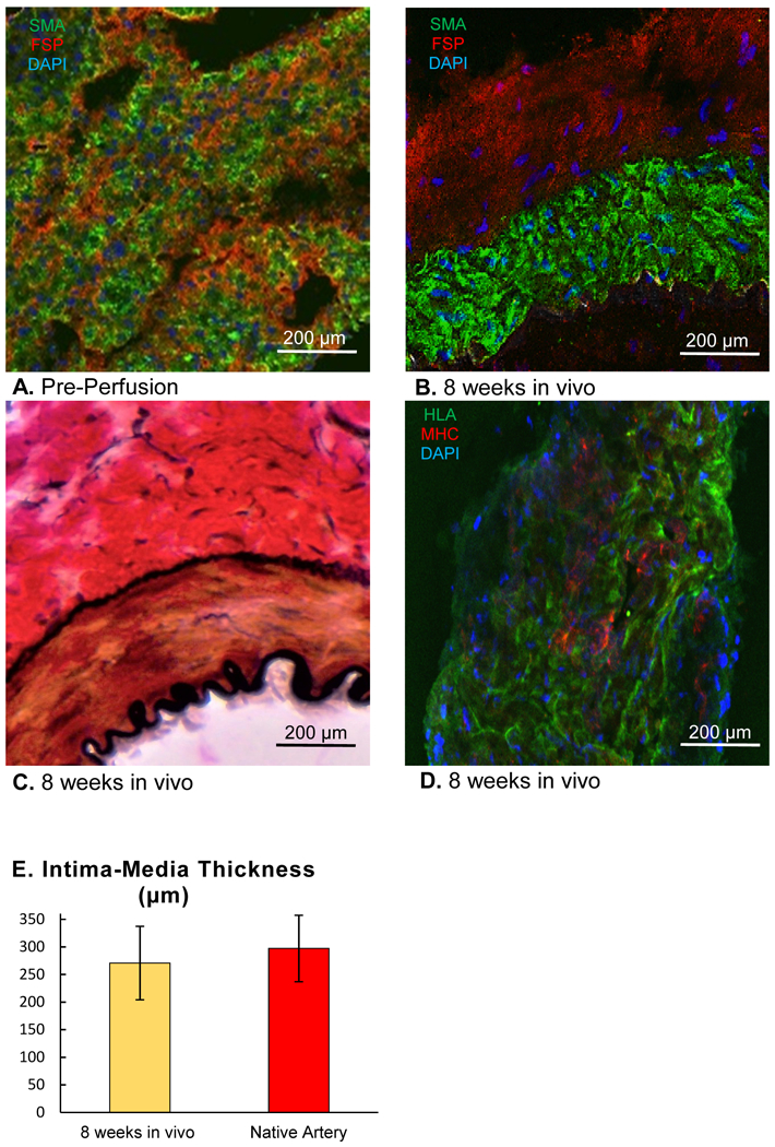
Results: Compared with engineered vascular conduits formed without external stabilization, glue membrane-stabilized conduits reached maturity in the bioreactor in one-fifth the time. After only 2 weeks of perfusion, the matured conduits exhibited flow dynamics similar to that of control arteries, as well as physiological responses to vasoconstricting and vasodilating drugs. The matured conduits had burst pressures exceeding 500 mm Hg and had sufficient mechanical stability for surgical anastomoses. The patency rate of implanted conduits at 8 weeks was 100%, with flow rate and hind-limb perfusion similar to those of sham controls. Grafts explanted after 8 weeks showed a histological structure resembling that of typical arteries, including intima, media, adventitia, and internal and external elastic membrane layers.

Conclusions: Our technique reduces the production time of self-assembled, cell sheet-derived engineered vascular conduits to 2 weeks, thereby permitting their use as bypass grafts within the clinical time window for elective cardiovascular surgery. Furthermore, our method uses only clinically approved materials and can be adapted to various cell sources, simplifying the path toward future clinical translation.

Keywords: bioengineering; bioreactors; coronary artery bypass; tissue adhesive; vascular grafting.


Figures

Figure 1.
Figure 1.
Engineering process of the vascular conduit. (A) Bi-level cell sheets are transferred into a 10 cm petri dish filled with smooth muscle growth media. (B) Cell Sheets are placed on top of a 22.5 G angiocath needle. (C) The cell sheets are wrapped around the needle to form a tube-like construct. (D) Tissue glue is added to a separate 10 cm petri dish and forms a flexible glue membrane on the surface of the media. (E) The glue membranes are wrapped around the cell sheet construct. (F) The final construct consists of an inner layer of cell sheets stabilized by a flexible tissue glue membrane.
Figure 2.
Figure 2.
(A) The engineered vascular conduit (EVC) is connected to a perfusion system consisting of a peristalsis pump, a media reservoir, two 22.5 G needles, and connection tubing. (B) The experimental timeline is illustrated. Immediately after construction of the EVC, burst pressure, flow dynamics, and histology were assessed. The EVC was perfused with human umbilical vein endothelial cells (HUVECs) for 1 day and subsequently with smooth muscle growth media (SMGM) for 13 days. Upon completion of the in vitro perfusion/maturation period, burst pressure, flow dynamics, histology, and vasoreactivity were assessed. The mature EVC was implanted as a femoral artery interposition graft in a rat model. Four weeks later, EVC histology was again analyzed. Finally, 8 weeks after implantation, the EVC was explanted and burst pressure, flow dynamics, histology, and vasoreactivity were analyzed.
Figure 3.
Figure 3.
(A) Time elapsed from construction of the engineered vascular conduit (EVC) until ready for in vitro perfusion, n=4 for both groups (Student’s t-test). (B) Time elapsed from construction of the EVC until ready for in vivo surgical implantation, n=4 for both groups (Student’s t-test). (C) Gross anatomy of the EVC immediately after construction and before in vitro perfusion. (D) Gross anatomy of the EVC after 14 days of in vitro perfusion and before in vivo implantation. (E) Proximal anastomosis between the EVC and the femoral artery of a nude rat, using 8 interrupted 10–0 nylon stitches. (F) Anastomotic strength of the EVC-femoral artery anastomosis at 1 and 3 days after in vivo implantation, compared to a femoral artery-femoral artery anastomosis using the contralateral femoral artery of the nude rat as an interposition graft, n=5 for all groups (one-way ANOVA with Tukey’s Test). *** indicates p<0.001.
Figure 4.
Figure 4.
(A) Pressure drop over a 1.5 cm engineered vascular conduit (EVC) segment at various time points after construction, n=5 for all samples. (B) Measurements of outer vessel diameter (n=5 for all samples) after 3 minutes wash in phosphate buffered salines (PBS), epinephrine (vasoconstrictor, 1 03BCM) or penicillamine (vasodilator, 1 μM) for EVCs pre-implantation, after 8 weeks in vivo, and for a native femoral artery from a nude rat. (C) Burst pressures for EVCs at various time points after construction, compared to a construct composed of glue membranes only, as well as a rat femoral artery. EVCs with burst pressures below 500 mmHg after 14 days of in vitro perfusion were excluded (i.e. not included in the pre-implantation sample), n=5 for all samples, p<0.01 for Pre-Implantation vs. all other samples, and p<0.01 for Scaffold only or Pre-Perfusion vs. 8 weeks in vivo or native femoral artery. N.S. = not statistically significant. (D) Percentage of EVCs that met the pre-defined criteria for implantation (burst pressure of 500 mmHg) after 14 days of in vitro perfusion vs. 14 days without perfusion, n=15 for both groups. (E) Young’s modulus from uniaxial tensile testing for EVCs immediately prior to implantation and after 8 weeks in vivo, compared to a native femoral artery. p<0.05 for Pre-Perfusion vs. both other groups, n=4 for all samples. One-way ANOVA followed by Tukey’s Test (A, B, C, E), Fisher’s exact test [95% confidence interval] (D). * indicates p<0.05. ** indicates p<0.01.
Figure 5.
Figure 5.
Engineered vascular conduits (EVCs) were tested as interposition grafts after femoral artery excision in rats. On the opposite side, animals were assigned to 2 groups: excision of the femoral artery followed by ligation of the proximal and distal ends without graft replacement, or sham surgery. (A) EVCs were anastomosed to the femoral artery as an interposition graft in end-to-end fashion (10–0 nylon suture, 5 stitches). (B) Time required to complete a femoral artery bypass grafting procedure utilizing the EVC or contralateral femoral artery as an interposition graft, n=5 for both groups (Student’s t-test). N.S. = not statistically significant. (C) Representative laser doppler images of hindlimbs at 1 day after sham surgery, ligation of the femoral artery, or EVC implantation. (D) Representative laser doppler images of hindlimbs at 8 weeks after sham surgery, ligation of the femoral artery, or EVC implantation. (E) Level of hindlimb perfusion following sham surgery, ligation of the femoral artery, or EVC implantation according to laser doppler imaging, n=20 animals (unitless doppler signal intensity, two-way ANOVA for repeated measures). (F) Blood flow in the EVC and native contralateral femoral artery was measured invasively using a flow probe immediately following graft implantation and after 8 weeks in vivo, n=5 for both groups (two-way ANOVA for repeated measures). * indicates p<0.05. N.S. = not statistically significant.
Figure 6.
Figure 6.
(A-F) Engineered vascular conduit (EVC) immediately before in vitro perfusion, immediately before implantation in vivo, and after 1 day, 4 weeks, and 8 weeks after implantation in vivo, compared to a native femoral artery of a nude rat. Human smooth muscle actin (SMA) antibody: green fluorescent protein; von Willebrand factor (vWF) antibody: Texas red; 4′,6-diamidino-2-phenylindole (DAPI): blue; confocal microscopy 20x. (G-L) Hematoxylin-Eosin staining of the EVC immediately before in vitro perfusion, immediately before implantation in vivo, and after 1 day, 4 weeks, and 8 weeks after implantation in vivo, compared to a native femoral artery of a nude rat. Light microscopy, 20x. Scale bar represents 500 μm.
Figure 7.
Figure 7.
(A) Immunohistochemical staining of a wall segment of the engineered vascular conduit (EVC) immediately after construction (pre-perfusion) with antibodies against smooth muscle actin (SMA, green fluorescent protein) and fibroblast surface protein (FSP, Texas Red), 4′,6-diamidino-2-phenylindole (DAPI): blue. (B) Immunohistochemical staining of a wall segment of the EVC after 8 weeks in vivo with antibodies against smooth muscle actin (SMA, green fluorescent protein) and fibroblast surface protein (FSP, Texas Red), DAPI: blue. (C) Van Gieson staining of a wall segment of the EVC after 8 weeks in vivo. Light Microscopy, 20x. (D) Immunohistochemical staining of a wall segment of the EVC after 8 weeks in vivo with antibodies against human leukocyte antigen A (HLA, green fluorescent protein) and rat-specific major histocompatibility complex I (MHC I, Texas Red). Confocal Microscopy, 20x (B, C, D). (E) Intima-media thickness of EVCs 8 weeks after implantation in vivo compared to that of native femoral arteries, n=5 for both groups. Student’s t-test.
Figure 8.
Figure 8.
(A) Engineered vascular conduits (EVCs) and native arteries were stained with a fluorescent dye (Basic Yellow 40) to label the cyanoacrylate scaffold at 1 day, 4 weeks, and 8 weeks after implantation in vivo. UV light, Basic Yellow 40: yellow-green fluorescent. (B) Percentage of the EVC surface covered with scaffold immediately before in vitro perfusion, immediately before implantation, and at 1 day, 4 weeks, and 8 weeks after implantation in vivo. p<0.05 for 4 weeks in vivo vs. all other groups, and p<0.01 for Pre-Perfusion, Pre-Implantation, and 1 day in vivo vs. 8 weeks in vivo and Native Artery, n=5 for all samples (one-way ANOVA followed by Tukey’s Test). (C) Fluorometric measurements of cumulative formaldehyde release after polymerization of N-butyl cyanoacrylate (Vetbond) and N-octyl cyanoacrylate (Dermabond, EVC scaffold), compared to phosphate buffered saline (PBS), n=5 for all samples (two-way ANOVA for repeated measures). (D) Percentage of cells staining positive for terminal deoxynucleotidyl transferase dUTP nick end labeling (TUNEL, cell death) and signal regulatory protein α (SIRP, granulocytes, macrophages) in standardized tissue sections (1 cm2 × 10 μm) at 1 day and 8 weeks after EVC implantation in vivo or after sham surgery, n=4 for all samples p= not statistically significant (Student’s t-test). * indicates p<0.05. ** indicates p<0.01. SD = standard deviation.

References

    1. Benjamin EJ, Virani SS, Callaway CW, Chamberlain AM, Chang AR, Cheng S, Chiuve SE, Cushman M, Delling FN, Deo R, de Ferranti SD, Ferguson JF, Fornage M, Gillespie C, Isasi CR, Jiménez MC, Jordan LC, Judd SE, Lackland D, Lichtman JH, Lisabeth L, Liu S, Longenecker CT, Lutsey PL, Mackey JS, Matchar DB, Matsushita K, Mussolino ME, Nasir K, O’Flaherty M, Palaniappan LP, Pandey A, Pandey DK, Reeves MJ, Ritchey MD, Rodriguez CJ, Roth GA, Rosamond WD, Sampson UKA, Satou GM, Shah SH, Spartano NL, Tirschwell DL, Tsao CW, Voeks JH, Willey JZ, Wilkins JT, Wu JH, Alger HM, Wong SS, Muntner P, American Heart Association Council on Epidemiology and Prevention Statistics Committee and Stroke Statistics Subcommittee. Heart Disease and Stroke Statistics-2018 Update: A Report From the American Heart Association. Circulation. 2018;137:e67–e492. - PubMed
    1. Eagle KA, Guyton RA, Davidoff R, Edwards FH, Ewy GA, Gardner TJ, Hart JC, Herrmann HC, Hillis LD, Hutter AM, Lytle BW, Marlow RA, Nugent WC, Orszulak TA, American College of Cardiology, American Heart Association. ACC/AHA 2004 guideline update for coronary artery bypass graft surgery: a report of the American College of Cardiology/American Heart Association Task Force on Practice Guidelines (Committee to Update the 1999 Guidelines for Coronary Artery Bypass Graft Surgery). Circulation. 2004;110:e340–437. - PubMed
    1. Alexander JH, Smith PK. Coronary-Artery Bypass Grafting. N Engl J Med. 2016;374:1954–1964. - PubMed
    1. Goldman S, Zadina K, Moritz T, Ovitt T, Sethi G, Copeland JG, Thottapurathu L, Krasnicka B, Ellis N, Anderson RJ, Henderson W, VA Cooperative Study Group #207/297/364. Long-term patency of saphenous vein and left internal mammary artery grafts after coronary artery bypass surgery: results from a Department of Veterans Affairs Cooperative Study. J Am Coll Cardiol. 2004;44:2149–2156. - PubMed
    1. Fitzgibbon GM, Kafka HP, Leach AJ, Keon WJ, Hooper GD, Burton JR. Coronary bypass graft fate and patient outcome: angiographic follow-up of 5,065 grafts related to survival and reoperation in 1,388 patients during 25 years. J Am Coll Cardiol. 1996;28:616–626. - PubMed

Publication types

MeSH terms