. 2019 Dec;8(1):31-45.
doi: 10.1080/21623945.2018.1551688. Epub 2018 Dec 11.

Breast cancer-released exosomes trigger cancer-associated cachexia to promote tumor progression

Affiliations

Breast cancer-released exosomes trigger cancer-associated cachexia to promote tumor progression

Qi Wu et al. Adipocyte. 2019 Dec.

Retraction in

  • Retraction.
    [No authors listed] [No authors listed] Adipocyte. 2023 Dec;12(1):2187568. doi: 10.1080/21623945.2023.2187568. Adipocyte. 2023. PMID: 36961293 Free PMC article. No abstract available.

Abstract

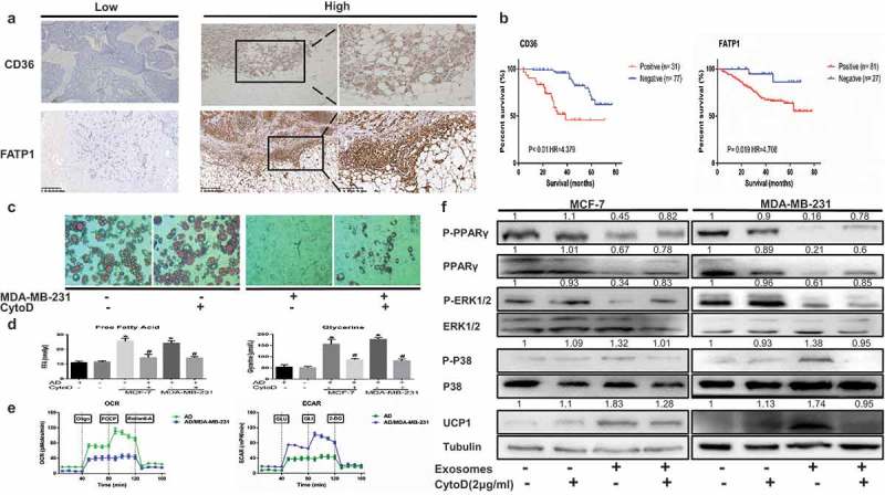
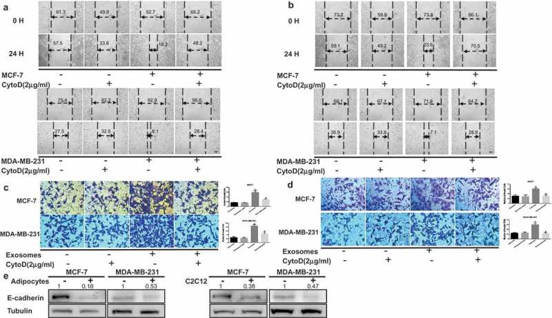
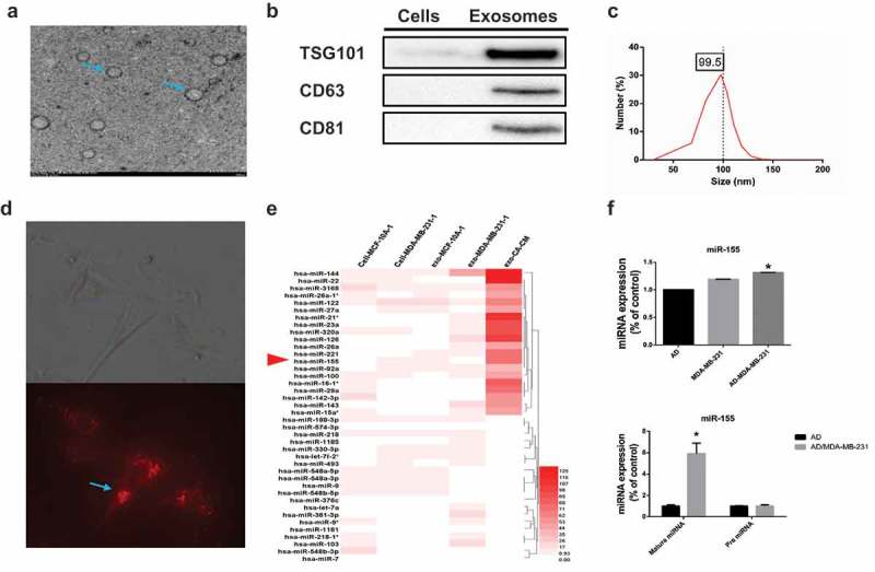
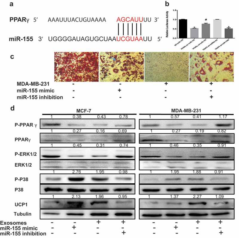
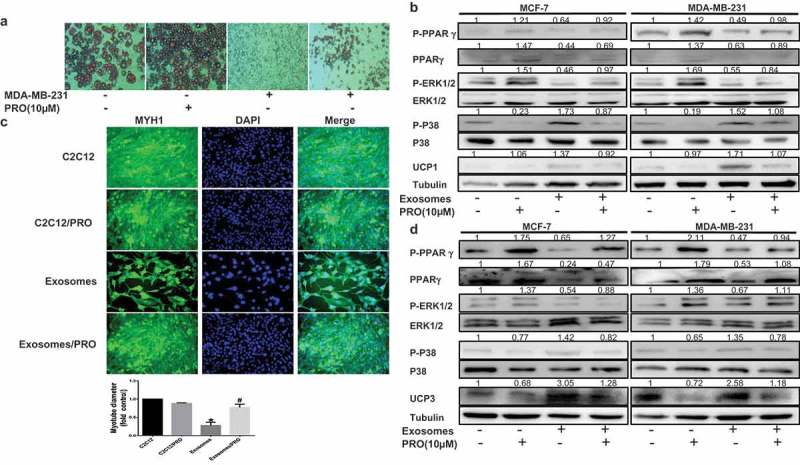
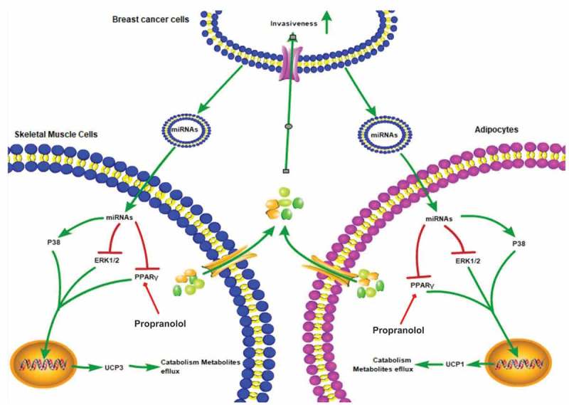
Cancer-secreted exosomes are emerging mediators of cancer-associated cachexia. Here, we show that miR-155 secreted by breast cancer cells is a potent role on the catabolism of adipocytes and muscle cells through targeting the PPARγ. After cocultivated with mature adipocytes or C2C12, tumour cells exhibit an aggressive phenotype via inducing epithelial-mesenchymal transition while breast cancer-derived exosomes increased catabolism and release the metabolites in adipocytes and muscle cells. In adipocytes, cancer cell-secreted miR-155 promotes beige/brown differentiation and remodel metabolism in resident adipocytes by downregulating the PPARγ expression, but does not significantly affect biological conversion in C2C12. Likewise, propranolol ameliorates tumour exosomes-associated cachectic wasting through upregulating the PPARγ expression. In summary, we have demonstrated that the transfer of miR-155 from exosomes acts as an oncogenic signal reprograming systemic energy metabolism and leading to cancer-associated cachexia in breast cancer.

Keywords: Breast cancer; cachexia; exosomes; tumour progression.


Figures

Figure 1.
Figure 1.
Breast cancer-derived exosomes reshape metabolic characteristics in adipocytes and metabolite biomarkers in breast cancer are linked to a poor prognosis. (A) Representative immunohistochemistry staining of CD36 and FATP1. The pictures also show positive staining for FATP1 and CD36 in breast cancer tissues located near stromal adipocytes. (B) Kaplan-Meier survival analysis of patients with biomarker-positive and biomarker-negative IHC staining. (C) Mature adipocytes after treated were stained with red oil. The adipocytes in the CytoD group were treated with cytochalasin D (final concentration, 2 μg/ml) and 50 μg of exosomes purified from cancer-associated conditioned medium (CA-CM) (enlarged 100X). (D) The levels of secreted metabolites (glycerol and free fatty acids) enriched in media were determined by colorimetric assay. AD: adipocytes. (E) Raw data for the oxygen consumption rate (OCR) and extracellular acidification rate (ECAR) as determined by the Seahorse XF24 analyzer. The ECAR was evaluated after the addition of 10 mM glucose to adipocytes in the presence or absence of exosomes. The OCR was measured in the presence of palmitate as described in the Methods. (F) Adipocytes were cocultivated in the presence or absence of exosomes. After 3 days, proteins were extracted for western blot analysis of the expression of the indicated proteins. The numbers represent the relatively quantitative results compared to the control group. Data are presented as the mean ± S.D. of at least three independent experiments. * P < 0.05 versus control values.
Figure 2.
Figure 2.
Skeletal muscle cells undergo extensive metabolism changes after treated breast cancer-secreted exosomes. (A) Immunofluorescence staining for myosin heavy chain 1 (MYH1) after treatment 24 h. Scale bars represent 50 μm. The C2C12 in the CytoD group were treated with cytochalasin D (final concentration, 2 μg/ml) and 50 μg of exosomes purified from cancer-associated conditioned medium (CA-CM). (B) The levels of secreted metabolites (pyruvate and lactate) enriched in media were determined by colorimetric assay. (C) Raw data for the oxygen consumption rate (OCR) and extracellular acidification rate (ECAR) as determined by the Seahorse XF24 analyzer. The ECAR was evaluated after the addition of 10 mM glucose to C2C12 in the presence or absence of exosomes. The OCR was measured in the presence of palmitate as described in the Methods. (D) C2C12 were cocultivated in the presence or absence of exosomes. After 24 h, proteins were extracted for western blot analysis of the expression of the indicated proteins. The numbers represent the relatively quantitative results compared to the control group. Data are presented as the mean ± S.D. of at least three independent experiments. * P < 0.05 versus control values, # P < 0.05 versus control values as positive group.
Figure 3.
Figure 3.
Tumor cells exhibit increased invasion capacities upon coculture with adipocytes or muscle cells. Cancer-associated conditioned medium (CA-CM) was collected from adipocytes cultivated with MCF-7 or MDA-MB-231 cells for 3 days or C2C12 cultivated with MCF-7 or MDA-MB-231 cells for 1 days, and adipocyte or C2C12-conditioned medium (AD-CM) were collected from cells cultivated alone as controls. All media contained 10% FBS. Wound healing assays were used to examine the effects of CA-CM from adipocytes (A) and C2C12 (B) on cell motility (Scale bar: 10 μm). Tumour cells were cultivated in control medium or CA-CM from adipocytes (C) and C2C12 (D). After 24 hours, the number of cells penetrating the membrane in Transwell invasion assays was analyzed. (E) E-cadherin protein expression was analyzed by western blot in extracts from tumor cells cocultivated in the presence or absence of adipocytes (3 days) or C2C12 (1 day). The numbers represent the relatively quantitative results compared to the control group. The bars represent the mean ± SD of triplicate datapoints (n = 3). * P < 0.05 versus control values, # P < 0.05 versus control values as positive group.
Figure 4.
Figure 4.
Breast cancer cells cocultured with adipocytes show altered exosomal miRNA expression profiles. (A) Exosomes originating from CA-CM viewed by electron microscopy (scale bar, 200 nm). (B) Exosomes from CA-CM were analyzed by western blot. (C) NanoSight analysis of exosomes derived from CA-CM. (D) Labelled tumor cell-secreted exosomes (red) were incubated with the indicated adipocytes. (E) miRNA microarray analysis of purified exosomes from control groups and CA-CM. The heatmap shows the top 40 differentially expressed miRNAs among different groups. (F) MDA-MB-231 were cocultivated in the presence or absence of adipocytes. After 3 days, exosomal miRNAs were further verified by qPCR. And RNA was extracted from the adipocytes and subjected to qPCR analysis with primers specific to mature miRNA. Data are presented as the mean ± S.D. of at least three independent experiments. * P < 0.05 versus control values.
Figure 5.
Figure 5.
ExomiR-155 mediates the adipocyte metabolism by downregulating PPARγ. The adipocytes in 50 μg of exosomes purified from cancer-associated conditioned medium (CA-CM). (A) The predicted miR-155 binding site in the 3ʹUTR of the PPARγ gene from TargetScan. (B) The GV272 vector containing the 3ʹUTR of the target gene harboring wild-type (wt) or mutated (mt) miRNA binding sites was transfected into HEK 293T cells stably expressing miRNA or empty vector (as a control). Luciferase activity was analyzed at 48 hours post-transfection, and the ratio of firefly luciferase activity to Renilla luciferase activity is shown. (C) Breast cancer cells were transfected with the control vector or miR-155 inhibitor, mature adipocytes were transfected with miR-155 mimic as the positive control and the control vector was applied as the negative control. Mature adipocytes cultured in the presence or absence of tumor exosomes for 3 days were stained with red oil (D), and Western blot analysis of related protein expression in different groups. The numbers represent the relatively quantitative results compared to the control group. Data are presented as the mean ± S.D. of at least three independent experiments. * P < 0.05 versus control values.
Figure 6.
Figure 6.
Propranolol ameliorates tumor exosomes-associated cachectic wasting in vitro. The adipocytes or C2C12 in 50 μg of exosomes purified from cancer-associated conditioned medium (CA-CM) and/or Propranolol (PRO, 10 μM). (A) Mature adipocytes in the presence or absence of exosomes and/or Propranolol were stained with red oil (enlarged 100X). (B) Adipocytes were cocultivated in the presence or absence of exosomes and/or Propranolol. After 3 days, proteins were extracted for western blot analysis of the expression of the indicated proteins. (C) Immunofluorescence staining for myosin heavy chain 1 (MYH1) after treatment 24 h. Scale bars represent 50 μm. (D) After C2C12 cells were treated for 1 days, and Western blot analysis of related protein expression in different groups. The numbers represent the relatively quantitative results compared to the control group. Data are presented as the mean ± S.D. of at least three independent experiments. * P < 0.05 versus control values, # P < 0.05 versus control values as positive group.
Figure 7.
Figure 7.
Working model for how breast-cancer-secreted exosomes reprogram metabolism in adipocytes and muscle cells to promote tumor progression. Breast cancer cells could secrete exosomes containing some special miRNAs including miR-155, which induce mitochondrial uncoupling and promote catabolism in adipocytes and muscle cells. High-energy metabolites released from adipocytes and muscle cells can be transported into breast cancer cells to remodel tumor metabolism and promote tumor progression. Moreover, propranolol ameliorates tumor exosomes-associated cachectic wasting through upregulating the PPARγ expression.

References

    1. Fearon KC, Glass DJ, Guttridge DC.. Cancer cachexia: mediators, signaling, and metabolic pathways. Cell Metab. 2012. August 8;16(2):153–166. PMID: 22795476. - PubMed
    1. Martinez-Outschoorn U, Sotgia F, Lisanti MP. Tumor microenvironment and metabolic synergy in breast cancers: critical importance of mitochondrial fuels and function. Semin Oncol. 2014. April;41(2):195–216. PMID: 24787293. - PubMed
    1. Tisdale MJ. Cachexia in cancer patients. Nat Rev Cancer. 2002. November;2(11):862–871. PMID: 12415256. - PubMed
    1. Bruggeman AR, Kamal AH, LeBlanc TW, Ma JD, Baracos VE, Roeland EJ. Cancer cachexia: beyond weight Loss. J Oncol Pract. 2016. November;12(11):1163–1171. PMID: 27858548. - PubMed
    1. Vigano AAL, Morais JA, Ciutto L, Rosenthall L, Di Tomasso J, Khan S, Olders H, Borod M, Kilgour RD. Use of routinely available clinical, nutritional, and functional criteria to classify cachexia in advanced cancer patients. Clin Nutr. 2017. October;36(5):1378–1390. PMID: 27793524. - PubMed

Publication types

MeSH terms