. 2018 Nov 24;19(1):411.
doi: 10.1186/s12891-018-2335-9.

Mismatch between femur and tibia coronal alignment in the knee joint: classification of five lower limb types according to femoral and tibial mechanical alignment

Affiliations

Mismatch between femur and tibia coronal alignment in the knee joint: classification of five lower limb types according to femoral and tibial mechanical alignment

Yu-Hsien Lin et al. BMC Musculoskelet Disord. .

Abstract

Background: Reasons for dissatisfaction with total knee arthroplasty (TKA) include unequal flexion or extension gap, soft tissue imbalance, and patella maltracking, which often occur with mismatch between femoral and tibial coronal bony alignment in the knee joint or extremely varus or valgus alignment. However, lower limb coronal alignment classification is based only on hip-knee-ankle angle (HKAA), leading to oversight regarding a mismatch between femoral and tibial coronal alignment. We aimed to classify alignment of the lower limbs according to the mechanical alignment of the femur and tibia in a healthy population.

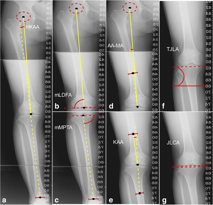
Methods: All 214 normal triple films were reviewed retrospectively. HKAA, mechanical lateral distal femoral angle (mLDFA), mechanical medial proximal tibial angle (mMPTA), angle between the femoral anatomical axis and the mechanical axis (AA-MA), and knee alignment angle (KAA) were measured. Subjects were categorized into one of five types based on the mechanical alignment of femur and tibia.

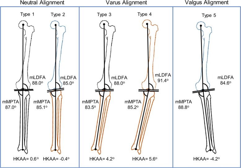
Results: Mean HKAA, mLDFA, and mMPTA of all subjects were 1.2°, 87.3°, and 85.8°, respectively. All subjects were classified into one of five types with significant differences (p < 0.001). About 61% of subjects showed neutral alignment, of which nearly 40% were type 2 (valgus of the femur and varus of the tibia with oblique joint line: mLDFA 85.0° ± 1.4°, mMPTA 85.1° ± 1.2°, TJLA 2.7° ± 2.4°) and 60% exhibited neutral alignment with a neutral femur and tibia (type 1). In varus and valgus types, mismatch between the mechanical angle of the femur and tibia was common. Varus alignment, including types 3 (varus of the tibia: mLDFA 88.0° ± 1.4°, mMPTA 83.5° ± 1.6°) and 4 (varus of both the tibia and femur: mLDFA 91.4° ± 1.4°, mMTPA 85.2° ± 2.0°), was found in 30% of subjects. Valgus alignment (type 5 valgus of femur: mLDFA 84.6° ± 1.6°, mMPTA 88.8° ± 2.0°) accounted for 8.9% of all subjects.

Conclusions: Mismatch between mechanical alignment of the femur and tibia was common in varus and valgus alignment types. Joint line obliquity was also observed in 40% of the neutral alignment population. This classification provides a quick, simple interpretation of femoral and tibial coronal alignment, and more detailed guidance for preoperative planning for TKA than the traditional varus-neutral-valgus classification.

Keywords: Coronal limb alignment; Mechanical alignment; Mismatch between femur and tibia; Normal knee; mLDFA; mMPTA.


Conflict of interest statement

Ethics approval and consent to participate

This study was approved by the institutional review board of Taichung Veterans General Hospital, Taichung, Taiwan (approval number CE16034B). A certificate of approval has been provided. Due to the retrospective nature of the study, formal informed consent from the participants is not required.

Consent for publication

Not applicable.

Competing interests

The authors declare that they have no competing interests.

Publisher’s Note

Springer Nature remains neutral with regard to jurisdictional claims in published maps and institutional affiliations.

Figures

Fig. 1
Fig. 1
Measured Coronal Alignment Parameters. The five angles were defined as the following: a Hip–knee–ankle angle (HKAA): the angle between the mechanical axis of the femur and the tibia. The value of HKAA was defined as positive if varus alignment was found and as negative if valgus alignment was found. b Mechanical lateral distal femoral angle (mLDFA): the lateral angle between the mechanical axis of the femur and the distal femur joint line, which defined the connection of the lowest points of the medial and lateral femoral condyle. c Mechanical medial proximal tibial angle (mMPTA): the medial angle between the mechanical axis of the tibia and the proximal tibia joint line, which defined the connection of the lowest points of the medial and lateral tibial plateau. d Angle between the femoral anatomical axis and the mechanical axis (AA-MA): the angle between the mechanical axis and the anatomical axis of the femur. e Knee alignment angle (KAA): the angle between the anatomical axis of the femur and the anatomical axis of the tibia in the short film of the knee. The value of KAA was defined as positive if varus alignment was found and as negative if valgus alignment was found in the short film of the knee. f Tibial joint line angle (TJLA): the angle formed between the parallel line to the floor and the proximal tibia joint line. If both lines intersect with an angle on the lateral side of the leg, it is a medial open angle. If both lines intersect with an angle on the medial side of the leg, it is a lateral open angle. Positive values represent a lateral open angle and negative values represent a medial open angle. g Joint line convergence angle (JLCA): the angle between the knee joint lines of the distal femur and proximal tibia
Fig. 2
Fig. 2
The Five Common Types of Normal Coronal Limb Alignment in a Taiwanese Population. The neutral alignment group consisted of two types (type 1 and type 2); the varus alignment group comprised 2 types (type 3, type 4); and the valgus alignment group consisted of one type (type 5). The black color indicates the femur or tibia in neutral alignment, the brown color indicates varus of the femur or tibia, and the blue color indicates valgus of the femur. HKAA, hip–knee–ankle angle; mLDFA, mechanical lateral distal femoral angle; mMPTA, mechanical medial proximal tibial angle

References

    1. Berend ME, Ritter MA, Meding JB, Faris PM, Keating EM, Redelman R, et al. Tibial component failure mechanisms in total knee arthroplasty. Clin Orthop Relat Res. 2004;428:26–34. doi: 10.1097/01.blo.0000148578.22729.0e. - DOI - PubMed
    1. Fang DM, Ritter MA, Davis KE. Coronal alignment in total knee arthroplasty: just how important is it? J Arthroplasty. 2009;24:39–43. doi: 10.1016/j.arth.2009.04.034. - DOI - PubMed
    1. Collier MB, Engh CA, Jr, McAuley JP, Engh GA. Factors associated with the loss of thickness of polyethylene tibial bearings after knee arthroplasty. J Bone Joint Surg Am. 2007;89:1306–1314. doi: 10.2106/00004623-200706000-00020. - DOI - PubMed
    1. Liau JJ, Cheng CK, Huang CH, Lo WH. The effect of malalignment on stresses in polyethylene component of total knee prostheses--a finite element analysis. Clin Biomech (Bristol, Avon) 2002;17:140–146. doi: 10.1016/S0268-0033(01)00109-7. - DOI - PubMed
    1. Baker PN, van der Meulen JH, Lewsey J, Gregg PJ. The role of pain and function in determining patient satisfaction after total knee replacement. Data from the National Joint Registry for England and Wales. J Bone Joint Surg Br. 2007;89:893–900. doi: 10.1302/0301-620X.89B7.19091. - DOI - PubMed

LinkOut - more resources