. 2019 Jan;59(1):371-384.
doi: 10.1111/trf.14939. Epub 2018 Nov 26.

Antibody-mediated immunosuppression can result from RBC antigen loss independent of Fcγ receptors in mice

Affiliations

Antibody-mediated immunosuppression can result from RBC antigen loss independent of Fcγ receptors in mice

Amanda Mener et al. Transfusion. 2019 Jan.

Abstract

Background: Anti-RhD administration can prevent de novo anti-RhD formation following RhD+ red blood cell (RBC) exposure, termed antibody-mediated immunosuppression (AMIS). Recent studies suggest that AMIS may occur through target antigen alterations, known as antigen modulation. However, studies suggest that AMIS may occur independent of antigen modulation. In particular, AMIS to RBCs that transgenically express the fusion hen egg lysozyme-ovalbumin-Duffy (HOD) antigen have been shown to occur independent of activating Fcγ receptors (FcγRs) thought to be required for antigen modulation. Therefore, we sought to determine the mechanism behind AMIS following HOD RBC exposure.

Study design and methods: Following transfer of HOD RBCs into wild-type or FcγR-chain knockout recipients in the presence or absence of monoclonal anti-hen egg lysozyme (HEL) antibody, individually or in combination, HOD antigen levels and anti-HOD antibody formation were examined.

Results: Our results demonstrate that anti-HEL antibodies individually or in combination suppressed anti-HOD IgM, which correlated with the rate of detectable decrease in HEL on HOD RBCs. Furthermore, exposure to anti-HEL antibodies alone or in combination equally suppressed anti-HOD IgG formation. Unexpectedly, combination or individual anti-HEL antibodies induced AMIS and antigen modulation in an FcγR-independent manner. Pre-exposure of HOD RBCs to anti-HEL antibodies reduced antigen levels and suppressed anti-HOD antibody formation following HOD RBC exposure.

Conclusion: These results suggest that antibody-mediated antigen modulation may reflect a mechanism of AMIS that can occur independent of activating FcγRs and may provide a surrogate to identify antibodies capable of inducing AMIS against different RBC antigens.


Conflict of interest statement

CONFLICT OF INTEREST

The authors have disclosed no conflicts of interest.

Figures

Fig. 1.
Fig. 1.
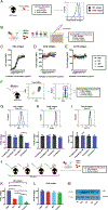
Anti-HEL monoclonal antibodies saturate HOD RBCs and dampen anti-HOD antibody response. (A) Antibody saturation of HOD RBCs individually or in combination was assessed by flow cytometry. (B) Following exposure of B6 mice to HOD RBCs in the presence (2F4, 4B7, or a combination of 2F4 and 4B7 [combo]) or absence (phosphate-buffered saline) of anti-HEL monoclonal antibodies, serum was assessed for the presence of anti-HOD IgM on Day 5 and anti-HOD IgG on Day 14 after transfusion by flow cross-match. Following exposure of HOD mice to HOD RBCs, anti-HOD IgM and IgG was assessed on Days 5 and 14 after transfusion, respectively, by flow-crossmatch. For B, **** = p < .0001, *** = p < .0002 and * = p < .05 by one-way analysis of variance with Tukey’s multiple comparisons test. Means ± SD shown. HEL = hen egg lysozyme; HOD = hen egg lysozyme-ovalbumin-Duffy; IgG = immunoglobulin G; IgM = immunoglobulin M; RBCs = red blood cells.
Fig. 2.
Fig. 2.
Anti-HEL monoclonal antibodies are detected on HOD RBCs following transfusion. (A) B6 mice were exposed to Dil-labeled HOD + RBCs and control DiO-labeled HOD RBCs in the presence (2F4, 4B7, or a combination of 2F4 and 4B7 [combo]) or absence (phosphate-buffered saline) of anti-HEL monoclonal antibodies. (B) The deposition of surface IgG antibody specifically on HOD+ RBCs was probed at 10 minutes (C), 2 hours (D), Day 1 (E), Day 2 (F), Day 3 (G) and Day 5 (H). For C-H, **** = p < .0001, *** = p < .0008, ** = p < .008, and * = p < .05 by one-way analysis of variance with Tukey’s multiple comparisons test. Means ± SD shown. HEL = hen egg lysozyme; HOD = hen egg lysozyme-ovalbumin-Duffy; IgG = immunoglobulin G; RBCs = red blood cells.
Fig. 3.
Fig. 3.
Anti-HEL antibodies induce a decrease in the level of detectable HEL antigen and fail to deposit complement or induce clearance. (A) B6 mice were exposed to a mixture of Dil-labeled HOD+ RBCs and control DiO-labeled HOD− RBCs in the presence (2F4, 4B7 or a combination of 2F4 and 4B7 [combo]) or absence (phosphate-buffered saline) of monoclonal anti-HEL antibodies. (B-G) Following transfusion, the level of HEL antigen on circulating HOD+ RBCs was assessed at 10 minutes (B), 2 hours (C), Day 1 (D), Day 2 (E), Day 3 (F), and Day 5 (G). Level of HEL antigen was measured by percentage of HEL antigen normalized to the HOD control group. (H) HOD + RBCs in the absence (phosphate-buffered saline) or presence of monoclonal anti-HEL antibodies (2F4 and 4B7) individually or in combination (combo) were examined for complement component 3 deposition following exposure in B6 mice at 10 minutes and 2 hours after transfusion. (I) Clearance of HOD+ RBCs in B6 mice exposed to DiI-labeled HOD+ RBCs and control DiO-labeled HOD− RBCs in the presence or absence of anti-HEL monoclonal antibodies (2F4 and 4B7) individually or in combination (combo). For B-G, **** = p < .0001, *** = p < .0006, ** = p < .002 and * = p < .05 by One-way analysis of variance with Tukey’s multiple comparisons test. For I, statistical analysis was performed using two-way analysis of variance with Dunnett’s multiple comparisons test. Means ± SD shown. HEL = hen egg lysozyme; HOD = hen egg lysozyme-ovalbumin-Duffy; RBCs = red blood cells.
Fig. 4.
Fig. 4.
Anti-HEL antibodies induce a decrease in the level of detectable HEL antigen in FcγR KO mice. (A-F) FcγR KO mice (Fcγ) were exposed to a mixture of Dil-labeled HOD+ RBCs and control DiO-labeled HOD− RBCs in the presence (2F4, 4B7 or a combination of 2F4 and 4B7 [combo]) or absence (phosphate-buffered saline) of monoclonal anti-HEL antibodies. RBCs were collected and the percentage of detectable HEL antigen, normalized to control HOD mice exposed to HOD+ RBCs, was measured at 10 minutes (A), 2 hours (B), Day 1 (C), Day 2 (D), Day 3 (E), and Day 5 (F). For A-F, **** = p < .0001, *** = p < .003, ** = p < .009 and * = p < .05 by one-way analysis of variance with Tukey’s multiple comparisons test. Means ± SD shown. FcγR = Fcγ receptor; HEL = hen egg lysozyme; HOD = hen egg lysozyme-ovalbumin-Duffy; RBCs = red blood cells.
Fig. 5.
Fig. 5.
HOD RBCs exposed to anti-HEL antibodies display decreased levels of bound antibody in FcγR KO mice. (A-F) FcγR KO mice (Fcγ) were exposed to a mixture of Dil-labeled HOD+ RBCs and control DiO-labeled HOD− RBCs in the presence (2F4, 4B7 or a combination of 2F4 and 4B7 [combo]) or absence (phosphate-buffered saline) of monoclonal anti-HEL antibodies. Following transfusion, RBCs were assessed for IgG deposition via the direct antiglobulin test at 10 minutes (A), 2 hours (B), Day 1 (C), Day 2 (D), Day 3 (E) and Day 5 (F). For A-F, **** = p < 0001, *** = p < .0006, ** = p < .002 and * = p < .05 by one-way analysis of variance with Tukey’s multiple comparisons test. Means ± SD shown. FcγR = Fcγ receptor; HEL = hen egg lysozyme; HOD = hen egg lysozyme-ovalbumin-Duffy; IgG = immunoglobulin G; RBCs = red blood cells.
Fig. 6.
Fig. 6.
HOD RBCs exposed to anti-HEL antibodies in FcγR KO mice results in dampening of the anti-HOD antibody response without complement deposition nor clearance of HOD RBCs. (A) Serum was assessed for the presence of anti-HOD IgM on Day 5 and anti-HOD IgG on Day 14 after transfusion of HOD+ RBCs in FcγR KO mice (Fcγ) by flow cross-match. (B) HOD RBCs in the absence (phosphate-buffered saline) or presence of anti-HEL monoclonal antibodies (2F4 and 4B7) individually or in combination (combo) were examined for C3 deposition following exposure in FcγR KO mice (Fcγ) at 10 minutes and 2 hours after transfusion. (C) Clearance of HOD RBCs in FcγR KO mice (Fcγ) exposed to DiI-labeled HOD+ RBCs and control DiO-labeled HOD− RBCs in the presence or absence of anti-HEL monoclonal antibodies (2F4 and 4B7) individually or in combination (combo). For A, **** = p < .0001, *** = p < 0003 and ** = p < .003 by one-way analysis of variance with Tukey’s multiple comparisons test. For C, statistical analysis was performed using two-way analysis of variance with Dunnett’s multiple comparisons test. Means ± SD shown. FcγR = Fcγ receptor; HEL = hen egg lysozyme; HOD = hen egg lysozyme-ovalbumin-Duffy; IgG = immunoglobulin G; IgM = immunoglobulin M; RBCs = red blood cells. [Color figure can be viewed at wileyonlinelibrary.com]
Fig. 7.
Fig. 7.
Re-transfusion of HOD RBCs with varying levels of HEL antigen correlates with dampening of the anti-HOD antibody response without clearance of HOD RBCs. (A) HEL antigen on homozygous and heterozygous HOD mice was assessed by flow cytometry. (B) B6 mice were exposed to HOD+ RBCs in the presence (2F4, 4B7 or a combination of 2F4 and 4B7 [combo]) or absence (phosphate-buffered saline) of monoclonal anti-HEL antibodies. The level of detectable HEL antigen was then assessed followed by re-transfusion of RBCs into naive B6 recipients. (C-F) Following re-transfusion, the level of HEL antigen was detected at 10 minutes (C), Day 1 (D), Day 3 (E), and Day 5 (F). (G) Serum was collected from retransfused recipients, and the development of anti-HOD IgM and IgG was assessed on Day 5 and Day 14 after transfusion, respectively, by flow cross-match. (H) Clearance of anti-HEL monoclonal antibody-antigen modulated HOD+ RBCs following re-transfusion into naive B6 mice. For B-G, **** = p < .0001, *** = p < .0002 and * = p < .05 significant by one-way analysis of variance with Tukey’s multiple comparisons test. For H, statistical analysis was performed using two-way analysis of variance with Dunnett’s multiple comparisons test. Means ± SD shown. HEL = hen egg lysozyme; HOD = hen egg lysozyme-ovalbumin-Duffy; IgG = immunoglobulin G; IgM = immunoglobulin M; RBCs = red blood cells. [Color figure can be viewed at wileyonlinelibrary.com]
Fig. 8.
Fig. 8.
HEL antigen decreases over time on HOD RBCs as they age but not with protease treatment in vitro. (A) HOD+ RBCs stained for the level of detectable HEL and Duffy antigens, followed by overlay of histograms. (B) HOD+ RBCs were treated with proteases in the presence or absence of monoclonal anti-HEL antibodies. Following treatment, the level of detectable HEL (C), Duffy (D), and Ter-119 (E) antigens were measured by flow cytometry. (F) HOD mice were biotinylated via intravenous injection of NHS-sulfo-biotin, followed by tracking biotin positive RBCs in vivo over time. (G-I) RBCs were harvested and evaluated for levels of HEL (G), Duffy (H), and Ter-119 (I) by flow cytometry on Day 0 (D0) or Day 14 (D14) after biotinylation. (J) B6 mice were either not immunized (phosphate-buffered saline) or passively immunized with anti-HEL monoclonal antibodies (2F4, 4B7 or a combination of 2F4 and 4B7 [combo]), followed by exposure to HOD+ RBCs. Two days after transfusion, HOD+ RBCs were harvested. The level of detectable HEL antigen (K) and Duffy antigen (L) were then measured by flow cytometry. RBCs were also subjected to western blot analysis for HEL (M). For C-E, statistical analysis was performed using two-way analysis of variance with Tukey’s multiple comparison’s test. For G-L, **** = p < .0001 and *** = p < .0003 by one-way analysis of variance with Tukey’s multiple comparisons test. Means ± SD shown. HEL = hen egg lysozyme; HOD = hen egg lysozyme-ovalbumin-Duffy; RBCs = red blood cells.

Similar articles

Cited by

References

    1. Stowell SR, Henry KL, Smith NH, et al. Alloantibodies to a paternally derived RBC KEL antigen lead to hemolytic disease of the fetus/newborn in a murine model. Blood 2013;122: 1494–504. - PMC - PubMed
    1. Heard DH, Hinde IT, Mynors LS. An experimental study of haemolytic disease of the newborn due to isoimmunization of pregnancy. J Hyg 1949;47:119–31. - PMC - PubMed
    1. Moise KJ. Fetal anemia due to non-Rhesus-D red-cell alloimmunization. Semin Fetal Neonatal Med 2008;13:207–14. - PubMed
    1. Moise KJ Jr. Management of rhesus alloimmunization in pregnancy. Obstet Gynecol 2008;112:164–76. - PubMed
    1. Clarke CA, Finn R, Lehane D, et al. Dose of anti-D gamma-globulin in prevention of Rh-haemolytic disease of the new- born. Br Med J 1966;1:213–4. - PMC - PubMed

Publication types