Review
. 2019 Jan 1;99(1):665-706.
doi: 10.1152/physrev.00067.2017.

Wound Healing: A Cellular Perspective

Affiliations
Review

Wound Healing: A Cellular Perspective

Melanie Rodrigues et al. Physiol Rev. .

Abstract

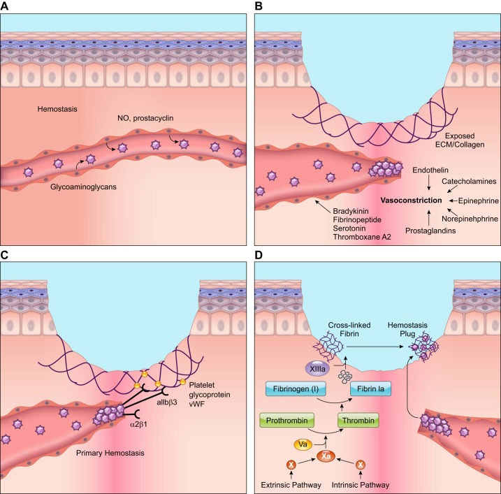
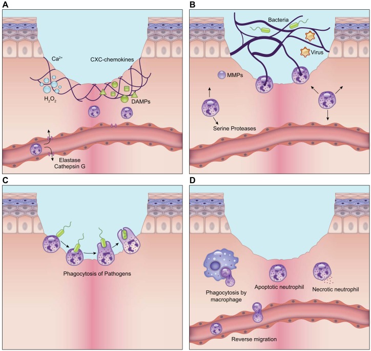
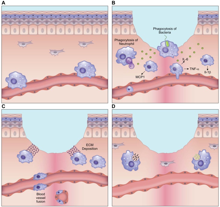
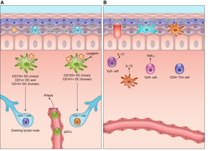
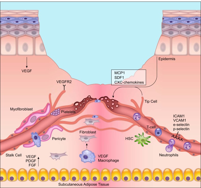
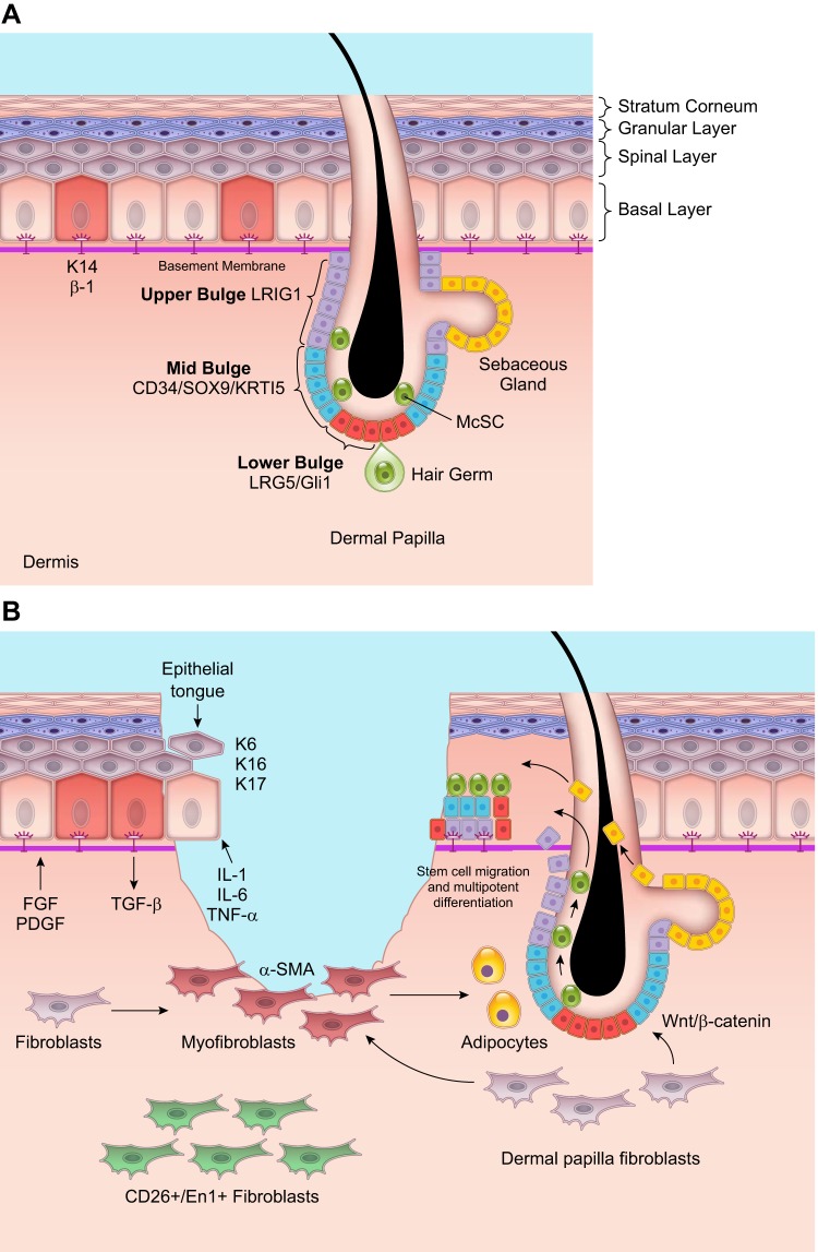
Wound healing is one of the most complex processes in the human body. It involves the spatial and temporal synchronization of a variety of cell types with distinct roles in the phases of hemostasis, inflammation, growth, re-epithelialization, and remodeling. With the evolution of single cell technologies, it has been possible to uncover phenotypic and functional heterogeneity within several of these cell types. There have also been discoveries of rare, stem cell subsets within the skin, which are unipotent in the uninjured state, but become multipotent following skin injury. Unraveling the roles of each of these cell types and their interactions with each other is important in understanding the mechanisms of normal wound closure. Changes in the microenvironment including alterations in mechanical forces, oxygen levels, chemokines, extracellular matrix and growth factor synthesis directly impact cellular recruitment and activation, leading to impaired states of wound healing. Single cell technologies can be used to decipher these cellular alterations in diseased states such as in chronic wounds and hypertrophic scarring so that effective therapeutic solutions for healing wounds can be developed.


Conflict of interest statement

No conflicts of interest, financial or otherwise, are declared by the authors.

Figures

FIGURE 1.
FIGURE 1.
Cellular responses during the hemostasis phase of wound healing. A: during hemostasis, platelets circulate in close proximity to the vessel wall. However, anti-thrombotic agents such as nitric oxide (NO) and prostacyclin released from endothelial cells prevent platelet attachment to the endothelial lining and platelet aggregation. B: wounding stimulates injured cells to rapidly release vasoconstrictors that cause reflexive contracture of the smooth muscle and temporary stoppage of bleeding. C: blood vessel rupture during wound healing exposes the subendothelial matrix. Platelets bind this subendothelial matrix and to each other using G protein-coupled receptors, integrins, and glycoproteins on their surface. von Willebrand factor (vWF) released by platelets also attaches to the subendothelial matrix. Platelets bind extracellular vWF through their surface receptors, strengthening the platelet plug. D: the extrinsic and intrinsic pathways lead to the activation of Factor X, which ultimately results in the cleavage of fibrinogen to fibrin. Cross-linked fibrin binds the aggregated platelet plug to form the thrombus that stops blood flow and provides a provisional matrix for healing. The illustration is a simplified rendering based on current knowledge.
FIGURE 2.
FIGURE 2.
Role of neutrophils in wound healing. A: neutrophils are recruited to the wound in response to calcium waves, damage-associated molecular patterns (DAMPs), hydrogen peroxide, lipid mediators, and chemokines that are released by resident cells immediately after wounding. B: neutrophils combat pathogens through release of proteases from their intracellular granules. They also produce neutrophil extracellular traps (NETs) that capture pathogens through a process called NETosis. In this process, neutrophils extend chromatin filaments coated with proteases outside of the cell to aid in the elimination of pathogens. C: neutrophils also perform phagocytosis in the wound. They probe antigens using surface receptors and integrins and form a phagocytic cup that engulfs antigens. Internalized antigens are degraded by proteases within the neutrophil granules. D: timely clearance of neutrophils is critical for the resolution of inflammation. They are either engulfed by macrophages through efferocytosis or they can re-enter the circulation and leave the wound through a process called reverse migration. The illustration is a simplified rendering based on current knowledge.
FIGURE 3.
FIGURE 3.
Macrophage phenotypes in wound healing. A: in the uninjured skin, circulating monocytes from the bone marrow are constantly rolling over the inner endothelial wall within the vessel lumen and surveying for damage. The few macrophages that are resident in the skin are prevalent in the perivascular space and can be from embryonic sources. B: following skin injury, during the inflammatory phase of healing, macrophages release pro-inflammatory cytokines such as interleukin (IL)-6, tumor necrosis factor (TNF)-α, and IL-1β to fight infection. Early macrophages in the wound release monocyte chemoattractant protein (MCP)-1 to draw in more monocytes from the bone marrow and heighten the macrophage response. These macrophages also actively participate in phagocytosis of pathogens. At the end of the inflammatory phase, macrophages engulf dying neutrophils, which marks the end of the inflammatory phase of wound healing. C: during the growth stage of wound healing, as granulation tissue forms, macrophages release growth factors such as vascular endothelial growth factor (VEGF) and platelet-derived growth factor (PDGF) that are used to signal and activate endothelial cells to perform angiogenesis. Some macrophages deposit extracellular matrix (ECM) at this stage. Unregulated deposition of ECM can lead to fibrosis, and such scar-forming macrophages are called fibrocytes. D: during wound remodeling, macrophages again take on a phagocytic role where they engulf both cell debris and excessive ECM, to bring the healed skin to a homeostatic state. The illustration is a simplified rendering based on current knowledge.
FIGURE 4.
FIGURE 4.
Dendritic cells (DCs) and T cells in the wound healing response. A: dendritic cells within the murine dermis can be CD103+, which correspond to CD141+ DCs in humans, or CD11b+, which correspond to the CD1c+ and CD14+ subset in humans. The CD103+ DCs mainly activate CD8+ T cells, and the CD11b+ DCs mainly activate the CD4+ T cells in the draining lymph nodes. The wounded skin also contains plasmacytoid dendritic cells (pDCs) that are activated to release interferon (IFN)-α/β that is important to the acute inflammatory response. Within the epidermis, Langerhans cells with high expression of Langerin (CD207) are the main antigen presenting cells. Langerhans cells are derived from early myeloid progenitors in the embryo and persist in adult skin. B: T cells in the skin can either be γδ+ or αβ+. γδ+ T cells of the epidermis are also called dendritic epithelial cells (DETCs). These cells survey the epidermis for infection and produce growth factors that are critical for signaling epidermal cells during wound healing. γδ+ T cells of the dermis are either Vγ5-positive that release tumor necrosis factor (TNF)-α and activate dermal DCs to release interleukin (IL)-12, or they are Vγ5-negative that produce IL-17 following infection. These γδ+ T cells can migrate to the draining lymph nodes and continue to activate DCs. The αβ+ T cells in the dermis mainly consist of CD4+ subsets, while the αβ+ T cells of the epidermis are the CD8+ killer cells. The illustration is a simplified rendering based on current knowledge.
FIGURE 5.
FIGURE 5.
Angiogenesis during wound healing. New blood vessel formation is one of the most important stages of wound healing. Endothelial cells at the leading edge or tip branch out or “sprout” to form new capillaries in response to vascular endothelial growth factor (VEGF) and other growth factor signals from epidermal cells, macrophages, and the subcutaneous adipose tissue. The endothelial cells during angiogenesis are leaky to allow for immune cells and other circulating cells to extravasate from the blood vessel lumen into the wound. Pro-angiogenic macrophages release growth factors for endothelial cell growth and fuse newly forming capillaries. Activated endothelial cells upregulate surface markers intercellular adhesion molecule (ICAM)-1, vascular cell adhesion molecule (VCAM)-1, E-selectin, and P-selectin that help with cell-cell interactions with leukocytes. Deletion of these surface markers during wound healing impairs wound repair. The illustration is a simplified rendering based on current knowledge.
FIGURE 6.
FIGURE 6.
Re-epithelialization and fibroblast-epidermal cell interactions during wound healing. A: in the uninjured skin, the epidermis is multi-layered with the lowermost basal layer attached to the basement membrane. This layer contains K14+/β1-integrin positive stem cells that divide and differentiate to form the keratinocytes of the spinal and granular layers. The uppermost layer is the stratum corneum that contains cornified and impermeable cells. The epidermis also contains appendages of which the hair follicle and sebaceous gland are of particular interest since they contain several stem cell subsets with high activity even during homeostasis. The base of the hair follicle and hair germ, which is adjacent to the dermal papilla, contains LRG5+/ Gli1+ stem cells. The mid-bulge contains CD34+/Sox9+/keratin 15+ stem cells, and the upper bulge contains LRIG1+ stem cells. Melanocyte stem cells (McScs) are also dispersed in the hair bulge and germ. There are also stem cells that reconstitute the sebaceous glands. B: following wounding, unipotent stem cells within the hair follicle move in a linear fashion and take on a multipotent differentiation potential to reconstitute various cell types of the epidermis. Fibroblasts in the dermal papilla can signal these stem cells in the hair follicle through the Wnt/β-catenin pathway. In return, the epidermal stem cells signal the fibroblasts, converting them to myofibroblasts, to help in wound contraction. A subset of myofibroblasts become adipocytes, and this switch has been found to reduce scar formation. Another subset of fibroblasts with expression of CD26/En1 preferentially deposit higher levels of ECM and are responsible for fibrosis. Interfollicular stem cells in the basement membrane of the epidermis also proliferate to generate keratinocytes that lose their cell-cell junctions and migrate into the wound forming the epithelial tongue. The proliferation and migration of differentiated keratinocytes and stem cells into the wound, together lead to re-epithelialization of the wound. The illustration is a simplified rendering based on current knowledge.

References

    1. Aarabi S, Bhatt KA, Shi Y, Paterno J, Chang EI, Loh SA, Holmes JW, Longaker MT, Yee H, Gurtner GC. Mechanical load initiates hypertrophic scar formation through decreased cellular apoptosis. FASEB J 21: 3250–3261, 2007. doi:10.1096/fj.07-8218com. - DOI - PubMed
    1. Abbott JD, Huang Y, Liu D, Hickey R, Krause DS, Giordano FJ. Stromal cell-derived factor-1alpha plays a critical role in stem cell recruitment to the heart after myocardial infarction but is not sufficient to induce homing in the absence of injury. Circulation 110: 3300–3305, 2004. doi:10.1161/01.CIR.0000147780.30124.CF. - DOI - PubMed
    1. Abe M, Kurosawa M, Ishikawa O, Miyachi Y. Effect of mast cell-derived mediators and mast cell-related neutral proteases on human dermal fibroblast proliferation and type I collagen production. J Allergy Clin Immunol 106: S78–S84, 2000. doi:10.1067/mai.2000.106058. - DOI - PubMed
    1. Achauer BM, VanderKam VM, Celikoz B, Jacobson DG. Augmentation of facial soft-tissue defects with Alloderm dermal graft. Ann Plast Surg 41: 503–507, 1998. doi:10.1097/00000637-199811000-00009. - DOI - PubMed
    1. Adzick NS, Longaker MT. Scarless fetal healing. Therapeutic implications. Ann Surg 215: 3–30, 1992. doi:10.1097/00000658-199201000-00004. - DOI - PMC - PubMed

Publication types