. 2018 Nov 26;13(11):e0208024.
doi: 10.1371/journal.pone.0208024. eCollection 2018.

Analysis of the bond interface between self-adhesive resin cement to eroded dentin in vitro

Affiliations

Analysis of the bond interface between self-adhesive resin cement to eroded dentin in vitro

Mariana Dias Moda et al. PLoS One. .

Abstract

The purpose of this study was to evaluate the bonding interface between a self-adhesive resin cement to in vitro eroded dentin. Seventy-two third molars were used and divided into two groups: sound dentin and in vitro eroded dentin. The in vitro erosion was performed following a demineralization protocol, in which the specimens were immersed in a demineralizing solution for 2 minutes per cycle and remineralizing solution for 10 minutes per cycle for 9 days. Both groups were submitted to four dentin surface treatments: control group (without any treatment), 2% chlorhexidine, 20% polyacrylic acid, and 0.1 M EDTA (n = 9). Blocks of resin-based composite were bonded with RelyX U200 self-adhesive resin cement applied on the pretreated dentin surfaces. The teeth were sectioned into beams (1mm2) and submitted to microtensile bond strength testing to evaluate the bond strength of self-adhesive resin cement to dentin after 24 hours and 8 months of immersion in artificial saliva. Three specimens of each group were longitudinally cut and evaluated using confocal laser scanning microscopy to analyze the dentin/cement interface. Eroded dentin showed higher bond strength values when compared to sound dentin for the 2% chlorhexidine group (p = 0.03), 24 hours after adhesion. When considering eroded dentin, the 0.1M EDTA group showed higher bond strength values with a statistically significant difference only for the control group (p = 0.002). After 8 months of storage, the present results showed that there was no statistically significant difference between the two substrates for all experimental groups (p>0.05). Analysis of the microscopy confocal showed different types of treatments performed on dentin generally increased tags formation when compared to the control group. The eroded dentin showed a significant increase in density and depth of resinous tags when compared to sound dentin. The storage of samples for 8 months seems to have not caused significant degradation of the adhesive interface.


Conflict of interest statement

The authors have declared that no competing interests exist.

Figures
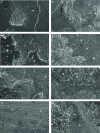
Fig 1
Fig 1. Scanning electron microscopy analysis of the different groups in sound and eroded dentin, 24 hours after the adhesive process.
(A) Representative specimen of the control group–sound dentin. Underlying dentin without exposure of the dentinal tubules (asterisk). (B) Representative specimen of the control group–eroded dentin, where it is possible to observe a greater bonding of the cement to the eroded substrate (asterisks). (C) Representative specimen 2% chlorhexidine sound dentin, it is to observe the presence of the resin cement (asterisks) on the smear layer (arrow). (D) Representative specimen 2% chlorhexidine eroded dentin, there is a remarkable exposure of the dentinal tubules compared to sound dentin (arrows). (E) Representative specimen 20% polyacrylic acid sound dentin, presence of the dentinal tubules partially occluded by the smear plug (arrows). (F) Representative specimen 20% polyacrylic acid eroded dentin, evidencing the presence of cement on the dentin (asterisks). (G) Representative specimen 0.1 M EDTA–sound dentin, it is noted that both the smear layer and the smear plug were removed, with great exposure of the dentinal tubules (arrows). (H) Representative specimen 0.1 M EDTA–eroded dentin, the presence of cement on the left with adjacent smear layer (asterisks), and evident increase in the opening of the dentinal tubules in the regions of exposed dentin (arrows), compared to the sound dentin of the same group. Magnification 1000x.
Fig 2
Fig 2. Scanning electron microscopy analysis of the different groups in sound and eroded dentin, 8 months after the adhesive process.
(A) Control group–sound dentin. Few or no presence of cement on the dentin, without exposure of the dentinal tubules (asterisks). (B) Control group–eroded dentin, showing the presence of mixed failure, with smear layer areas (asterisks) surrounded by resin cement (arrows). (C) 2% Chlorhexidine group–sound dentin. Presence of the resin cement on the dentin surface (asterisks), without exposure of the dentinal tubules. (D) 2% Chlorhexidine group–eroded dentin. Presence of the resin cement on the dentin surface (asterisks), whit few exposure of the dentinal tubules (arrows). (E) 20% Polyacrylic acid group–sound dentin. Few exposure of the dentinal tubules (asterisks). (F) 20% Polyacrylic acid group–eroded dentin. It is evidenced the presence of resinous material on the smear layer (asterisks) and many dentinal tubules exposed on the surface, with exposure of the collagen fibers (arrows); most of the exposed tubules are obliterated. (G) 0.1 M EDTA group–sound dentin, evidencing the presence of resin cement (asterisks), with few exposure of the dentinal tubules (arrow). (H) 0.1 M EDTA group–eroded dentin, evidencing the presence of a few exposed tubules on the surface and others filled by the resin cement (asterisks). Magnification 1000x.
Fig 3
Fig 3. Incidence (%) of failures mode among treatment groups, 24 hours after the adhesion process.
Fig 4
Fig 4. Incidence (%) of failures mode among treatment groups, 8 months after the adhesion process.
Fig 5
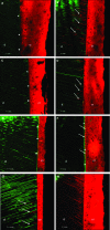
Fig 5. Analysis in confocal microscopy of sound and eroded dentin submitted to different surface treatments.
(A) Control group–sound dentin. It is possible to observe a continuous interface, without cracks (asterisks) and without the formation of tags. (B) Control group–eroded dentin. Adhesive interface without hydrid layer formation, however, an intense formation of resin tags (arrows) was observed in relation to the sound dentin. (C) 2% Chlorhexidine group–sound dentin. Note the absence of cracks in the adhesive interface as well as hybrid layer formation (asterisks). (D) 2% Chlorhexidine group–eroded dentin. Absence of cracks in the adhesive interface, with formation of some visible tags (arrows). (E) 20% Polyacrylic acid group–sound dentin. Pronounced hybrid layer formation throughout the adhesive interface (asterisks). (F) 20% Polyacrylic acid group–eroded dentin. Hybrid layer formation throughout the adhesive interface, with increased thickness compared to sound dentin (asterisks) and tag formation ≤ 3 μm in almost all interface (arrows). (G) 0.1 M EDTA group–sound dentin. Regarding the quality of the adhesive interface, it is possible to note the absence of cracks, with the formation of some resin tags measuring from 9 to 15 μm (arrows). (H) 0.1 M EDTA group–eroded dentin. Absence of cracks in the adhesive interface, without hybrid layer, but with the very dense formation of resin tags as well as in depth. *d = dentin / *rc = resin cement. Asterisks: hybrid layer formation; arrows: resin tags formation.

Similar articles

Cited by

References

    1. Scaramucci T, Hara AT, Zero DT, Ferreira SS, Aoki IV, Sobral MA. Development of an orange juice surrogate for the study of dental erosion. Braz Dent J. 2011; 22(6): 473–478. - PubMed
    1. Barbosa CS, Kato MT, Buzalaf MA. Effect of supplementation of soft drinks tea extract on their erosive potential against dentine. Aust Dent J. 2011; 56(3): 317–321. 10.1111/j.1834-7819.2011.01338.x - DOI - PubMed
    1. Hara AT, Livengood SV, Lippert F, Eckert GJ, Ungar PS. Dental surface texture characterization based on erosive tooth wear processes J Dent Res. 2016; 95(5): 537–542. 10.1177/0022034516629941 - DOI - PubMed
    1. Moretto MJ, Magalhães AC, Sassaki KT, Delbem AC, Martinhon CC. Effect of different fluoride concentrations of experimental dentifrices on enamel erosion and abrasion. Caries Res. 2010; 44(2): 135–140. 10.1159/000302902 - DOI - PubMed
    1. Buzalaf MA, Hannas AR, Kato MT. Saliva and dental erosion. J Appl Oral Sci. 2012; 20(5): 493–502. 10.1590/S1678-77572012000500001 - DOI - PMC - PubMed

Publication types