. 2018 Nov 26;14(11):e1007397.
doi: 10.1371/journal.ppat.1007397. eCollection 2018 Nov.

The RNA helicase DDX3X is an essential mediator of innate antimicrobial immunity

Affiliations

The RNA helicase DDX3X is an essential mediator of innate antimicrobial immunity

Daniel Szappanos et al. PLoS Pathog. .

Abstract

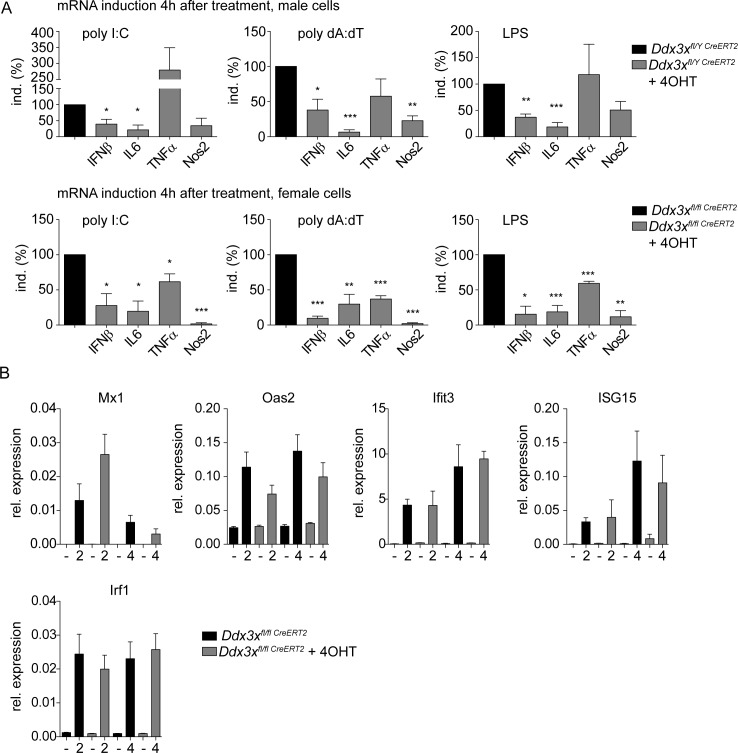
DExD/H box RNA helicases, such as the RIG-I-like receptors (RLR), are important components of the innate immune system. Here we demonstrate a pivotal and sex-specific role for the heterosomal isoforms of the DEAD box RNA helicase DDX3 in the immune system. Mice lacking DDX3X during hematopoiesis showed an altered leukocyte composition in bone marrow and spleen and a striking inability to combat infection with Listeria monocytogenes. Alterations in innate immune responses resulted from decreased effector cell availability and function as well as a sex-dependent impairment of cytokine synthesis. Thus, our data provide further in vivo evidence for an essential contribution of a non-RLR DExD/H RNA helicase to innate immunity and suggest it may contribute to sex-related differences in resistance to microbes and resilience to inflammatory disease.


Conflict of interest statement

The authors have declared that no competing interests exist.

Figures

Fig 1
Fig 1. Generation of Ddx3xfl/fl mice, analysis of DDX3X and DDX3Y activity in fibroblasts from gene-targeted mice.
A Targeting strategy. Black triangles indicate loxP sites, black bars the position of probes used to confirm deletion by Southern blot. B Western blot showing the complete absence of DDX3X protein in bone marrow-derived macrophages from Ddx3xfl/fl CreERT2 female and Ddx3xfl/y CreERT2 male mice after treatment with 4-OHT during mCSF-mediated differentiation. C Mouse embryonic fibroblasts (MEFs) derived from female Ddx3xfl/fl CreERT2 mice were treated with 4-OHT to delete Ddx3x. The cells were transiently transfected with 1 μg IRF3, 1 μg TBK1 and increasing amounts of DDX3Y and DDX3X (numbers denote μg transfected plasmid). One day after transfection, RNA was isolated and Ifnb expression was determined by qPCR. Bars represent the mean value +/-SD of technical replicates. Due to variations in transfection efficiencies across different experiments one representative experiment of at least three biological replicates is shown. D Mouse embryonic fibroblasts (MEFs) derived from male Ddx3xfl/y CreERT2 mice were treated with 4-OHT to remove DDX3X, or with Ddx3y siRNA as indicated. Subsequently the cells were transfected with 10 μg of poly (dA:dT). 4h after transfection, RNA was isolated and Ifnb expression was determined by qPCR. Bars represent the mean value +/-SD of 3 biological replicates. E Tbk1/Ikkε-deficient MEFs were transfected with 1 μg constitutively active IRF7-M15 and with increasing amounts of DDX3Y and DDX3X as indicated (right). Expression levels were normalized to Gapdh and to the expression level of cells transfected with empty vector. Bars represent the mean value +/-SD of technical replicates. Due to variations in transfection efficiencies across different experiments one representative experiment of at least three biological replicates is shown. F HEK293 cells were transfected with an NFκB reporter gene and the NFκB pathway was stimulated by co-transfecting different amounts of the adapter protein MAVS. The effect of DDX3X was assessed by additional co-transfection of a DDX3X expression plasmid. Bars represent the mean value +/-SD of technical replicates. Due to variations in transfection efficiencies across different experiments one representative experiment of at least three biological replicates is shown. Statistical analysis was carried out using unpaired, two-tailed Student's t-test. Mean and standard error of the mean (SEM) are indicated in the graphs. *P<0.05, **P<0.01, ***P<0.005.
Fig 2
Fig 2. Innate immunity of DDX3X-deficient cells and of Ddx3xfl/y Vav-iCre mice to VSV and Listeria monocytogenes infection.
A, B WT MEFs, or MEFs derived from Ddx3xfl/y CreERT2 mice and treated with 4-OHT to delete Ddx3x were infected with VSV at an MOI of 0,1. After the indicated periods, RNA was isolated and pan-Ifna (A) or Ifnb (B) mRNA expression was determined by qPCR. Lines represent the mean +/- standard deviation (SD). Statistical significance was calculated using the unpaired, two-tailed Student's t-test. *P<0.05, **P<0.01, ***P<0.005. C Supernatants from cells infected with VSV at an MOI of 0.1 were collected and viral loads at the indicated times were measured by plaque-forming unit (pfu) assay. Lines represent the mean +/- standard deviation (SD). Statistical significance was calculated using the unpaired, two-tailed Student's t-test. *P<0.05, **P<0.01, ***P<0.005. D Ddx3xfl/y and Ddx3xfl/y Vav-iCre mice (n = 7) were infected intravenously with 106 pfu of VSV. Survival was monitored for the indicated period of time. Statistical significance was calculated using the Log-rank (Mantel-Cox) test and Gehan-Breslow-Wilcoxon test. Differences between mouse genotypes did not reach statistical significance. E Ddx3xfl/y and Ddx3xfl/y Vav-iCre mice (n = 10) were injected intraperitoneally with 1,6x10⁵ CFU L. monocytogenes (strain EGD). Survival was monitored daily for the indicated period. Statistical significance was calculated using the Log-rank (Mantel-Cox) test (P-value: <0,0001) and Gehan-Breslow-Wilcoxon Test (P-value: <0,0001). F Ddx3xfl/y mice (n = 14) and Ddx3xfl/y Vav-iCre mice (n = 13) were injected intraperitoneally with 8x10⁴ CFU L. monocytogenes (strain EGD). After 72 h, mice were sacrificed and the bacterial load was determined in spleen and liver. Pooled data from three independent experiments is shown. Lines represent the mean +/- standard error of the mean (SEM). P-values were calculated using the unpaired, two-tailed Student's t-test. *P<0.05, **P<0.01, ***P<0.005.
Fig 3
Fig 3. Ddx3xfl/y Vav-iCre mice show altered cellular compositions in bone marrow and spleen.
A Bone marrow cells were isolated from uninfected Ddx3xfl/y and Ddx3xfl/y Vav-iCre mice and stained for indicated surface markers. Total cell numbers (referring to one femur and one tibia) are shown. n = 17 Ddx3xfl/y and 20 Ddx3xfl/y Vav-iCre mice for total, analyzed in 5 independent experiments. n = 12/13 for CD19+, Ter119+, CD3+ and CD11b+, analyzed in 4 independent experiments. B Spleens were isolated from uninfected Ddx3xfl/y and Ddx3xfl/y Vav-iCre mice and stained for indicated surface markers. Total cell numbers per organ are shown. Ddx3xfl/y (n = 20) and Ddx3xfl/y Vav-iCre mice (n = 23) were analyzed for total and CD3+ cell counts in 6 independent experiments. B220+ cells were analyzed in 5 independent experiments (n = 16/19). CD4+ and CD8+ (both pre-gated on CD3+), CD11b+F4/80+ and Ly6G+Ly6Clo and Ly6ChiLy6G-/lo (both pre-gated on CD11b+), were analyzed in 4 independent experiments (n = 12/13). NK1.1+ cells (pre-gated on CD3-), were analyzed in 4 independent experiments (n = 16/17). CD1d-tetramer+ (pre-gated on CD3+) and CD11c+cells were analyzed in 3 independent experiments (n = 8/9). C Bone marrow cells were isolated from uninfected Ddx3xfl/y and Ddx3xfl/y Vav-iCre mice and stained with a lineage mix (consisting of Gr1, Mac1, Ter119, B220 and CD3) and antibodies against cKit, Sca1 and CD127. Total cell numbers (referring to one femur and one tibia) for lineage- Sca1+ cKit+ (LSK), lineage- Sca1- cKit+ (LK), as well as lineage- Sca1/cKitdouble-dim CD127+ common lymphoid progenitor (CLP) cells are shown (n = 16 for Ddx3xfl/y and 20 for Ddx3xfl/y Vav-iCre mice for LSK and LK cells, analyzed in 4 independent experiments; n = 11/14 for CLP cells, analyzed in 4 independent experiments).
Fig 4
Fig 4. Loss of DDX3X affects hematopoiesis.
A Bone marrow cells were isolated from uninfected Ddx3xfl/y and Ddx3xfl/y Vav-iCre mice and stained for B220, CD43, BP1, CD24 and IgM. Total cell numbers (referring to one femur and one tibia) of CD43+ B220+ CD24- BP1- pre-pro B cells, CD43+ B220+ CD24+ BP1- early pro B cells, CD43+ B220+ CD24lo BP1+ late pro B cells, CD43+ B220+ CD24hi BP1+ large pre B cells, CD43- B220+ small pre B cells, B220lo IgM+ immature B cells and B220hi IgM+ mature B cells are shown (n = 9 for Ddx3xfl/y and 11 for Ddx3xfl/y Vav-iCre mice, analyzed in 2 independent experiments). B Bone marrow cells were isolated from uninfected Ddx3xfl/y and Ddx3xfl/y Vav-iCre mice and stained with a lineage mix (consisting of Gr1, Ter119, B220 and CD3) and antibodies against CD122, NK1.1 and Dx5. Total cell numbers (referring to one femur and one tibia) of lineage- CD122+ NK1.1- Dx5- NK progenitors (NKp), lineage- CD122+ NK1.1+ immature NK cells (iNK) and lineage- CD122+ NK1.1+ Dx5+ mature NK cells (mNK) are shown (n = 13 for Ddx3xfl/y and 15 for Ddx3xfl/y Vav-iCre mice, analyzed in 3 independent experiments).C Thymi were isolated from uninfected Ddx3xfl/y and Ddx3xfl/y Vav-iCre mice and stained for CD4 and CD8. Total cell numbers per organ of CD4+ CD8+ (DP), CD4+ CD8- (CD4SP) and CD4- CD8+ (CD8SP) are shown (n = 9 for Ddx3xfl/y and 11 for Ddx3xfl/y Vav-iCre mice, analyzed in 2 independent experiments). D Thymi were isolated from uninfected Ddx3xfl/y and Ddx3xfl/y Vav-iCre mice and stained with PBS-57 loaded CD1d-tetramer as well as antibodies against CD24, CD44 and NK1.1. Total numbers per organ of CD1d-tet+ CD24- CD44- NK1.1- (stage 1), CD1d-tet+ CD24- CD44+ NK1.1- (stage 2) and CD1d-tet+ CD24- CD44+ NK1.1+ (stage 3) NKT cells are shown (n = 9 Ddx3xfl/y and 11 Ddx3xfl/y Vav-iCre mice, analyzed in 2 independent experiments).
Fig 5
Fig 5. Absence of DDX3X increases hematopoietic cell death.
Spleens were isolated from uninfected Ddx3xfl/y and Ddx3xfl/y Vav-iCre mice and stained with fluorochrome-coupled Annexin V plus the indicated surface markers, followed by staining with 7-AAD. Percentage of Annexin V+ and/or 7AAD+ cells are shown, as indicated (n = 9 for Ddx3xfl/y and 11 for Ddx3xfl/y Vav-iCre mice, analyzed in 2 independent experiments). Statistical analysis was carried out using unpaired, two-tailed Student's t-test. Mean and standard error of the mean (SEM) are indicated in the graphs. *P<0.05, **P<0.01, ***P<0.005.
Fig 6
Fig 6. Ddx3xfl/y Vav-iCre mice show alterations in spleen and peritoneum upon Listeria monocytogenes infection.
A Ddx3xfl/y and Ddx3xfl/y Vav-iCre mice were infected intraperitoneally with 1x10⁵ CFU Listeria monocytogenes (strain EGD) for 24 h, spleens were isolated, homogenized and stained with the indicated surface markers, followed by flow cytometry. Total cell numbers per organ are shown (n = 16 for Ddx3xfl/y and 20 for Ddx3xfl/y Vav-iCre mice for total, B220+ and CD3+ cells, analyzed in 5 independent experiments; n = 4/8 for CD4+, CD8+ and CD1d-tetramer+ cells, all pre-gated on CD3+, analyzed in 2 independent experiments; n = 4/8 for NK1.1+ cells (pre-gated on CD3-), analyzed in two independent experiments; n = 12/12 for CD11c+, CD11b+F4/80+ cells, analyzed in 3 independent experiments; n = 12/12 for Ly6G+Ly6C-/lo and Ly6GloLy6Chi cell populations, both pre-gated on CD11b+and analyzed in 3 independent experiments). B Ddx3xfl/y mice and Ddx3xfl/y Vav-iCre mice were infected intraperitoneally with 1x10⁵ CFU L. monocytogenes (strain EGD) for 24h or treated with PBS as a control. Peritoneal exudate cells were collected via peritoneal lavage and stained with the indicated surface markers. Total cell numbers of B220+ B cells, CD3+ T cells, Ly6G+Ly6Clo neutrophils and Ly6G-/lo Ly6Chi monocytes (both pre-gated on CD11b+) and CD11b+ F4/80+ macrophages are shown. n = 7 (Ddx3xfl/y mice) and 5 (Ddx3xfl/y Vav-iCre mice) for PBS controls and n = 12/11 for infected animals. P-values were calculated using the unpaired, two-tailed Student's t-test. Mean and standard error of the mean (SEM) are indicated in the graphs. *P<0.05, **P<0.01, ***P<0.005.
Fig 7
Fig 7. Reduced NK-mediated IFNγ production in Ddx3xfl/y Vav-iCre mice infected with Listeria monocytogenes.
A Ddx3xfl/y and Ddx3xfl/y Vav-iCre mice were infected intraperitoneally with 1x10⁵ CFU L. monocytogenes (strain EGD) for the indicated periods of time. n ≥ 3 for each genotype. Mice were sacrificed and serum was collected. Cytokine levels were analyzed by flow cytometry-based bead array. B Ddx3xfl/y and Ddx3xfl/y Vav-iCre mice were infected intraperitoneally with 1x10⁵ CFU L. monocytogenes (strain EGD) for 24 h, spleens were isolated, homogenized and splenocytes were stained for indicated surface markers followed by fixation, permeabilization and intracellular staining for IFNγ. Data show percent of IFNγ-producing CD3ε-NK1.1+NKp46+ (NK), CD3ε+CD8+ (CD8+) and CD3ε+CD1d-tet+ (iNKT) cells. n = 7 for Ddx3xfl/y and n = 11 for Ddx3xfl/y Vav-iCre mice, analyzed in 3 independent experiments. C After depleting NK-cells with an anti-NK1.1 antibody, Ddx3xfl/y and Ddx3xfl/y Vav-iCre mice were infected intraperitoneally with 1x 10⁵ CFU L. monocytogenes (strain EGD) for 24 h. Mice were sacrificed, serum was collected and IFNγ levels were determined by ELISA. n = 6 for Ddx3xfl/y and 7 for Ddx3xfl/y Vav-iCre mice, analyzed in 2 independent experiments. D After depleting NK-cells with an anti-NK1.1 antibody, wt mice were infected intraperitoneally with 1x 10⁵ CFU L. monocytogenes (strain EGD) for 72 h. The mice were sacrificed and the bacterial load was determined in spleen and liver by CFU assay. Lines in all panels represent the mean with standard error of the mean (SEM). Statistical significance was calculated using the unpaired, two-tailed Student's t-test. *P<0.05, **P<0.01, ***P<0.005.
Fig 8
Fig 8. Loss of DDX3X compromises the immune response of macrophages to Listeria monocytogenes.
A Bone marrow-derived macrophages (BMDMs) of Ddx3xfl/y CreERT2 male and Ddx3xfl/fl CreERT2 female mice were infected with L. monocytogenes (strain LO28) with a multiplicity of infection (MOI) of 10. Colony-forming units (CFU) were determined 1, 2, 4 or 8 h after infection by plating serial dilutions of lysed cells on brain–heart infusion (BHI) agar plates. The last two columns in the right panel represent cells pretreated with IFNγ over night. n = 6, analyzed in 2 independent experiments, for male BMDMs (left panel). n = 12, analyzed in 4 independent experiments, for female BMDMs (right panel). Data are represented as mean values +/- standard deviations (SD). Statistical analysis was performed using the unpaired, two-tailed Student's t-test. * P<0.05. B BMDM of mice with the indicated genotypes were left untreated or infected with L. monocytogenes at MOI 10 for 4 hours as described below. RNA isolated from the infected cells was isolated and subjected to RNA-Seq. Left panel: heat map of log 2-fold gene expression changes between Listeria-treated and untreated samples for genes differentially expressed upon treatment in WT samples of either sex (padj <0.01, abs(logFC)>1; DeSeq2 v1.18.1). Genes assigned to the GO category GO:0045087 innate immune response are indicated on the left. From left to right, genes from female knock-out, female wild-type, male knock-out and male wild-type cells (as indicated below the lanes) are compared. Upper right panel: functional enrichment analysis of the differentially expressed genes using DAVID v6.8; the top 10-enriched GO terms ranked by p-value for the category GOTERM_BP_3 are shown. Lower right panel: Venn diagram of gene sets differentially expressed upon Lm infection in the four experimental conditions displayed in the heat map (DESeq2 v 1.18.1; absolute log2 fold change > 1; FDR < 0.01). Solid blue: Lm-infected versus uninfected in macrophages representing a male wildtype genotype (DDX3Xfl/y); light blue: Lm-infected versus uninfected in macrophages representing a male DDX3X-deficient genotype (DDX3X-/y); Solid red: Lm-infected versus uninfected in macrophages representing a female wildtype genotype (DDX3Xfl/fl); light red: Lm-infected versus uninfected in macrophages representing a female DDX3X-deficient genotype (DDX3X-/-). C Bone marrow-derived macrophages (BMDMs) of Ddx3xfl/y CreERT2 male and Ddx3xfl/fl CreERT2 female mice with or without 4_OHT-mediated DDX3X deletion, were infected with L. monocytogenes (strain LO28) with a multiplicity of infection (MOI) of 10. After 4 h of infection total RNA was isolated and analyzed by qPCR for expression of the indicated genes. Upper panels show results from female cells, lower panels show results from male cells, normalized to their respective controls (male and female macrophages without 4-OHT treatment = 100%). Bars represent mean values +/- standard deviations (SD) of at least three independent experiments. Statistical significance was calculated using the paired, two-tailed Student's t-test. *P<0.05, **P<0.01, ***P<0.005. D Primers flanking intron sequences were chosen to analyze the primary unprocessed transcripts in untreated or 4-OHT treated BMDMs of Ddx3xfl/fl CreERT2 female mice. Cells were infected with L. monocytogenes (strain LO28) with a multiplicity of infection (MOI) of 10. After 2 and 4 h of infection total RNA was isolated and analyzed by qPCR for expression of the indicated primary transcripts. Bars represent mean values +/- standard deviations (SD) of at least three independent experiments, normalized to male and female controls (macrophages without 4-OHT treatment = 100%). Statistical significance was calculated using the paired, two-tailed Student's t-test. *P<0.05, **P<0.01, ***P<0.005.
Fig 9
Fig 9. DDX3X deficient macrophages show compromised response to TLR ligands, but respond normally to IFNs.
A Bone marrow-derived macrophages (BMDMs) of Ddx3xfl/y CreERT2 male and Ddx3xfl/fl;CreERT2 female mice with or without 4_OHT-mediated DDX3X deletion, were treated with the indicated pathogen-associated molecular patterns (PAMPs). After 4 h of treatment total RNA was isolated and analyzed by qPCR for expression of the indicated genes. Upper panel shows results from male cells, lower panel shows results from female cells. Bars represent mean values +/- standard deviations (SD) of at least three independent experiments, normalized to their respective controls (male and female macrophages without 4-OHT treatment = 100%). Statistical significance was calculated using the paired, two-tailed Student's t-test. *P<0.05, **P<0.01, ***P<0.005. B Bone marrow-derived macrophages (BMDMs) of Ddx3xfl/fl CreERT2 female mice with (grey bars) or without (black bars) 4-OHT-mediated DDX3X deletion, were treated with IFNβ (250U/ml) or IFNγ (5ng/ml) for 2 h or 4 h, as indicated. Total RNA was isolated and expression levels of the indicated genes was analyzed by qPCR. Bars represent mean values +/- standard error of the mean (SEM) of three independent experiments.

References

    1. Goubau D, Deddouche S, Reis E Sousa C. Cytosolic sensing of viruses. Immunity. 2013;38: 855–869. 10.1016/j.immuni.2013.05.007 - DOI - PMC - PubMed
    1. Paludan SR, Bowie AG. Immune sensing of DNA. Immunity. 2013;38: 870–880. 10.1016/j.immuni.2013.05.004 - DOI - PMC - PubMed
    1. Stavrou S, Aguilera AN, Blouch K, Ross SR. DDX41 Recognizes RNA/DNA Retroviral Reverse Transcripts and Is Critical for In Vivo Control of Murine Leukemia Virus Infection. MBio. American Society for Microbiology; 2018;9: e00923–18. 10.1128/mBio.00923-18 - DOI - PMC - PubMed
    1. Soulat D, Burckstummer T, Westermayer S, Goncalves A, Bauch A, Stefanovic A, et al. The DEAD-box helicase DDX3X is a critical component of the TANK-binding kinase 1-dependent innate immune response. EMBO J. 2008; 27: 2135–2146. 10.1038/emboj.2008.126 - DOI - PMC - PubMed
    1. Schröder M, Baran M, Bowie AG. Viral targeting of DEAD box protein 3 reveals its role in TBK1/IKKepsilon-mediated IRF activation. EMBO J. 2008; 27: 2147–2157. 10.1038/emboj.2008.143 - DOI - PMC - PubMed

Publication types

MeSH terms