Review
. 2019 May 1;43(3):273-303.
doi: 10.1093/femsre/fuy042.

Extracellular membrane vesicles in the three domains of life and beyond

Affiliations
Review

Extracellular membrane vesicles in the three domains of life and beyond

Sukhvinder Gill et al. FEMS Microbiol Rev. .

Abstract

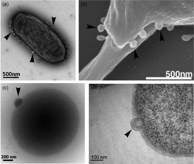
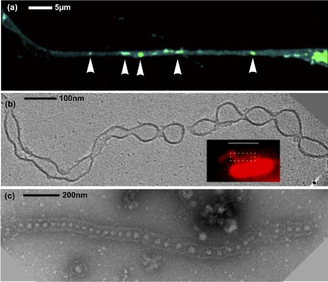
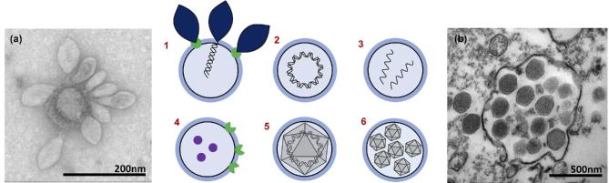
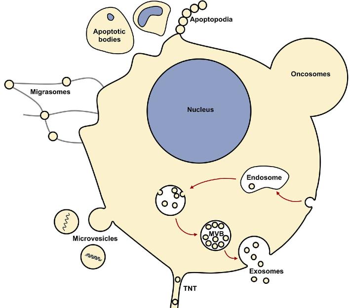
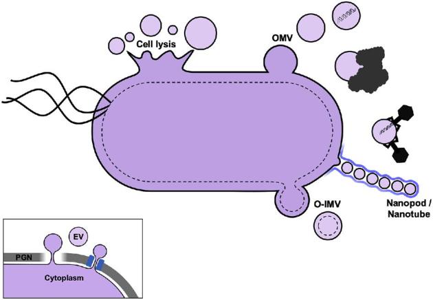
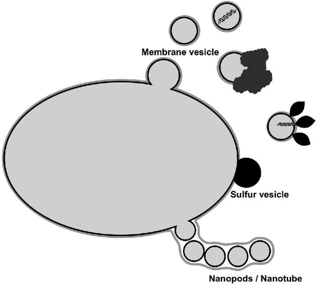
Cells from all three domains of life, Archaea, Bacteria and Eukarya, produce extracellular vesicles (EVs) which are sometimes associated with filamentous structures known as nanopods or nanotubes. The mechanisms of EV biogenesis in the three domains remain poorly understood, although studies in Bacteria and Eukarya indicate that the regulation of lipid composition plays a major role in initiating membrane curvature. EVs are increasingly recognized as important mediators of intercellular communication via transfer of a wide variety of molecular cargoes. They have been implicated in many aspects of cell physiology such as stress response, intercellular competition, lateral gene transfer (via RNA or DNA), pathogenicity and detoxification. Their role in various human pathologies and aging has aroused much interest in recent years. EVs can be used as decoys against viral attack but virus-infected cells also produce EVs that boost viral infection. Here, we review current knowledge on EVs in the three domains of life and their interactions with the viral world.

Keywords: Archaea; LUCA; evolution; extracellular vesicles; nanotubes; virus.


Figures

Figure 1.
Figure 1.
Biogenesis of extracellular vesicles in the three domains of life. Vesicle budding indicated with arrows. (a) TEM showing hypervesiculation in the bacterium S. typhimurium. Image kindly provided by Mario F. Feldman (University of Alberta, Canada). (b) SEM showing microvesicles budding from the eukaryote Leishmania donovani. Image reprinted from Silverman et al. (2008). (c) Cryo-TEM of vesicle budding from the archaeon T. kodakaerensis. The protrusion of the S layer can also be observed clearly. (d) TEM of ultrathin cell sections of vesicle budding from T. kodakaerensis. Figures (c) and (d) provided by the authors.
Figure 2.
Figure 2.
Nanotube production in the three domains of life. (a) TNT connecting eukaryotic (human) cells, with labeled vesicles indicated by arrows. Adapted with permission from Keller et al. (2017): image cropped and arrow style altered. (b) 'Nanotubes' produced by the bacteria S. oneidensis form outer membrane extensions with regular constrictions forming vesicles. Adapted with permission from Subramanian et al. (2018). Image courtesy of Poorna Subramanian (California Institute of Technology, USA). (c) 'Nanopods' produced by the archaeon T. prieurii. Discrete vesicles are surrounded by the cellular S-layer forming a tubular structure. Image kindly provided by Aurore Gorlas (Institute for Integrative Biology of the Cell, Université Paris-Saclay, France).
Figure 3.
Figure 3.
EVs and viruses interact in multiple ways. 1 and (a): Virus receptors on vesicles could act as decoys protecting the host from infection. (a) TEM showing several Sulfolobus spindle-shaped virus 1 (SSV1), from the Fuselloviridae family, attached to a membrane vesicle. 2 and 3: Encapsulated DNA/ RNA can be infectious as in pleolipoviruses or plasmidions. 4: Virus receptors and effectors can transfer between cells, promoting infection of non-susceptible hosts. 5: Membrane-bound viruses resist human attack. 6 and (b): VPVs allow for high MOI and 'Trojan horse’-style infection. Image (a) kindly provided by Virginija Krupovic, Institut Pasteur, France. Image (b) kindly provided by Jônatas Santos Abrahão, Institute of Biological Sciences, Universidade Federal de Minas Gerais, Brazil and obtained by the Center of Microscopy of UFMG, Brazil.
Figure 4.
Figure 4.
EV production in Eukaryotes. Multiple types of EVs originate through many complex and varied pathways. Eukaryotic EV functions include protein sorting/trafficking, intercellular communication, host adaptation during infection, metastatic niche adaptation, immune evasion and pathogenesis.
Figure 5.
Figure 5.
EV production in Bacteria. Two main types of EVs originate from diderm bacteria (OMVs and O-IMVs); however, cell lysis and nanotubes also produce EVs. Functions include intercellular communication, HGT, biofilm formation/maintenance, biomineralization, pathogenesis, viral defense, disposal/detoxification and relief of envelope stress. Inset: EVs in Firmicutes are produced from the single cytoplasmic membrane and must cross the thick PGN layer either by degradation of PGN or through pores.
Figure 6.
Figure 6.
TEM of ultrathin sections of EVs from three bacterial species. Both OMVs and O-IMVs are observed in these EV preparations, with features of O-IMVs indicated. O-IMVs are surrounded by an external bilayer, probably corresponding to the outer membrane (OM) of the cell, and an inner membrane (IM), probably corresponding to the cytoplasmic membrane, which entraps electron dense material. In the image of O-IMVs from A. baumannii, the putative peptidoglycan layer (PG) can be seen. Images kindly provided by Elena Mercade and Carla Pérez-Cruz (Universitat de Barcelona, Spain).
Figure 7.
Figure 7.
EV production in Archaea. EVs originate from membrane budding and nanotubes. Functions include HGT, intercellular competition, disposal/detoxification, biomineralization and possibly viral defense.

References

    1. Abels ER, Breakefield XO. Introduction to extracellular vesicles: biogenesis, RNA cargo selection, content, release, and uptake. Cell Mol Neurobiol 2016;36:301–12. - PMC - PubMed
    1. Acehan D, Santarella-Mellwig R, Devos DP. A bacterial tubulovesicular network. J Cell Sci 2014;127:277–80. - PubMed
    1. Acosta JC, Banito A, Wuestefeld T et al.. A complex secretory program orchestrated by the inflammasome controls paracrine senescence. Nat Cell Biol 2013;15:978–90. - PMC - PubMed
    1. Albers SV, Meyer BH. The archaeal cell envelope. Nat Rev Microbiol 2011;9:414–26. - PubMed
    1. Albuquerque PC, Nakayasu ES, Rodrigues ML et al.. Vesicular transport in Histoplasma capsulatum: an effective mechanism for trans-cell wall transfer of proteins and lipids in ascomycetes. Cell Microbiol 2008;10:1695–710. - PMC - PubMed

Publication types