Review
. 2019 Jan 1;60(1):69-79.
doi: 10.1093/jrr/rry096.

Clustered DNA double-strand break formation and the repair pathway following heavy-ion irradiation

Affiliations
Review

Clustered DNA double-strand break formation and the repair pathway following heavy-ion irradiation

Yoshihiko Hagiwara et al. J Radiat Res. .

Abstract

Photons, such as X- or γ-rays, induce DNA damage (distributed throughout the nucleus) as a result of low-density energy deposition. In contrast, particle irradiation with high linear energy transfer (LET) deposits high-density energy along the particle track. High-LET heavy-ion irradiation generates a greater number and more complex critical chromosomal aberrations, such as dicentrics and translocations, compared with X-ray or γ irradiation. In addition, the formation of >1000 bp deletions, which is rarely observed after X-ray irradiation, has been identified following high-LET heavy-ion irradiation. Previously, these chromosomal aberrations have been thought to be the result of misrepair of complex DNA lesions, defined as DNA damage through DNA double-strand breaks (DSBs) and single-strand breaks as well as base damage within 1-2 helical turns (<3-4 nm). However, because the scale of complex DNA lesions is less than a few nanometers, the large-scale chromosomal aberrations at a micrometer level cannot be simply explained by complex DNA lesions. Recently, we have demonstrated the existence of clustered DSBs along the particle track through the use of super-resolution microscopy. Furthermore, we have visualized high-level and frequent formation of DSBs at the chromosomal boundary following high-LET heavy-ion irradiation. In this review, we summarize the latest findings regarding the hallmarks of DNA damage structure and the repair pathway following heavy-ion irradiation. Furthermore, we discuss the mechanism through which high-LET heavy-ion irradiation may induce dicentrics, translocations and large deletions.


Figures

Fig. 1.
Fig. 1.
Scale diagrams of heavy-ion–induced DNA damage. The scale diagrams of heavy-ion–induced complex DNA lesions and chromatin fiber. Heavy-ion radiation causes complex DNA lesions, which contain DSBs, and SSBs and/or base damage within 1–2 helical turns, along the particle track. The scale of a chromatin fiber is less than ~30 nm.
Fig. 2.
Fig. 2.
3D-SIM analysis reveals clustered DSB formation following heavy-ion irradiation. (A) Representative images for 3D γH2AX polygon rendering using 3D-SIM analysis. 1BR hTERT cells were fixed at 2 h after 1 Gy carbon-ion irradiation (290 MeV/n, Mono, LET 60 keV/μm), and fixed cells were stained with antibodies for γH2AX, RPA and CENPF and with DAPI. To detect RPA foci, which show clear and sharp signals for SIM analysis, G2 cells were examined. G2 cells were identified by CENPF (the original data are shown in [12]). The raw image of γH2AX is shown in the left panel. γH2AX polygon rendering, which is generated by surface mode in Imaris 8.1.2 (Bitplane, Zurich, Switzerland), is shown in the right panel. γH2AX foci and DAPI are shown as yellow and blue polygons, respectively. The direction of the carbon ion radiation is indicated by a white arrow. (B) 3D polygon rendering of γH2AX and the spot signal of RPA in (A) are shown. Clustered RPA foci are identified within the γH2AX signal in carbon ion–irradiated G2 cells. γH2AX the signal is shown by the polygon. RPA foci are shown as green spots. (C) A diagram for DSB distribution after high-LET particle irradiation. Super resolution imaging allows visualization of the distribution of γH2AX along the chromatin loop. RPA forms a single focus representing a DSB unless >2 DSBs are formed within 100 nm.
Fig. 3.
Fig. 3.
Formation of clustered DSBs at the chromosome the boundary following heavy-ion irradiation. (A) The frequency of γH2AX foci formation at chromosome boundary after high-LET carbon ion irradiation is greater than after X-rays. A representative image of γH2AX and chromosome 1 using combination staining of immunofluorescence (IF) and fluorescence in situ hybridization (FISH) is shown. 1BR hTERT G1 cells were synchronized by contact inhibition. Cells were fixed at 15 min after 3 Gy carbon-ion irradiation (290 MeV/n, Mono, LET 70 keV/μm). γH2AX foci, chromosome 1 and DAPI are shown by red, green and blue, respectively. Enlarged images are shown in the right panel. Images from different angles are shown in the right bottom panel. The percentage of γH2AX foci formation at the chromosome boundary after carbon ion irradiation (8.64%) is 4-fold greater than that after X-rays (2.23%) [29]. The direction of the carbon ion radiation is indicated by a white arrow. (B) The representative image of clustered γH2AX foci formation following carbon ion irradiation. Clustered γH2AX foci in 1BR hTERT cells are also observed by high-resolution microscopy involving deconvolution, not 3D-SIM [18]. Cells were stained with γH2AX and DAPI at 30 min after 1 Gy carbon-ion irradiation (290 MeV/n, Mono, LET 60 keV/μm). Enlarged γH2AX foci are shown in the right panel. γH2AX foci and DAPI are shown by green and blue. Although it is technically not feasible to obtain high-resolution or super-resolution Grade IF samples after sample preparation for FISH, combining evidence strongly suggests that γH2AX foci formation at the chromosome boundary after high-LET carbon ion irradiation contains multiple DSBs. The direction of the carbon ion radiation is indicated by a white arrow. (C) A diagram of the formation of chromosome rearrangement via mis-rejoining between two distinct chromosomes following high-LET heavy-ion irradiation. High-LET heavy-ion radiation causes multiple DSB formation within a limited area. Furthermore, high-LET heavy-ion radiation can cause multiple DSBs at chromosome boundaries. If two or more DSBs are induced at the chromosome boundary between different chromosomes A and B, interchromosomal exchanges such as dicentrics or translocations may occur.
Fig. 4.
Fig. 4.
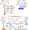
DSB repair pathway following heavy-ion radiation. (A) The model of the DSB repair pathway in G2 cells following X-ray irradiation. Recent studies have demonstrated that ~70% of DSBs are repaired by NHEJ in human G2 cells, whereas ~30% of DSBs are repaired by HR in G2 cells. Ku70/80 and DNA-PKcs complex bind all the DSB ends. Approximately 70% of DSBs are rapidly rejoined using XLF, XRCC4 and LIG4 in c-NHEJ. For HR, DNA end resection is initiated by MRE11 endonuclease, which is stimulated by CtIP. MRE11 endonuclease activity creates nicks, followed by exonucleases digesting ssDNA either 3′ to 5′ or 5′ to 3′. MRE11 digests ssDNA by its 3′ to 5′ exonuclease activity, proceeding to the DSB terminal, whereas EXO1 exonuclease digests ssDNA from 5′ to 3′ with BLM and DNA2. Extension of resection is promoted by BRCA1. The process of this pathway is summarized in [53]. Following resection, ssDNA is rapidly coated with RPA, which is then displaced by RAD51. RAD51 promotes recombination with the sister chromatid, and HR is completed. (B) The model of the DSB repair pathway following heavy-ion irradiation. Approximately 90% of DSBs induced by high-LET heavy-ion irradiation are repaired by resection-mediated pathway, i.e. mainly HR and others, e.g. SSA or alt-NHEJ. The increased percentage of HR usage after high-LET heavy-ion irradiation can be explained by the speed of DSB repair. In human G1 cells, which primarily use NHEJ but not other pathways, DSB repair after high-LET particle irradiation shows significantly slower kinetics than that after X- or γ-ray irradiation [31]. This suggests that DSB end complexity influences the speed of DSB repair. The current model proposes that, in G2 phase, NHEJ factor initially binds to DSB ends; however, when rapid NHEJ does not ensue, DSB end resection and HR occur [31, 45]. Thus, when DSB end complexity is induced by high-LET particle irradiation, as shown in (2) and (3), these DSBs showing the delay of DSB repair are repaired by HR. At DSB ends induced by high-LET particle irradiation, the resection is both CtIP-dependent and -independent. We postulate four pathways leading to resection-mediated DSB repair in heavy-ion dependent resection. (1) MRE11 endonuclease initiates resection with CtIP. After the incision, EXO1/BLM/DNA2 promote extension of resection as described above. (2) When SSBs are generated close to DSBs, it may bypass the step of MRE11/CtIP-dependent resection. (3) When base damage is generated close to DSBs, the damaged base is removed by DNA glycosylases and a nick is generated by AP endonucleases. Afterwards, the generated nick may trigger EXO1/BLM/DNA2-dependent extension of resection without MRE11/CtIP-dependent endonuclease activity. (4) Ku binding on the DSB end may be prevented by base damage or SSB repair or BER proteins in the presence of complex DNA lesions. EXO1/BLM exonucleases may readily promote resection due to the structure of the DSB terminal, to which Ku cannot bind. It has not been fully investigated whether all the resected DSB ends are repaired by a precise HR pathway or other error prone pathways, e.g. alt-NHEJ, SSA or other pathways.
Fig. 5.
Fig. 5.
c-NHEJ is the major DSB repair pathway in G1 phase following high-LET heavy-ion irradiation. Olaparib (10 μM KU-0059436) was added 30 min before IR. 1BR hTERT, human normal fibroblasts, were irradiated with 3 Gy carbon ions (290 MeV/n, Mono, 70 keV/μm), and cells were fixed at indicated time points. G1 cells were identified as CENPF-/EdU-(39). A box plot of a single experiment is shown. Similar results were obtained in two independent experiments. *P < 0.05, **P < 0.01, ***P < 0.001.
Fig. 6.
Fig. 6.
Hallmarks of DNA damage following heavy-ion irradiation. Schematic diagram for how heavy-ion irradiation induced complex DNA lesions and chromosome rearrangements led to cell death or senescence-like growth arrest (SLGA). (1) High-LET heavy-ion radiation causes clustered DSBs along the particle track. (2) In addition, high-LET heavy-ion radiation causes complex DNA lesions, which contain DSBs, SSBs and/or base damage within 1–2 helical turns. These types of damage are rarely observed in X-ray–irradiated cells. (3) We identified the formation of clustered DSBs at the chromosome boundaries along the particle track. The formation of clustered DSBs at the chromosome boundaries can be a critical risk for chromosomal rearrangements such as dicentrics, translocations and large deletions. The two major types of deletion (i.e. interstitial deletion and terminal deletion) are induced by IR. Small chromosome fragments are produced following interstitial deletion if multiple DSBs in close proximity occur in a single chromosome. Because these fragments can be recognized as DSBs and are not readily repaired, the persistent fragments activate cell cycle checkpoint arrest, apoptosis, or SLGA. High-LET radiation produces such interstitial deletion-derived fragments more frequently than X-ray irradiations, because clustered DSBs are generated along the particle track. In general, chromosome rearrangements including dicentrics, translocations and large deletions induce cell death or SLGA in the first mitosis or after the mitosis. In contrast, interstitial deletions, followed by the formation of fragments, cause cell death and/or SLGA in non-dividing cells. In future work, it will be important to elucidate the impact of complex DNA lesions on cell fate, i.e. whether a high level of resection or the existence of unrepairable damage due to complex DNA lesions affects chromosome rearrangement, cell death and SLGA.

Similar articles

Cited by

References

    1. Ritter S, Durante M. Heavy-ion induced chromosomal aberrations: a review. Mutat Res 2010;701:38–46. - PubMed
    1. Durante M, Cucinotta FA. Heavy ion carcinogenesis and human space exploration. Nat Rev Cancer 2008;8:465–72. - PubMed
    1. Durante M, Orecchia R, Loeffler JS. Charged-particle therapy in cancer: clinical uses and future perspectives. Nat Rev Clin Oncol 2017;14:483–95. - PubMed
    1. Veldeman L, Madani I, Hulstaert F et al. . Evidence behind use of intensity-modulated radiotherapy: a systematic review of comparative clinical studies. Lancet Oncol 2008;9:367–75. - PubMed
    1. Loeffler JS, Durante M. Charged particle therapy—optimization, challenges and future directions. Nat Rev Clin Oncol 2013;10:411–24. - PubMed

MeSH terms