Review
. 2019 Jan;143(1):26-35.
doi: 10.1016/j.jaci.2018.11.015. Epub 2018 Nov 23.

The microbiome in patients with atopic dermatitis

Affiliations
Review

The microbiome in patients with atopic dermatitis

Amy S Paller et al. J Allergy Clin Immunol. 2019 Jan.

Erratum in

  • Corrigendum.
    [No authors listed] [No authors listed] J Allergy Clin Immunol. 2019 Apr;143(4):1660. doi: 10.1016/j.jaci.2019.01.022. J Allergy Clin Immunol. 2019. PMID: 30954089 No abstract available.

Abstract

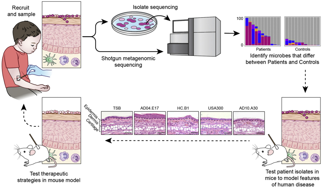
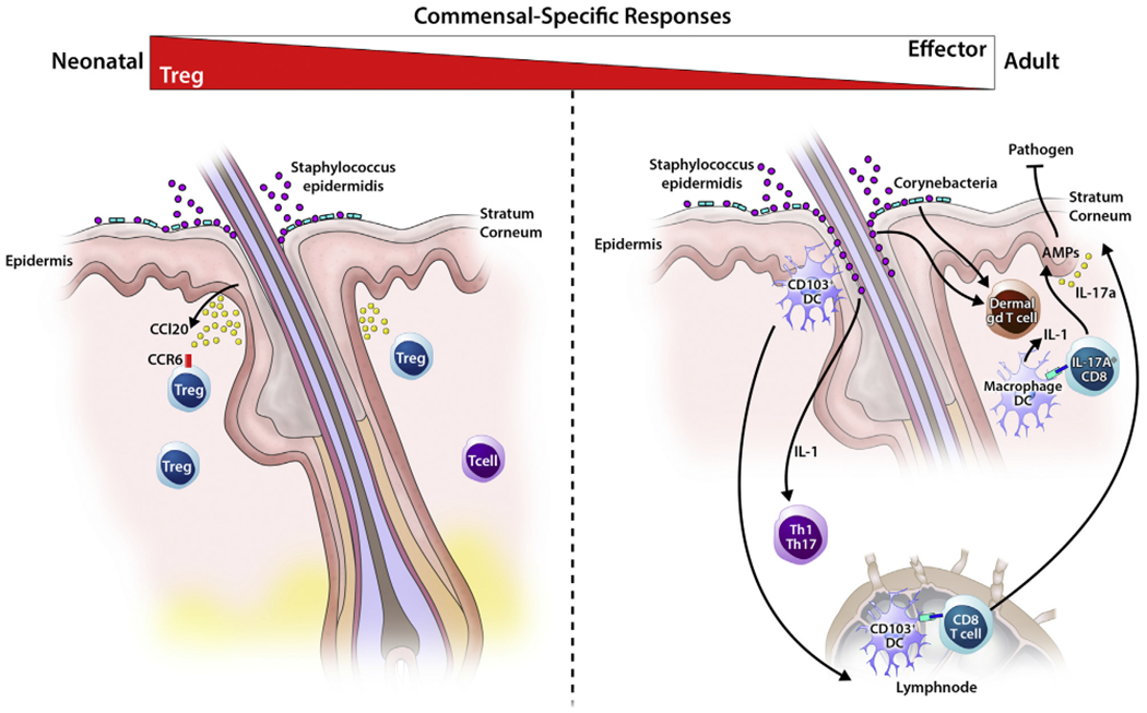
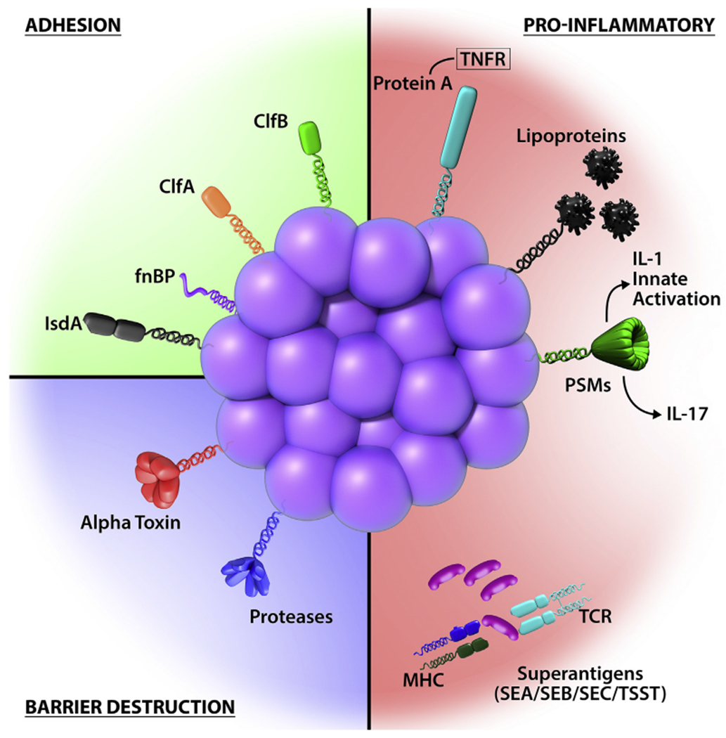
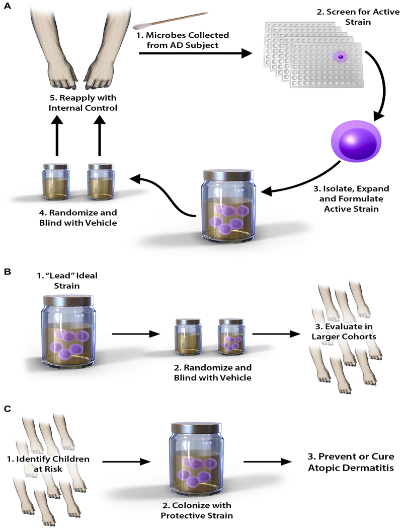
As an interface with the environment, the skin is a complex ecosystem colonized by many microorganisms that coexist in an established balance. The cutaneous microbiome inhibits colonization with pathogens, such as Staphylococcus aureus, and is a crucial component for function of the epidermal barrier. Moreover, crosstalk between commensals and the immune system is now recognized because microorganisms can modulate both innate and adaptive immune responses. Host-commensal interactions also have an effect on the developing immune system in infants and, subsequently, the occurrence of diseases, such as asthma and atopic dermatitis (AD). Later in life, the cutaneous microbiome contributes to the development and course of skin disease. Accordingly, in patients with AD, a decrease in microbiome diversity correlates with disease severity and increased colonization with pathogenic bacteria, such as S aureus. Early clinical studies suggest that topical application of commensal organisms (eg, Staphylococcus hominis or Roseomonas mucosa) reduces AD severity, which supports an important role for commensals in decreasing S aureus colonization in patients with AD. Advancing knowledge of the cutaneous microbiome and its function in modulating the course of skin disorders, such as AD, might result in novel therapeutic strategies.

Keywords: Atopic dermatitis; Staphylococcus aureus; biotherapy; commensal; host-microbiome interaction; immune regulation; microbiome.


Figures

FIG 1.
FIG 1.
An example of a pipeline for skin microbiome studies in patients with AD. The pipeline can begin by posing a scientific question, with subsequent recruitment of patients and control subjects, phenotyping of patients with AD, and collection of clinical samples (top left). Microbiota from clinical samples can either be directly sequenced to study the complex communities of microbiota (the microbiome) or first cultivated to investigate individual clinical isolates through whole-genome sequencing (and/or with model systems). Shotgun metagenomic sequencing refers to the sequencing of all genetic material in a complex sample and can provide information on bacteria, fungi, and viruses within the sample; the functional potential of the mixed microbial communities; and the different microbial strains. Sequencing both cultivated isolates (whole-genome sequencing) and the complex communities of microbes (shotgun metagenomics sequencing) can provide complementary information. The bioinformatics analyses of microbial sequencing data can identify the microbiome differences between patients with AD and healthy control subjects, which is graphically represented by bar charts that indicate the relative abundances of different staphylococci shown as different colors. The y-axis of bar charts represents 0% to 100% relative abundance. Differences in the microbiota found on patients versus control subjects can be studied in mouse models using clinically relevant isolates to examine microbial strain-level differences. For example, differences in host responses can be observed in the histology (lower panels) from mice who undergo application of different patient-associated staphylococcal strains (AD10.A30, USA300, HC.B1, AD04.E17) versus tryptic soy broth (TSB) control; comparisons can be made in epidermal thickening and immune cell infiltrates elicited by different strains. Further studies might provide insight into the role of the skin microbiome in disease pathogenesis, which could lead to development of microbiome-targeted therapeutics for patients.
FIG 2.
FIG 2.
Age-dependent immune-commensal crosstalk in skin. Left panel, Some cutaneous immune cells, in particular CD4+ T cells generated in response to the commensal S epidermidis, are notably age dependent. Neonatal colonization by S epidermidis yields a population of antigen-specific CD4+ T cells dominated by Treg cells. In contrast, when exposure is delayed until adulthood, cytokine-producing effector CD4+ T cells predominate. Only early-life exposure is conducive to the establishment of antigen-specific immune tolerance and protection against skin inflammation on subsequent exposure. One factor accounting for this age-dependent response is the high density of Treg cells found in neonatal skin, which conditions the skin for tolerogenic interactions with the microbiota through yet undefined mechanisms. Neonatal skin Treg cells are preferentially localized around hairfollicles, a dense niche for commensal skin microbes. Right panel, Colonization of adult skin with specific microbiota results in an IL-1–meditated homeostatic effector immune response, including TH1 and TH17 cells, as well as dermal IL-17A+ γδ T cells. Certain strains of S epidermidis also induce commensal-specific populations of IL-17A+CD8+ T cells (TC17) through CD103+ dendritic cells.
FIG 3.
FIG 3.
S aureus has highly evolved multiple cell-wall proteins and secreted factors that enable adhesion to human skin and barrier disturbance by using physical, chemical, and inflammatory mechanisms. Adhesion, S aureus has developed several surface molecules to adhere to the human stratum corneum, including clumping factors A and B (ClfA and ClfB), fibronectin-binding protein (fnBP), and iron-regulated surface determinant A (IsdA). Barrier destruction, S aureus α-toxin, a water-soluble cytotoxin, forms a heptameric β-barrel pore in host cell membranes. In the epidermis it directlyforms pores in keratinocytes, which erodes the integrity of the epidermal barrier. S aureus produces at least 10 proteases, a number of which facilitate dissolution of the stratum corneum. In addition to secreted proteases, S aureus can directly stimulate endogenous keratinocyte proteases, including KLK6, KLK13, and KLK14, highlighting an additional mechanism toward barrier destruction. Proinflammatory mechanisms, Cell-wall bound protein A, when solubilized, triggers inflammatory responses from keratinocytes through TNF receptor (TNFR). Staphylococcal superantigens, such as SEA, SEB, SEC, and toxic shock syndrome toxin-1 (TSST-1), trigger B-cell expansion and cytokine release. S aureus secretes PSMs, which are direct proinflammatory drivers with compartment-specific effects. In the epidermal compartment PSMs stimulate IL-36α-driven γδ T cell–mediated inflammation, whereasinthe dermal compartmentthey stimulate IL-1β-driven TH17 inflammation.
FIG 4.
FIG 4.
Potential role of biotherapy in patients with AD. Biotherapy for AD takes advantage of the natural antipathogen properties of human CoNS species. A, As a first step, microbes are grown out from swabs of an affected subject, with the most biologically active strains then selected for expansion. This generates the lead ideal strain, which is trialed back on the donor subject and compared with a randomized placebo vehicle. B, Ifsuccessful atthis stage,the lead strain istrialed in a largercohortof affected subjects. C, Finally, the strain is applied to high-risk children as a preventative measure.

References

    1. Byrd AL, Belkaid Y, Segre JA. The human skin microbiome. Nat Rev Microbiol 2018;16:143–55. - PubMed
    1. Findley K, Oh J, Yang J, Conlan S, Deming C, Meyer JA, et al. Topographic diversity of fungal and bacterial communities in human skin. Nature 2013;498: 367–70. - PMC - PubMed
    1. Grice EA, Kong HH, Conlan S, Deming CB, Davis J, Young AC, et al. Topographical and temporal diversity of the human skin microbiome. Science 2009;324:1190–2. - PMC - PubMed
    1. Oh J, Byrd AL, Deming C, Conlan S, NISC program, Kong HH, et al. Biogeography and individuality shape function in the human skin metagenome. Nature 2014;514:59–64. - PMC - PubMed
    1. Costello EK, Lauber CL, Hamady M, Fierer N, Gordon JI, Knight R. Bacterial community variation in human body habitats across space and time. Science 2009;326:1694–7. - PMC - PubMed

Publication types