

Protein identification strategies in MALDI imaging mass spectrometry: a brief review
- PMID: 30476689
- PMCID: PMC6382520
- DOI: 10.1016/j.cbpa.2018.10.023
Protein identification strategies in MALDI imaging mass spectrometry: a brief review
Abstract
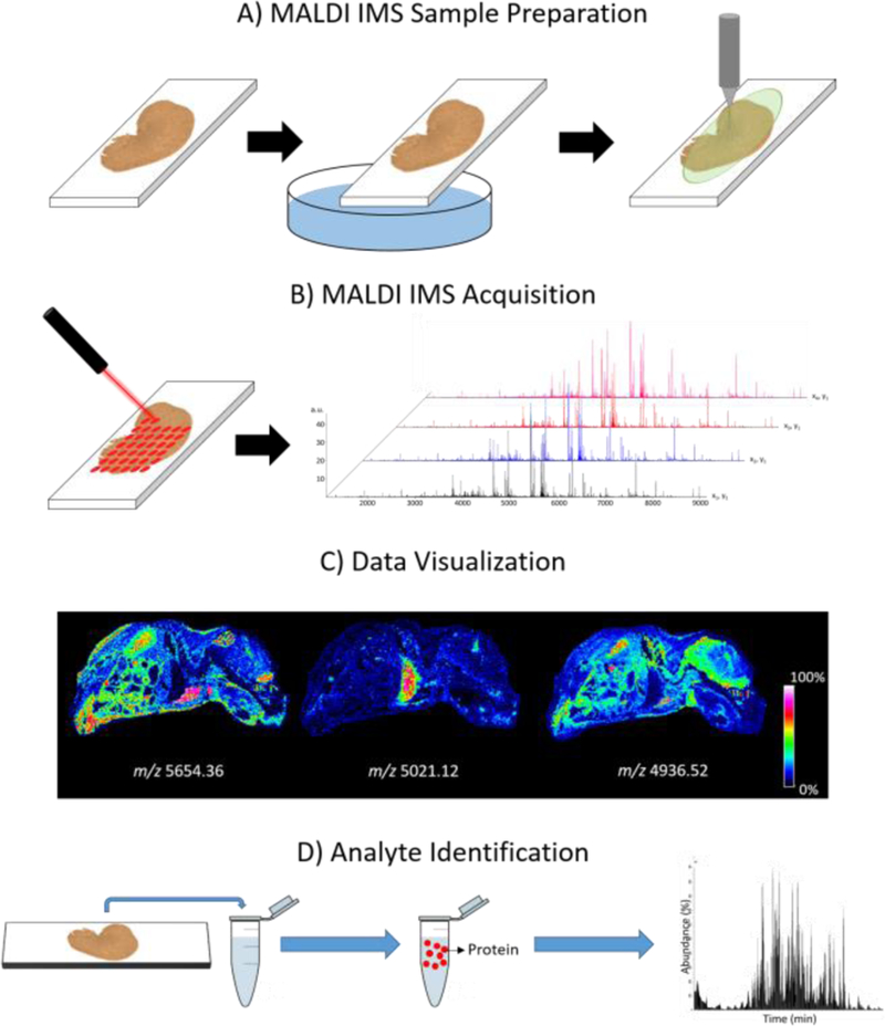
Matrix assisted laser desorption/ionization (MALDI) imaging mass spectrometry (IMS) is a powerful technology used to investigate the spatial distributions of thousands of molecules throughout a tissue section from a single experiment. As proteins represent an important group of functional molecules in tissue and cells, the imaging of proteins has been an important point of focus in the development of IMS technologies and methods. Protein identification is crucial for the biological contextualization of molecular imaging data. However, gas-phase fragmentation efficiency of MALDI generated proteins presents significant challenges, making protein identification directly from tissue difficult. This review highlights methods and technologies specifically related to protein identification that have been developed to overcome these challenges in MALDI IMS experiments.
Copyright © 2018 Elsevier Ltd. All rights reserved.
Figures

References
-
- Caprioli RM, Farmer TB, Gile J: Molecular imaging of biological samples: Localization of peptides and proteins using MALDI-TOF MS. Analytical Chemistry 1997, 69:4751–4760. - PubMed
-
-
Kompauer M, Heiles S, Spengler B: Atmospheric pressure MALDI mass spectrometry imaging of tissues and cells at 1.4-mu m lateral resolution. Nature Methods 2017, 14:90–96.
Using a modified MALDI source, the authors present an atmospheric pressure MALDI MSI setup capable of 1.4 µm lateral resolution and a mass resolution greater than 100,000. They achieve this by using a focusing objective with a numerical aperature of 0.9 at 337 nm and a free working distance of 18 mm in coaxial geometry, coupled to an orbitrap mass spectrometer.
-
-
- Qin L, Zhang YW, Liu YQ, He HX, Han MM, Li YY, Zeng MM, Wang XD: Recent advances in matrix-assisted laser desorption/ionisation mass spectrometry imaging (MALDI-MSI) for in situ analysis of endogenous molecules in plants. Phytochemical Analysis 2018, 29:351–364. - PubMed
-
- Pratavieira M, Menegasso ARD, Esteves FG, Sato KU, Malaspina O, Palma MS: MALDI Imaging Analysis of Neuropeptides in Africanized Honeybee (Apis mellifera) Brain: Effect of Aggressiveness. Journal of Proteome Research 2018, 17:2358–2369. - PubMed
-
- Ly A, Buck A, Balluff B, Sun N, Gorzolka K, Feuchtinger A, Janssen KP, Kuppen PJK, van de Velde CJH, Weirich G, et al. : High-mass-resolution MALDI mass spectrometry imaging of metabolites from formalin-fixed paraffin-embedded tissue. Nature Protocols 2016, 11:1428–1443. - PubMed
Publication types
MeSH terms
Substances
Grants and funding
LinkOut - more resources
Full Text Sources
Research Materials
