Review
. 2018 Dec:53:178-186.
doi: 10.1016/j.sbi.2018.11.001. Epub 2018 Nov 23.

Structural gymnastics of RAG-mediated DNA cleavage in V(D)J recombination

Affiliations
Review

Structural gymnastics of RAG-mediated DNA cleavage in V(D)J recombination

Heng Ru et al. Curr Opin Struct Biol. 2018 Dec.

Abstract

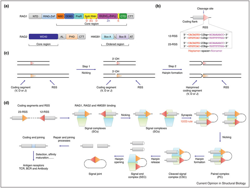
A hallmark of vertebrate immunity is the diverse repertoire of antigen-receptor genes that results from combinatorial splicing of gene coding segments by V(D)J recombination. The (RAG1-RAG2)2 endonuclease complex (RAG) specifically recognizes and cleaves a pair of recombination signal sequences (RSSs), 12-RSS and 23-RSS, via the catalytic steps of nicking and hairpin formation. Both RSSs immediately flank the coding end segments and are composed of a conserved heptamer, a conserved nonamer, and a non-conserved spacer of either 12 base pairs (bp) or 23 bp in between. A single RAG complex only synapses a 12-RSS and a 23-RSS, which was denoted the 12/23 rule, a dogma that ensures recombination between V, D and J segments, but not within the same type of segments. This review recapitulates current structural studies to highlight the conformational transformations in both the RAG complex and the RSS during the consecutive steps of catalysis. The emerging structural mechanism emphasizes distortion of intact RSS and nicked RSS exerted by a piston-like motion in RAG1 and by dimer closure, respectively. Bipartite recognition of heptamer and nonamer, flexibly linked nonamer-binding domain dimer relatively to the heptamer recognition region dimer, and RSS plasticity and bending by HMGB1 together contribute to the molecular basis of the 12/23 rule in the RAG molecular machine.


Conflict of interest statement

Conflict of interest

There are no conflicts of interest to declare.

Figures

Figure 1
Figure 1
Overview of RAG-mediated V(D)J recombination pathway. (a) Domain organization of RAG1, RAG2 and HMGB1 proteins. NTD: N-terminal domain; RING-ZnF: RING domain and zinc finger domain; NBD: nonamer binding domain; DDBD: dimerization and DNA binding domain; PreR: pre-RNase H domain; RNH: RNase H-like domain; ID: insertion domain, which can be further divided into two parts, ZnC2 and ZnH2; CTD: C-terminal domain; CTT: C-terminal tail; WD40: tryptophan-aspartic acid repeat domain; AL: acidic linker; PHD: plant homeodomain; L: linker; AT: acidic tail. Potential catalytic residues are indicated as red dots in RNH and the zinc ion is indicated as a slate dot in ID. Core or ordered regions in RAG1, RAG2 and HMGB1 are indicated. (b) Schematic representation of RSSs. The consensus sequences of heptamer and nonamer are shown in red and magenta respectively. (c) Schematic representation of RAG mediated catalysis that involves two consecutive reactions, nicking and hairpin formation. (d) Overview of the V(D)J recombination process. The coding regions and RSSs are shown as rectangles and triangles respectively. The dimeric (RAG1-RAG2)2 complex is shown as stacked cyan and light green trapezoids. Briefly, RAG binds a single RSS in the presence of HMGB1 to form signal complexes (SCs), either 12-SC or 23-SC, which can undergo nicking (shown by a yellow pentagram) at the coding flank–RSS junction in the presence of Mg2+. Synapsis of one 12-RSS and one 23-RSS in the same RAG dimer forms the paired complex (PC), followed by generation of the cleaved signal complex (CSC) with hairpin coding end and cleaved signal end. Hairpin release produces the signal end complex (SEC). Further processing by enzymes in the non-homologous end-joining (NHEJ) DNA repair pathway results in ligation of the coding ends and circularization of the signal ends. This panel is adapted from Figure 1a of Ru et al [39]
Figure 2
Figure 2
Overview of the RAG complex structures and the structural basis of the 12/23 rule. (a) Overall structure of the Apo-RAG dimer (PDB: 4WWX). The domains are colored as in Figure 1(a). (b) Overall structure of the RAG dimer in complex with nicked 12- and 23-RSS intermediates (PDB: 6DBI). The domains are colored as in Figure 1(a). The 12-RSS and 23-RSS intermediates are shown in light slate with heptamer and nonamer are highlighted in red and magenta respectively. (c) Schematic representation of the interactions between RAG dimer and RSSs based on the synaptic RAG complex with nicked RSS intermediates. The dimer axes of the catalytic region and the NBD dimer do not coincide. (d) Orthogonal views of the superposition between Apo-RAG (green), RAG dimer singly bound to intact 12-RSS (cyan), and RAG dimer doubly bound to intact 12- and 23-RSS substrates (magenta) by aligning one RAG1-RAG2 monomer (PDB: 4WWX, 6DBX, and 6DBT). The NBDs are highlighted in darker green, cyan and magenta respectively. The intact 12-RSS from the singly bound RAG complex is omitted. Tilt of NBD dimer upon substrate RSS engagement is obvious. (e) Bottom view of the NBD dimer movement upon nicking of the intact RSS substrates. NBD dimers from RAG bound to intact RSSs (PDB: 6DBT) and nicked RSSs (PDB: 6DBI) are colored in magenta and orange respectively.
Figure 3
Figure 3
DNA distortions and RAG movements in the consecutive catalytic steps. (a) Cartoon representation of the DNAs that are bound to the RAG complex at different states. Left: unmelted RSS (PDB: 6DBU); middle: melted RSS (PDB: 6DBR); right: nicked RSS (PDB: 6DBJ). The heptamer of the RSS is shown in red and scissile phosphate in the nucleotide is highlighted in cyan. The catalytic metal ions shown as orange spheres indicate the location of the active site. (b) DNA distortion and base flipping in the nicked RSS intermediate facilitate hairpin formation. The heptamer in the RSS is shown in red and the flipped base and scissile phosphate are highlighted in cyan. The location of the nucleophile 3’–OH is indicated. The RNH domain is shown in yellow ribbon, and the catalytic residues and coordinated metal ions are shown in green sticks and orange spheres respectively. (c) DNA melting in the intact RSS substrate facilitates the nicking step. The color scheme is the same as in (b). (d) Different location of the α15 helices in the IDs of RAG1 from Apo-RAG (light green, PDB: 4WWX), RAG bound to unmelted RSS (light magenta, PDB: 6DBT), RAG bound to melted RSS (blue, PDB: 6DBV) and RAG bound to nicked RSS (orange, PDB: 6DBI) when RAG1-RAG2 monomers are aligned. Only the IDs are shown and the location of Zn2+ is indicated. (e) α15–α16 loop from the first RAG monomer and the ordered β4–β5 loop from the second RAG monomer stabilizes the melted RSS (PDB: 6DBR). The α15–α16 region is shown as blue ribbon and the β4–β5 loop from the second RAG (RNH’ in yellow) is highlighted in green. The heptamer in the RSS is shown in red and the nucleotide to be nicked is highlighted in cyan. (f) Closure of the RAG dimer upon binding of nicked RSS intermediates. The Apo-RAG (PDB: 4WWX) and synaptic RAG (PDB: 6DBI) bound to nicked 12- and 23-RSS (red and blue) are shown as green and orange ribbons. (g) Opening of the RAG dimer upon intact RSS substrates binding. The Apo-RAG (PDB: 4WWX) and synaptic RAG (PDB: 6DBI) that bound to intact 12- and 23-RSS (red and blue) are shown as green and magenta ribbons. (h) Structure-derived insights on RAGmediated cleavage pathway in V(D)J recombination. RAG1 (light green and light cyan), RAG2 (green and cyan), and HMGB1 (orange) are represented as cartoons. Coding segments, heptamers, nonamers, and 3’ extension DNAs are shown by semi-transparent blue, red, magenta, and light slate rectangles, respectively. 12 and 23 bp spacers are shown as black lines. Briefly, without binding to an RSS, apo-RAG is in an open conformation with the flexibly attached NBD dimer. When bound to a singly intact RSS (e.g. 12-RSS) or simultaneously bound to 12- and 23-RSS intact DNA in the presence of HMGB1, an even open conformation of RAG dimer is induced. The NBD dimer is tilted through the interaction with nonamer(s) from the RSS(s). Then, the heptamer in the bound RSS(s) will undergo the melting step, which induces the rotation of the coding flank and the localization of the scissile phosphate into the active site to facilitate nicking. When both 12- and 23-RSS are synapsed and nicked in the same RAG dimer, the PC is formed, which assumes a fully closed conformation with flipped bases in the coding flank (blue filled circle) and the RSS (red filled circle) on both nicked RSS intermediates to facilitate the hairpin formation. The coding ends are then linked into hairpin DNA to form the CSC. NHEJ factors are recruited to dissociate the hairpin coding ends, leaving the SEC with only the bound signal ends. However, the steps of how RAG captures the other RSS after nicking on the first RSS is not known.
Figure 4
Figure 4
Molecular ruler supervises the accurate positioning and cleavage on the RSS. (a) Cartoon representation of the first RAG monomer that performs catalysis. The first RAG monomer is shown in gray surface. In the superimposed ribbon, ID and RNH are in magenta and yellow with the remaining RAG1 in gray, and RAG2 is in blue. The heptamer and nonamer of bound RSS is highlighted in red and magenta. Of note, the conserved CAC sequence in the heptamer lacks recognition by RAG. (b) Cartoon representation of the second RAG monomer that recognizes the last three positions of the heptamer, the spacer and the nonamer of the RSS. The second RAG monomer is shown in gray surface with superimposed ribbon. The heptamer and nonamer of bound RSS is highlighted in red and magenta. The NBD dimer (orange), DDBD (blue) and CTD (green) from the second RAG recognize the nonamer, spacer and last three positions of the heptamer and thus serve as a molecular ruler that positions the CAC of the heptamer into the active site of the first RAG.

Similar articles

Cited by

References

    1. Fanning L, Connor A, Baetz K, Ramsden D, Wu GE: Mouse RSS spacer sequences affect the rate of V(D)J recombination. Immunogenetics 1996, 44:146–150. - PubMed
    1. Tonegawa S: Somatic generation of antibody diversity. Nature 1983, 302:575–581. - PubMed
    1. Early P, Huang H, Davis M, Calame K, Hood L: An immunoglobulin heavy chain variable region gene is generated from three segments of DNA: VH, D and JH. Cell 1980, 19:981–992. - PubMed
    1. Taccioli GE, Rathbun G, Oltz E, Stamato T, Jeggo PA, Alt FW: Impairment of V(D)J recombination in double-strand break repair mutants. Science 1993, 260:207–210. - PubMed
    1. Sakano H, Maki R, Kurosawa Y, Roeder W, Tonegawa S: Two types of somatic recombination are necessary for the generation of complete immunoglobulin heavy-chain genes. Nature 1980, 286:676–683. - PubMed

Publication types

LinkOut - more resources