Sequential Neuroimaging of the Fetus and Newborn With In Utero Zika Virus Exposure
- PMID: 30476967
- PMCID: PMC6583436
- DOI: 10.1001/jamapediatrics.2018.4138
Sequential Neuroimaging of the Fetus and Newborn With In Utero Zika Virus Exposure
Abstract
Importance: The evolution of fetal brain injury by Zika virus (ZIKV) infection is not well described.
Objectives: To perform longitudinal neuroimaging of fetuses and infants exposed to in utero maternal ZIKV infection using concomitant magnetic resonance imaging (MRI) and ultrasonography (US), as well as to determine the duration of viremia in pregnant women with ZIKV infection and whether the duration of viremia correlated with fetal and/or infant brain abnormalities.
Design, setting, and participants: A cohort of 82 pregnant women with clinical criteria for probable ZIKV infection in Barranquilla, Colombia, and Washington, DC, were enrolled from June 15, 2016, through June 27, 2017, with Colombian women identified by community recruitment and physician referral and travel-related cases of American women recruited from a Congenital Zika Program.
Interventions and exposures: Women received 1 or more MRI and US examinations during the second and/or third trimesters. Postnatally, infants underwent brain MRI and cranial US. Blood samples were tested for ZIKV.
Main outcomes and measures: The neuroimaging studies were evaluated for brain injury and cerebral biometry.
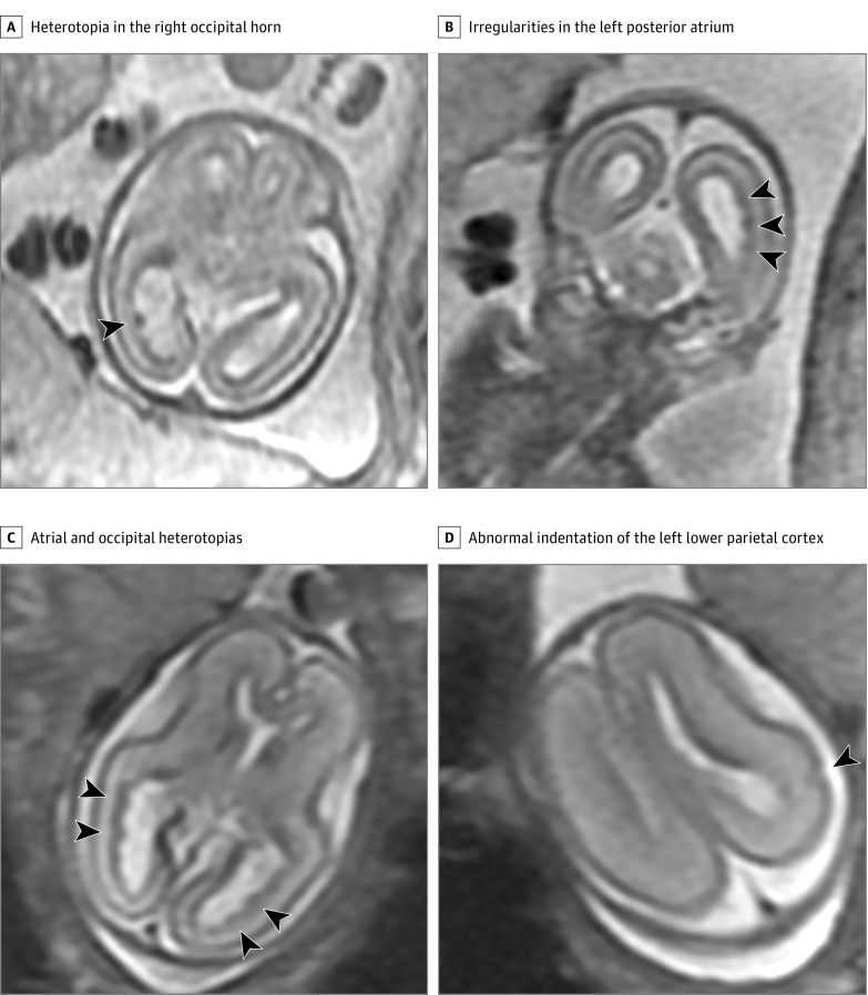
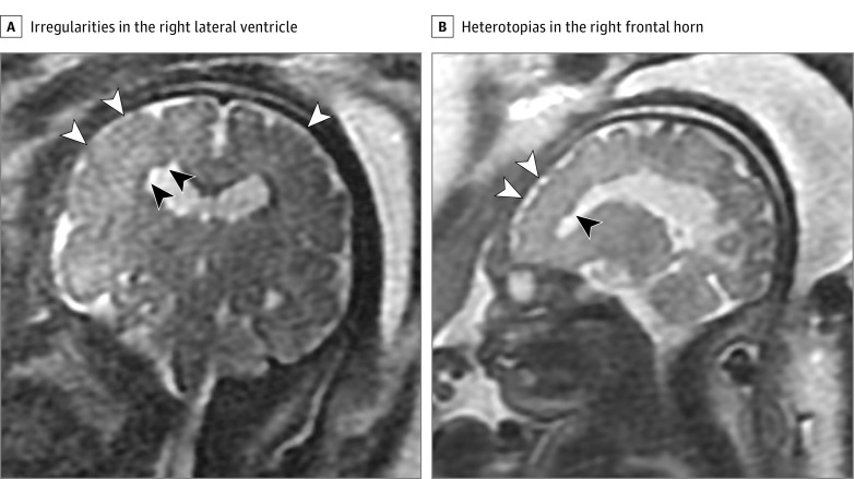
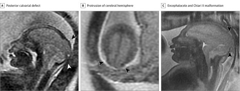
Results: Of the 82 women, 80 were from Colombia and 2 were from the United States. In 3 of 82 cases (4%), fetal MRI demonstrated abnormalities consistent with congenital ZIKV infection. Two cases had heterotopias and malformations in cortical development and 1 case had a parietal encephalocele, Chiari II malformation, and microcephaly. In 1 case, US results remained normal despite fetal abnormalities detected on MRI. Prolonged maternal polymerase chain reaction positivity was present in 1 case. Of the remaining 79 cases with normal results of prenatal imaging, postnatal brain MRI was acquired in 53 infants and demonstrated mild abnormalities in 7 (13%). Fifty-seven infants underwent postnatal cranial US, which detected changes of lenticulostriate vasculopathy, choroid plexus cysts, germinolytic/subependymal cysts, and/or calcification in 21 infants (37%).
Conclusions and relevance: In a cohort of pregnant women with ZIKV infection, prenatal US examination appeared to detect all but 1 abnormal fetal case. Postnatal neuroimaging in infants who had normal prenatal imaging revealed new mild abnormalities. For most patients, prenatal and postnatal US may identify ZIKV-related brain injury.
Conflict of interest statement
Figures
Comment in
-
Revealing the Effects of Zika-Detection of Brain Abnormalities and Other Disabilities Associated With Congenital Infection.JAMA Pediatr. 2019 Jan 1;173(1):16-18. doi: 10.1001/jamapediatrics.2018.4164. JAMA Pediatr. 2019. PMID: 30476947 Free PMC article. No abstract available.
References
Publication types
MeSH terms
Substances
Grants and funding
LinkOut - more resources
Full Text Sources
Medical
Molecular Biology Databases