. 2018 Nov 23;9(12):570.
doi: 10.3390/genes9120570.

UrdA Controls Secondary Metabolite Production and the Balance between Asexual and Sexual Development in Aspergillus nidulans

Affiliations

UrdA Controls Secondary Metabolite Production and the Balance between Asexual and Sexual Development in Aspergillus nidulans

Sandesh S Pandit et al. Genes (Basel). .

Abstract

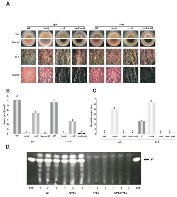
The genus Aspergillus includes important plant pathogens, opportunistic human pathogens and mycotoxigenic fungi. In these organisms, secondary metabolism and morphogenesis are subject to a complex genetic regulation. Here we functionally characterized urdA, a gene encoding a putative helix-loop-helix (HLH)-type regulator in the model fungus Aspergillus nidulans. urdA governs asexual and sexual development in strains with a wild-type veA background; absence of urdA resulted in severe morphological alterations, with a significant reduction of conidial production and an increase in cleistothecial formation, even in the presence of light, a repressor of sex. The positive effect of urdA on conidiation is mediated by the central developmental pathway (CDP). However, brlA overexpression was not sufficient to restore wild-type conidiation in the ΔurdA strain. Heterologous complementation of ΔurdA with the putative Aspergillus flavus urdA homolog also failed to rescue conidiation wild-type levels, indicating that both genes perform different functions, probably reflected by key sequence divergence. UrdA also represses sterigmatocystin (ST) toxin production in the presence of light by affecting the expression of aflR, the activator of the ST gene cluster. Furthermore, UrdA regulates the production of several unknown secondary metabolites, revealing a broader regulatory scope. Interestingly, UrdA affects the abundance and distribution of the VeA protein in hyphae, and our genetics studies indicated that veA appears epistatic to urdA regarding ST production. However, the distinct fluffy phenotype of the ΔurdAΔveA double mutant suggests that both regulators conduct independent developmental roles. Overall, these results suggest that UrdA plays a pivotal role in the coordination of development and secondary metabolism in A. nidulans.

Keywords: Aspergillus nidulans; UrdA; brlA; central developmental pathway; epistasis; morphological development; mycotoxin; secondary metabolism; sterigmatocystin; transcription factor; veA.


Conflict of interest statement

The authors declare no conflict of interest.

Figures

Figure 1
Figure 1
Multiple Sequence Alignment of UrdA orthologs from Aspergillus species. Sequences from orthologs of (A) A. nidulans (UrdA), A. Calidoustus (Acal), A. rambellii (Aram), A. ochraceoroseus (Aoch), A. sydowii (Asyd) and A. versicolor (Aver) were aligned using Clustal Omega and visualized using Genedoc (see Materials and Methods). The extension of the predicted N-terminal, central and C-terminal regions, as well as the putative nuclear localization signal (NLS) and helix-loop-helix (HLH) domains are indicated by black lines. (B) Hydrophobic cluster analysis (HCA) of UrdA. Residues in green indicate hydrophobic clusters and, thus, possible secondary structures. Residues in blue are positively charged amino acids while those in red are negatively charged. Proline, serine, glycine and threonine are designated by symbols. See Materials and Methods for additional details.
Figure 2
Figure 2
3D modeling of UrdA sequence. (A) Sequence alignment of AN4394/AnUrdA and its A. flavus ortholog KOC16320/AfUrdA. The position and extension of the three predicted α-helices of the putative HLH transcriptional regulatory domain are indicated by blue lines while orange lines delimit the extension of the loops. (B) Swiss-model images of a hypothetic dimer formed by the HLH domains of AnUrdA and AfUrdA. Helices are shown in blue and loops in orange.
Figure 3
Figure 3
urdA promotes asexual development. (A) Wild type (WT), ΔurdA and urdA-complementation (urdA-com) strains were point-inoculated on glucose minimal medium (GMM) plates and incubated for 7 days in light and dark. Micrographs were taken with a Leica MZ75 dissecting microscope attached to a Leica DC50LP camera at 50× magnification after spraying the plates with 70% ethanol to enhance visualization of cleistothecia. The white arrows indicates the cleistothecia. (B) Quantification of conidiophores in 36 h and 48 h cultures grown on solid GMM in light and dark. Top-agar inoculated cultures were used to analyze the expression of brlA (C) abaA (D) and wetA (E). Values are means of three replicates and Error bars indicate standard errors. Different letters above the bar graphs represent significantly different values (p ≤ 0.05).
Figure 4
Figure 4
Overexpression of brlA is not sufficient to induce conidiation in the absence of urdA. (A) Aspergillus nidulans wild type (TRV50) and ΔurdA strains were transformed with an overexpression plasmid containing A. nidulans brlA coding region driven by the inducible promoter alcA. Diagnostic PCR was used to confirm the incorporation of the overexpression plasmid into the genome of the ΔurdA host strain after transformation, using primers AN_alcA(P)_F & ANbrlA_R (Table S2), labeled in this figure as F and R respectively. The expected 3 kb PCR product was obtained. (B) A. nidulans wild type (WT), ΔurdA, OEbrlA and ΔurdA-OEbrlA strains were inoculated with 106 conidia/mL in liquid GMM at 250 rpm for 18 h and then shifted onto solid GMM or TMM. Micrographs were taken after 12 days with a Leica MZ75 dissecting microscope attached to a Leica DC50LP camera at 50× magnification. (C) One gram of mycelium was also inoculated in 50 mL of liquid TMM and grown at 37 °C for 18 h after shift to observed possible formation of conidia in submerged cultures. Black arrows show formation of conidia from the tip of hyphae.
Figure 5
Figure 5
urdA represses sexual development. (A) Quantification of cleistothecia (both immature and mature) in 36 h and 48 h cultures grown on solid GMM in light and dark. Asterisks indicate not detected. Top-agar inoculated cultures were used to analyze the expression of nsdD (B) steA (C) and veA (D). Values are means of three replicates and error bar indicates standard errors. Different letters above the bar graphs represent significantly different values (p ≤ 0.05).
Figure 6
Figure 6
Heterologous complementation of A. flavus urdA in the A. nidulans ΔurdA strain. (A) A. nidulans and A. flavus urdA complementation strains (urdA-com and AflurdA-com respectively), together with the ΔurdA and wild type (WT) control were point-inoculated on GMM and incubated for 4 days at 37 °C in the light. (B) Quantification of conidia. Cores (7 mm diameter) were collected 1 cm from the colony center, homogenized in water and counted under the microscope. (C) Quantification of cleistothecia. Cores (16 mm diameter) were harvested 1 cm from the colony center and sprayed with 70% ethanol to improve visualization of fruiting bodies. Asterisks indicate not detected. Values are means of three replicates and error bars indicate standard errors. Different letters above the bar graphs represent significantly different values (p ≤ 0.05).
Figure 7
Figure 7
Effect of urdA on sterigmatocystin (ST) production in A. nidulans. (A) Thin-layer chromatography (TLC) analysis of ST produced by wild type (WT) and ΔurdA cultures grown on top-agar inoculated solid GMM at 37 °C in light and dark conditions for 36 h, and 48 h. ST, a commercial ST standard from SIGMA. Broken arrows indicate other metabolites whose synthesis is also affected by urdA. (B) Densitometry of the ST bands using ImageJ software [51]. The ST bands were normalized to wild-type levels in the light, considered as 100 percent. (C) Effect of urdA on aflR and stcU expression analyzed by RT-qPCR. Values are means of three replicates and error bar indicates standard errors. Different letters above the bar graphs represent significantly different values (p ≤ 0.05).
Figure 8
Figure 8
urdA influences accumulation and location of VeA in fungal cells. A. nidulans wild type (WT) and a ΔurdA strain containing a veA::gfp::pyrG cassette were cultured in the dark for 6 h and then shifted to light. (A) Micrographs showing green fluorescence, DAPI stained nuclei, and merged images. (B) Nuclear to cytoplasmic ratio of the fluorescence in each compartments were calculated using the pixel intensity values. The mean were taken from 40 measurements at different nuclear and cytoplasmic compartments. Different letters above the bar graphs represent significantly different values (p ≤ 0.05).
Figure 9
Figure 9
Epistatic relation between veA and urdA. (A) Wild type, ΔurdA, ΔveA and ΔveA ΔurdA were point-inoculated and cultivated in dark and light conditions for 6 days. Micrographs were taken with a Leica MZ75 dissecting microscope attached to a Leica DC50LP camera at 50× magnification. (B) Quantification of conidiospores as described in Material and Methods (C) Quantification of cleistothecia after spraying the plates with 70% ethanol to enhance visualization. (D) TLC analysis of ST. Cores (16 mm) were collected 1.5 cm from the center of the colony after 6 days and ST was extracted as described in Material and Methods. Asterisks indicate not detected. Std., Standard. Values are means of three replicates and error bars indicate standard errors. Different letters above the bar graphs represent significantly different values (p ≤ 0.05).
Figure 10
Figure 10
Model for UrdA activity. The upstream developmental activators (UDA) signaling pathway controls UrdA [27], and UrdA controls the central developmental pathway (CDP) pathway and conidiation (in both veA1 [27] and in veA wild-type background, as shown in the present study). UrdA also negatively affects NsdD, SteA and VeA, inhibiting sexual development. In addition, UrdA represses the production of ST (by negatively influencing AflR) and of other known secondary metabolites. Protein codes are used for simplicity.

References

    1. Adrio J.L., Demain A.L. Fungal biotechnol. Int. Microbiol. 2003;6:191–199. doi: 10.1007/s10123-003-0133-0. - DOI - PubMed
    1. Reverberi M., Ricelli A., Zjalic S., Fabbri A.A., Fanelli C. Natural functions of mycotoxin and control of their biosynthesis in fungi. Appl. Microbiol. Biotechnol. 2010;87:899–911. doi: 10.1007/s00253-010-2657-5. - DOI - PubMed
    1. Brakhage A.A., Schroeckh V. Fungal secondary metabolites-strategies to activate silent gene clusters. Fungal Gen. Biol. 2011;48:15–22. doi: 10.1016/j.fgb.2010.04.004. - DOI - PubMed
    1. Bennett J.W., Klich M. Mycotoxins. Clin. Microbiol. Rev. 2003;16:497–516. doi: 10.1128/CMR.16.3.497-516.2003. - DOI - PMC - PubMed
    1. Woloshuk C.P., Shim W.B. Aflatoxins, fumonisins, and trichothecenes: A convergence of knowledge. FEMS Microbiol. Rev. 2013;37:94–109. doi: 10.1111/1574-6976.12009. - DOI - PubMed

LinkOut - more resources