. 2018 Nov 26;18(1):75.
doi: 10.1186/s12896-018-0482-6.

Myogenic differentiation of primary myoblasts and mesenchymal stromal cells under serum-free conditions on PCL-collagen I-nanoscaffolds

Affiliations

Myogenic differentiation of primary myoblasts and mesenchymal stromal cells under serum-free conditions on PCL-collagen I-nanoscaffolds

Aijia Cai et al. BMC Biotechnol. .

Abstract

Background: The creation of functional skeletal muscle via tissue engineering holds great promise without sacrificing healthy donor tissue. Different cell types have been investigated regarding their myogenic differentiation potential under the influence of various media supplemented with growth factors. Yet, most cell cultures include the use of animal sera, which raises safety concerns and might lead to variances in results. Electrospun nanoscaffolds represent suitable matrices for tissue engineering of skeletal muscle, combining both biocompatibility and stability. We therefore aimed to develop a serum-free myogenic differentiation medium for the co-culture of primary myoblasts (Mb) and mesenchymal stromal cells derived from the bone marrow (BMSC) and adipose tissue (ADSC) on electrospun poly-ε-caprolacton (PCL)-collagen I-nanofibers.

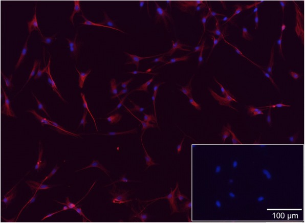
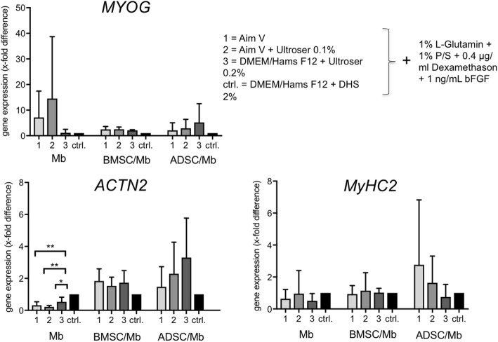
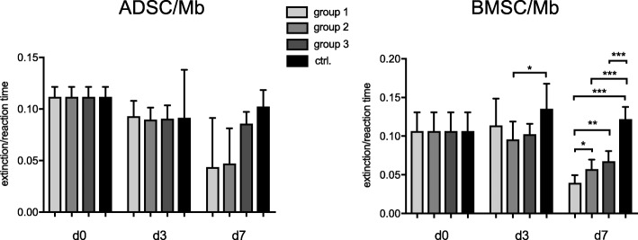
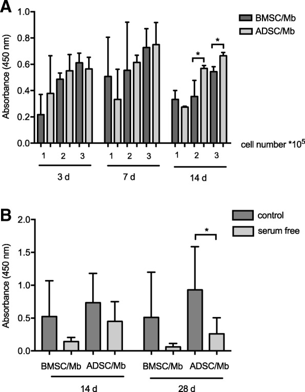
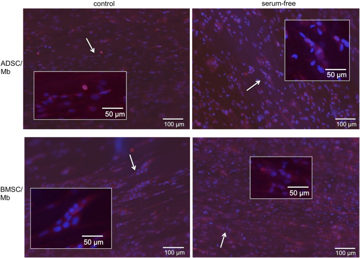
Results: Rat Mb were co-cultured with rat BMSC (BMSC/Mb) or ADSC (ADSC/Mb) two-dimensionally (2D) as monolayers or three-dimensionally (3D) on aligned PCL-collagen I-nanofibers. Differentiation media contained either AIM V, AIM V and Ultroser® G, DMEM/Ham's F12 and Ultroser® G, or donor horse serum (DHS) as a conventional differentiation medium. In 2D co-culture groups, highest upregulation of myogenic markers could be induced by serum-free medium containing DMEM/Ham's F12 and Ultroser® G (group 3) after 7 days. Alpha actinin skeletal muscle 2 (ACTN2) was upregulated 3.3-fold for ADSC/Mb and 1.7-fold for BMSC/Mb after myogenic induction by group 3 serum-free medium when compared to stimulation with DHS. Myogenin (MYOG) was upregulated 5.2-fold in ADSC/Mb and 2.1-fold in BMSC/Mb. On PCL-collagen I-nanoscaffolds, ADSC showed a higher cell viability compared to BMSC in co-culture with Mb. Myosin heavy chain 2, ACTN2, and MYOG as late myogenic markers, showed higher gene expression after long term stimulation with DHS compared to serum-free stimulation, especially in BMSC/Mb co-cultures. Immunocytochemical staining with myosin heavy chain verified the presence of a contractile apparatus under both serum free and standard differentiation conditions.

Conclusions: In this study, we were able to myogenically differentiate mesenchymal stromal cells with myoblasts on PCL-collagen I-nanoscaffolds in a serum-free medium. Our results show that this setting can be used for skeletal muscle tissue engineering, applicable to future clinical applications since no xenogenous substances were used.

Keywords: ADSC; BMSC; Electrospun PCL-collagen I-nanofibers; Mesenchymal stromal cells; Myoblasts; Myogenic differentiation; Nanoscaffolds; Serum-free media.


Conflict of interest statement

Ethics approval and consent to participate

Animal experiments were carried out following the German regulations for the care of laboratory animals at all times. Experiments were approved by the Animal Care Committee of the University of Erlangen and the Government of Mittelfranken, Germany (DMS-2532-2-161).

Consent for publication

Not applicable.

Competing interests

The authors declare that they have no competing interests.

Publisher’s Note

Springer Nature remains neutral with regard to jurisdictional claims in published maps and institutional affiliations.

Figures

Fig. 1
Fig. 1
Fluorescence microscopy of desmin-positive rat primary myoblasts. Myoblasts were enriched with a preplate-technique by seeding the supernatant of isolated cells into new flasks after two, 24, and 48 h. The third preplate was further passaged until passage 3. Merge of DAPI (blue) and desmin (red, with Alexa Fluor 647 as secondary antibody) showed that nearly all cells were myoblasts (desmin-positive). Insert shows fibroblasts isolated from rat skin negative for desmin
Fig. 2
Fig. 2
Characterization of adipose derived stem cells (ADSC). a ADSC were analyzed for cell surface markers in passage 6 (P6) and passage 11 (P11). Over 90% of cells were positive for CD90 and CD29 and negative for CD45 and CD11b/c in both passages. Paired t-test between P6 and P11 showed no differences in expression of surface markers. ADSC in passage 4 were differentiated into adipocytes (b), osteocytes (c), and chondrocytes (d). Lipid vacuoles were visualized with oil red O staining (b), calcium deposits were stained with Alizarin Red S (c), and proteoglycans of chondrogenic pellets were detected by Alcian blue staining (d). Inserts represent ADSC, cultured in proliferation medium as negative controls
Fig. 3
Fig. 3
Gene expression of myogenic markers in Mb, BMSC/Mb, and ADSC/Mb after serum-free myogenic differentiation. Expressions are demonstrated in x-fold difference compared with Mb, BMSC/Mb, ADSC/Mb stimulated with standard myogenic differentiation medium (ctrl. = control = 1) using the 2-ΔΔCt-method. Markers are presented as mean ± standard deviation. In Mb, serum-free differentiation led to a downregulation of ACTN2 (alpha actinin skeletal muscle 2). Statistical differences were tested with one-way ANOVA and Bonferroni‘s correction for multiple comparisons (n = 3). Levels of significance were * p ≤ 0.05, ** p ≤ 0.01
Fig. 4
Fig. 4
CK activity of ADSC/Mb and BMSC/Mb after (serum-free) myogenic differentiation. CK activity was measured as extinction during minute 2–6 of reaction time. Cells were allowed to differentiate for three (d3) and 7 days (d7) in serum-free media group 1, 2, 3, and standard myogenic differentiation medium (ctrl.) after 2 days of proliferation (d0). Values are presented as mean ± standard deviation. In BMSC/Mb, ctrl. Led to higher CK activity compared to all serum-free media. Highest CK activity was seen for group 3 of all serum-free media after 7 days of myogenic differentiation. Statistical differences were tested with one-way ANOVA and Bonferroni’s correction for multiple comparisons (n = 3). Levels of significance were * p ≤ 0.05, ** p ≤ 0.01, *** p ≤ 0.001
Fig. 5
Fig. 5
Cell viability on PCL-collagen I-nanoscaffolds. a BMSC/Mb and ADSC/Mb were seeded at different densities (1 × 105, 2 × 105, 3 × 105) on PCL-collagen I-nanoscaffolds and were allowed to proliferate for different time periods: 3 days (d), 7 d, or 14 d. Cell viability was determined by WST 8-assay. Absorbance at a wave length of 450 nm is expressed as mean ± standard deviation. At 14 days, ADSC showed a significantly higher cell viability for 2 × 105 and 3 × 105 cells in comparison to BMSC/Mb. b BMSC/Mb or ADSC/Mb were seeded at a density of 3 × 105 cells on PCL-collagen I-nanoscaffolds. Myogenic differentiation was induced by standard differentiation medium (control) or group 3 serum-free medium. WST 8-assay was repeated after 14 and 28 days of differentiation. Results showed decreased cell viability after serum free differentiation. ADSC/Mb showed higher cell viability compared to BMSC/Mb for all groups. Statistical differences were tested with repeated measures ANOVA for comparison between paired variables and Tukey’s multiple comparisons test as posthoc test at different time points. Pairwise comparison between BMSC/Mb and ADSC/Mb was done using unpaired t-test (n = 3). Level of significance was * p ≤ 0.05
Fig. 6
Fig. 6
Scanning electron microscopy of ADSC/Mb, BMSC/Mb, and C2C12 on PCL-collagen I-nanoscaffolds after long-term myogenic differentiation. After 28 days of myogenic differentiation, (a) ADSC/Mb were confluent, covering almost the entire surface of the scaffold. b BMSC/Mb were less densely spread on the scaffold. c C2C12 cells showed parallel alignment on the scaffolds with myotube-like structures
Fig. 7
Fig. 7
Myogenic differentiation of BMSC/Mb, ADSC/Mb, and C2C12 after long-term stimulation on PCL-collagen I-nanoscaffolds. Cells were stimulated with group 3 serum-free medium. Expressions are demonstrated in x-fold difference compared with BMSC/Mb and ADSC/Mb, stimulated with standard myogenic differentiation medium (control = 1) using the 2-ΔΔCt-method. Markers are presented as mean ± standard deviation. MyHC2 (myosine heavy chain 2), ACTN2 (alpha actinin skeletal muscle 2), and MYOG (myogenin) were downregulated after 28 days of serum free myogenic differentiation for BMSC/Mb compared to controls. Statistical differences were tested with paired t-test or Wilcoxon test, as appropriate (n = 3). Level of significance was * p ≤ 0.05
Fig. 8
Fig. 8
MHC expression after long-term myogenic differentiation on PCL-collagen I-nanoscaffolds. With fluorescence microscopy, the myogenic differentiation potential of BMSC/Mb and ADSC/Mb seeded on PCL-collagen I-nanoscaffolds was analyzed after 28 days of standard and group 3 serum-free myogenic differentiation. Both control and serum-free media led to positive expression of myosin heavy chain for BMSC/Mb and ADSC/Mb. Furthermore, multinucleated cells as possible myotube formation were found (representatives are marked with arrows and magnified in inserts)

References

    1. Lee KT, Mun GH. A systematic review of functional donor-site morbidity after latissimus dorsi muscle transfer. Plast Reconstr Surg. 2014;134(2):303–314. doi: 10.1097/PRS.0000000000000365. - DOI - PubMed
    1. Knox AD, Ho AL, Leung L, Tashakkor AY, Lennox PA, Van Laeken N, Macadam SA. Comparison of outcomes following autologous breast reconstruction using the DIEP and Pedicled TRAM flaps: a 12-year clinical retrospective study and literature review. Plast Reconstr Surg. 2016;138(1):16–28. doi: 10.1097/PRS.0000000000001747. - DOI - PubMed
    1. Cittadella Vigodarzere G, Mantero S. Skeletal muscle tissue engineering: strategies for volumetric constructs. Front Physiol. 2014;5:362. doi: 10.3389/fphys.2014.00362. - DOI - PMC - PubMed
    1. Mertens JP, Sugg KB, Lee JD, Larkin LM. Engineering muscle constructs for the creation of functional engineered musculoskeletal tissue. Regen Med. 2014;9(1):89–100. doi: 10.2217/rme.13.81. - DOI - PMC - PubMed
    1. Willett NJ, Krishnan L, Li MT, Guldberg RE, Warren GL. Guidelines for models of skeletal muscle injury and therapeutic assessment. Cells Tissues Organs. 2016;202(3–4):214–226. doi: 10.1159/000445345. - DOI - PubMed

Publication types

MeSH terms

LinkOut - more resources