Review
. 2018 Nov 26;17(1):168.
doi: 10.1186/s12943-018-0913-y.

Effects of tumor metabolic microenvironment on regulatory T cells

Affiliations
Review

Effects of tumor metabolic microenvironment on regulatory T cells

Yi-An Wang et al. Mol Cancer. .

Abstract

Recent studies have shown that on one hand, tumors need to obtain a sufficient energy supply, and on the other hand they must evade the body's immune surveillance. Because of their metabolic reprogramming characteristics, tumors can modify the physicochemical properties of the microenvironment, which in turn affects the biological characteristics of the cells infiltrating them. Regulatory T cells (Tregs) are a subset of T cells that regulate immune responses in the body. They exist in large quantities in the tumor microenvironment and exert immunosuppressive effects. The main effect of tumor microenvironment on Tregs is to promote their differentiation, proliferation, secretion of immunosuppressive factors, and chemotactic recruitment to play a role in immunosuppression in tumor tissues. This review focuses on cell metabolism reprogramming and the most significant features of the tumor microenvironment relative to the functional effects on Tregs, highlighting our understanding of the mechanisms of tumor immune evasion and providing new directions for tumor immunotherapy.

Keywords: Cancer metabolism microenvironment; Hypoxia; Low pH; Regulatory T cells; Signaling pathway.


Conflict of interest statement

Ethics approval and consent to participate

Not applicable.

Consent for publication

Not applicable.

Competing interests

The authors declare that they have no competing interests.

Publisher’s Note

Springer Nature remains neutral with regard to jurisdictional claims in published maps and institutional affiliations.

Figures
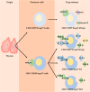
Fig. 1
Fig. 1
Differentiation of Tregs. nTregs: mature after thymus undergoes selection and exerts an immune function in the periphery. iTregs: peripheral T cells stimulated by antigen are converted to different Tregs subtypes by different inhibitory cytokines, secreting different cytokines to play immunosuppressive roles
Fig. 2
Fig. 2
Immunosuppression of Tregs. (a)Tregs secrete granzyme B and perforin that act on effector cells and cause apoptosis. (b)Tregs secrete inhibitory cytokines that bind to receptors on the surface of effector cells and inhibit the immune response. (c) CTLA-4 on the surface of Tregs competes with CD80/CD86 on the surface of effector cells to inhibit their immune function and promote the secretion of IDO; IDO degrades tryptophan in tissues to kynurenine; deletion of tryptophan leads to effector cell apoptosis; and kynurenine promotes effector cell apoptosis and acts on Tres’s AhR to promote its proliferation. ICOS on the surface of Tregs binds to ICOSL on the surface of effector cells, promoting the effector cell secretion inhibitory cytokine IL-10. (d) CD73/CD39 on the surface of Tregs convert ATP in tissues to adenine, which binds to receptors on the surface of effector cells and inhibit their immune function
Fig. 3
Fig. 3
Effect of glycolysis on Tregs. CD28 on the surface of Tregs is activated to promote GCK expression via PI3K/mTORC2, GCK, and actin, which can promote cytoskeletal remodeling, and GCK can promote glycolysis to provide energy. Upon stimulation of TLR1/2 or IL-2R on the Treg surface, activation of the PI3K/AKT/mTORC1 signaling pathway promotes Glut1 expression on the cell membrane surface and promotes glycolysis to promote proliferation. Akt inhibits the function of Tregs by inhibiting the expression of Foxp3, which in turn inhibits the transcription factor Foxo1/3. Foxp3 inhibits glycolysis by inhibiting the signaling pathway of PI3K/AKT/mTORC1 and the expression of Myc. AMPK in Tregs inhibits glycolysis by suppressing mTORC1
Fig. 4
Fig. 4
Effect of FAO and OXPHOS on Tregs. AMPK in Tregs promotes FAO by increasing the expression of CPT1A. PD-1 on the surface of Tregs also promotes FAO by increasing CPT1A expression. Foxp3 promotes OXPHOS by increasing the expression of mitochondria-associated proteins, ROS, a by-product of OXPHOS, stabilizes NFAT in the nucleus and binds to CNS2 to promote Foxp3 expression
Fig. 5
Fig. 5
The microenvironment recruits Tregs to tumor tissue by chemotaxis. Hypoxia promotes the expression of chemokines on tumor cells and Tregs surface through the HIF signaling pathway, and recruits Tregs by chemotaxis to the tumor tissue. Low pH microenvironment promotes the secretion of chemokine-containing exosomes from tumor cells that recruit Tregs to tumor tissue by chemotaxis
Fig. 6
Fig. 6
Hypoxia induces differentiation of T-cells to Tregs. T cells directly promote the expression of Foxp3 through the HIF signaling pathway. T cells promote the expression of TGF-β through HIF signaling, inhibit the key enzyme PHD2 for HIF-1α degradation, and indirectly promote the expression of Foxp3. Tumor cells promote the expression of TGF-β through the HIF signaling pathway, act on the receptors on the surface of T cells, activate the downstream SMAD signaling pathway, and promote the expression of Foxp3.Tumor cells promote the expression of PD-L1 on the cell surface through the HIF signaling pathway, bind PD-1 on the surface of T cells, inhibit the downstream AKT and mTOR pathways, and promote the expression of Foxp3
Fig. 7
Fig. 7
Low pH induces differentiation of T cell into Tregs. Tumor-derived exosomes contain TGF-β, which acts on the receptor on the surface of T cells to activate the SMAD signaling pathway and promote the expression of Foxp3. Tumor-derived exosomes contain IL-10, which acts on receptors on the T cell surface to activate the JAK/STAT signaling pathways and promote Foxp3 expression. Tumor-derived exosomes contain miR-214, which enters Tregs by endocytosis, inhibits the expression of PTEN protein, and then promotes the expression of the cycle-related transcription factor E2F by activating the PI3K signaling pathway and promote Tregs proliferation. PTEN maintains the stability of Tregs by inhibiting the activation of PI3K/Akt to stabilize the expression of CD25 and promotes the induction of the JAK-STAT signaling pathway to maintains the stability of Tregs
Fig. 8
Fig. 8
Effects of the metabolite, RA, on Tregs. RA in the microenvironment promotes the expression of Foxp3 by promoting the activation of the downstream JAK/STAT5 signaling pathway by IL-2 and the activation of the downstream SMAD signaling pathway by TGF-β, indirectly promoting the differentiation and proliferation of Tregs

References

    1. Wei Fang, Wu Yingfen, Tang Le, Xiong Fang, Guo Can, Li Xiayu, Zhou Ming, Xiang Bo, Li Xiaoling, Li Guiyuan, Xiong Wei, Zeng Zhaoyang. Trend analysis of cancer incidence and mortality in China. Science China Life Sciences. 2017;60(11):1271–1275. doi: 10.1007/s11427-017-9172-6. - DOI - PubMed
    1. Hanahan D, Weinberg RA. Hallmarks of cancer: the next generation. Cell. 2011;144(5):646–674. doi: 10.1016/j.cell.2011.02.013. - DOI - PubMed
    1. Tu C, Zeng Z, Qi P, Li X, Guo C, Xiong F, Xiang B, Zhou M, Liao Q, Yu J, et al. Identification of genomic alterations in nasopharyngeal carcinoma and nasopharyngeal carcinoma-derived Epstein-Barr virus by whole genome sequencing. Carcinogenesis. 2018; 10.1093/carcin/bgy108. - PubMed
    1. Zou Guoying, Ren Biqiong, Liu Yi, Fu Yin, Chen Pan, Li Xiayu, Luo Shudi, He Junyu, Gao Ge, Zeng Zhaoyang, Xiong Wei, Li Guiyuan, Huang Yumei, Xu Keqian, Zhang Wenling. Inhibin B suppresses anoikis resistance and migration through the transforming growth factor-β signaling pathway in nasopharyngeal carcinoma. Cancer Science. 2018;109(11):3416–3427. doi: 10.1111/cas.13780. - DOI - PMC - PubMed
    1. Tang Y, He Y, Zhang P, Wang J, Fan C, Yang L, Xiong F, Zhang S, Gong Z, Nie S, et al. LncRNAs regulate the cytoskeleton and related rho/ROCK signaling in cancer metastasis. Mol Cancer. 2018;17(1):77. - PMC - PubMed

Publication types

MeSH terms

LinkOut - more resources