. 2018 Dec 11;115(50):12728-12732.
doi: 10.1073/pnas.1803615115. Epub 2018 Nov 26.

Genetically encodable bioluminescent system from fungi

Affiliations

Genetically encodable bioluminescent system from fungi

Alexey A Kotlobay et al. Proc Natl Acad Sci U S A. .

Abstract

Bioluminescence is found across the entire tree of life, conferring a spectacular set of visually oriented functions from attracting mates to scaring off predators. Half a dozen different luciferins, molecules that emit light when enzymatically oxidized, are known. However, just one biochemical pathway for luciferin biosynthesis has been described in full, which is found only in bacteria. Here, we report identification of the fungal luciferase and three other key enzymes that together form the biosynthetic cycle of the fungal luciferin from caffeic acid, a simple and widespread metabolite. Introduction of the identified genes into the genome of the yeast Pichia pastoris along with caffeic acid biosynthesis genes resulted in a strain that is autoluminescent in standard media. We analyzed evolution of the enzymes of the luciferin biosynthesis cycle and found that fungal bioluminescence emerged through a series of events that included two independent gene duplications. The retention of the duplicated enzymes of the luciferin pathway in nonluminescent fungi shows that the gene duplication was followed by functional sequence divergence of enzymes of at least one gene in the biosynthetic pathway and suggests that the evolution of fungal bioluminescence proceeded through several closely related stepping stone nonluminescent biochemical reactions with adaptive roles. The availability of a complete eukaryotic luciferin biosynthesis pathway provides several applications in biomedicine and bioengineering.

Keywords: bioluminescence; fungal luciferase; fungal luciferin biosynthesis.


Conflict of interest statement

Conflict of interest statement: K.S.S. and I.V.Y. are shareholders of Planta LLC. Planta LLC filed patent applications related to the use of the enzymes of fungal bioluminescent system.

Figures

Fig. 1.
Fig. 1.
Luciferin biosynthesis pathway in fungal bioluminescence and gene cluster containing key enzymes in the clade of bioluminescent fungi. (A) Proposed pathway of fungal luciferin biosynthesis and recycling. Caffeic acid is converted to hispidin by hispidin synthase (HispS) and hydroxylated by H3H, yielding 3-hydroxyhispidin (fungal luciferin). The luciferase (Luz) adds molecular oxygen, producing an endoperoxide as a high-energy intermediate with decomposition that yields oxyluciferin (caffeylpyruvate) and light emission. Oxyluciferin can be recycled to caffeic acid by caffeylpyruvate hydrolase (CPH). (B) Schematic depiction of the genomic cluster of N. nambi containing luciferase, H3H, hispidin synthase, and caffeylpyruvate hydrolase (cph) genes. (C) Gene cluster in the clade of bioluminescent fungi. The species tree in Left is based on the comparison of protein-coding genes shared by most of the analyzed species. The red crosses mark the branches of the tree that eventually lost the ability to glow. Right shows the structure of the luciferase-containing gene cluster if such a cluster was found in the relevant genome. The genes coding for luciferase (luz), h3h, hispidin synthase (hisps), and caffeylpyruvate hydrolase (cph) are colored. The lighter blue and red colors of hisps and luz genes indicate that only a partial or truncated gene was found in Armillaria mellea and Guyanagaster necrorhiza, respectively. Other genes that might belong to the cluster are named from O1 to O4 (colored in gray). Green ticks represent a cytochrome P450-like gene (different shades of green indicate different orthologous groups), and black ticks indicate other genes.
Fig. 2.
Fig. 2.
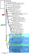
Phylogeny of Agaricales species in which genomes are sequenced. The rectangles with the gene names indicate where luz, h3h, and hisps genes emerged as a result of duplication. An oval in the bioluminescence (BL) clade indicates the common ancestor of all bioluminescent species. The red crosses mark the branches of the tree that eventually lost bioluminescence. The lineages of bioluminescent fungi are also shown in the same clade. The scale estimates the number of substitutions per site.
Fig. 3.
Fig. 3.
Fungal luciferase as a reporter gene. (A) Photo of P. pastoris cells expressing nnluz, nnh3h, nnhisps, and npgA genes growing in a medium containing caffeic acid. The photo was taken on a NIKON D800 camera, ISO 1600, exposure 8 s. (B) Human HEK293NT cells cotransfected with fungal luciferase (green channel) and red fluorescent protein Katushka (violet channel). Fungal luciferin was added to the medium to the final concentration of 650 μg/mL before image acquisition. (C) Image of a mouse with s.c. injected murine carcinoma cells CT26 expressing either nnluz (on the left) or P. pyralis luciferase (on the right) after i.p. administration of a mix of fungal (0.5 mg) and firefly (0.5 mg) luciferins. Color indicates the intensity of emitted light. (D) Expression of nnluz gene in an X. laevis embryo. The right embryo was microinjected with the mixture of rhodamine lysine dextran and nnluz mRNA at the two-cell stage, and then, it was microinjected with luciferin into the blastocoel cavity at the gastrula stage. As a control, the left embryo was microinjected with rhodamine lysine dextran only at the two-cell stage, and then, it was also microinjected with luciferin into the blastocoel cavity at the gastrula stage. The violet channel indicates rhodamine fluorescence, and the green channel indicates nnLuz bioluminescence.

References

    1. Shimomura O. Bioluminescence: Chemical Principles and Methods. World Scientific; Singapore: 2006.
    1. Wainwright PC, Longo SJ. Functional innovations and the conquest of the oceans by Acanthomorph fishes. Curr Biol. 2017;27:R550–R557. - PubMed
    1. Verdes A, Gruber DF. Glowing worms: Biological, chemical, and functional diversity of bioluminescent annelids. Integr Comp Biol. 2017;57:18–32. - PubMed
    1. Labella AM, Arahal DR, Castro D, Lemos ML, Borrego JJ. Revisiting the genus photobacterium: Taxonomy, ecology and pathogenesis. Int Microbiol. 2017;20:1–10. - PubMed
    1. Thouand G, Marks R. Bioluminescence: Fundamentals and Applications in Biotechnology. Springer; Berlin: 2016.

Publication types