. 2018 Dec 11;115(50):E11661-E11670.
doi: 10.1073/pnas.1805950115. Epub 2018 Nov 26.

Dual functions for OVAAL in initiation of RAF/MEK/ERK prosurvival signals and evasion of p27-mediated cellular senescence

Affiliations

Dual functions for OVAAL in initiation of RAF/MEK/ERK prosurvival signals and evasion of p27-mediated cellular senescence

Ben Sang et al. Proc Natl Acad Sci U S A. .

Erratum in

Abstract

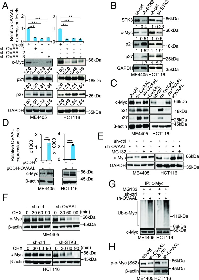
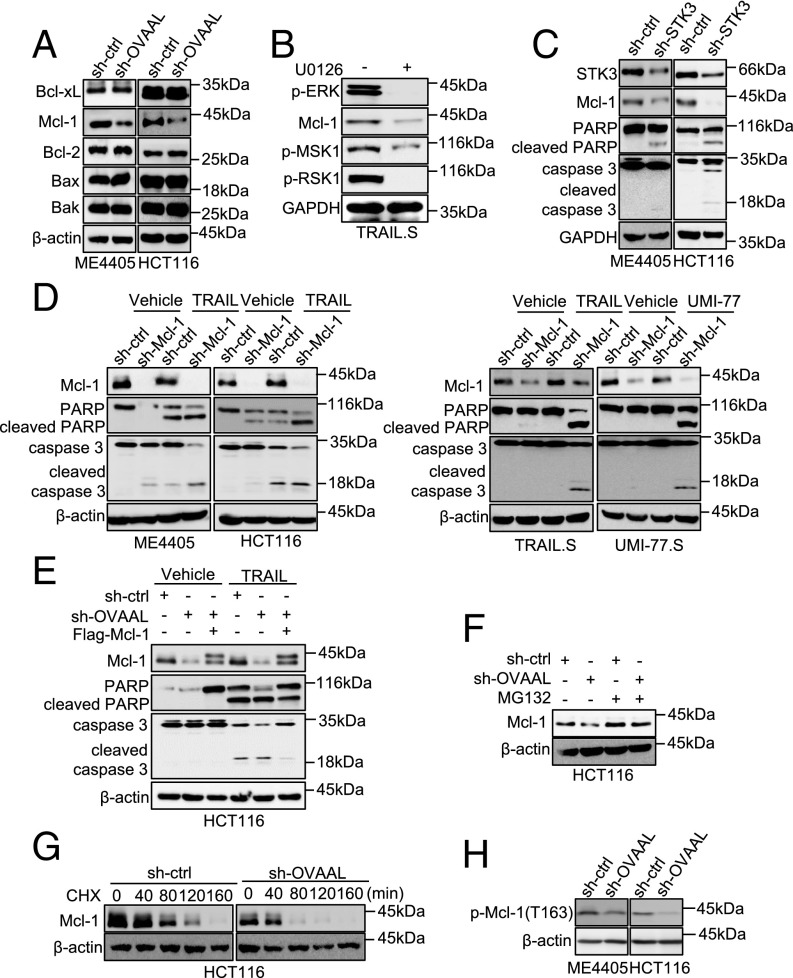
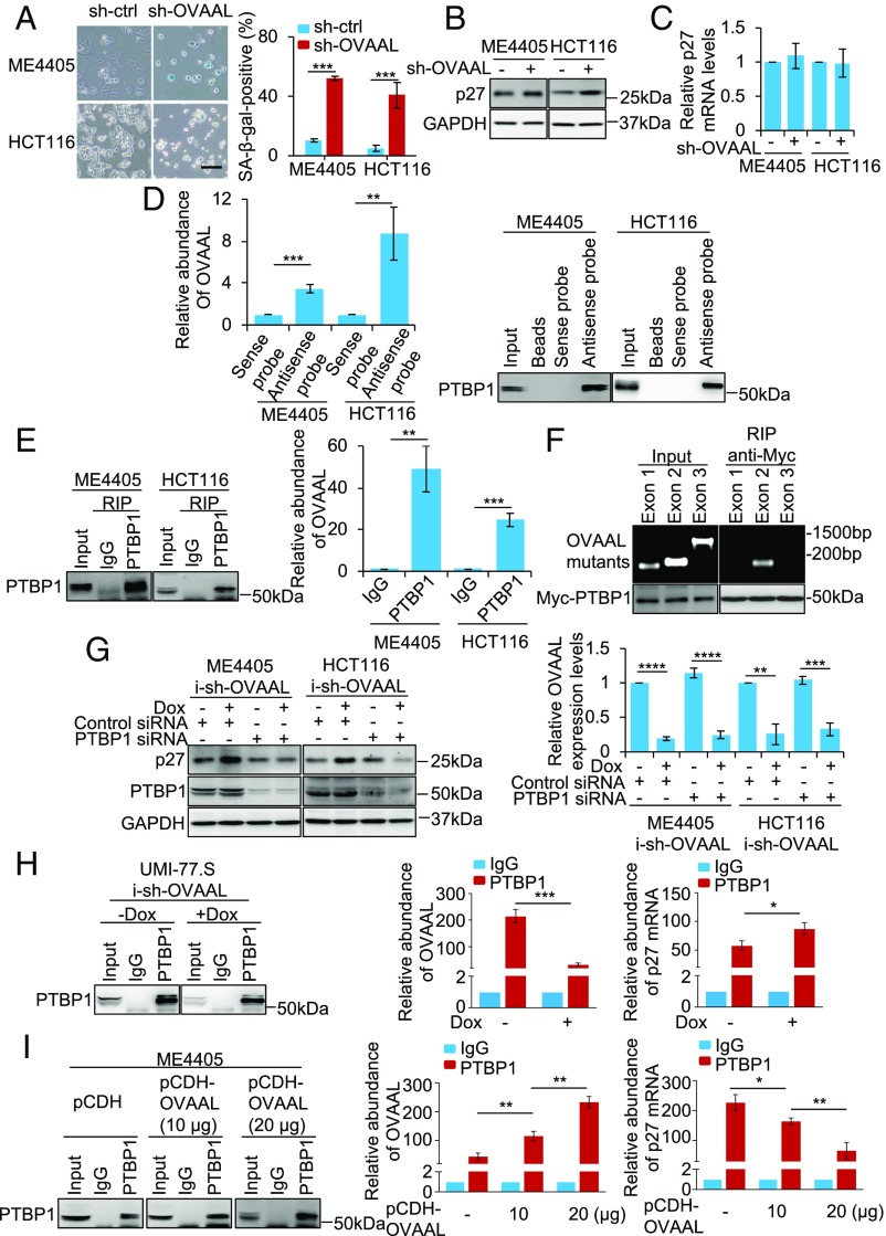
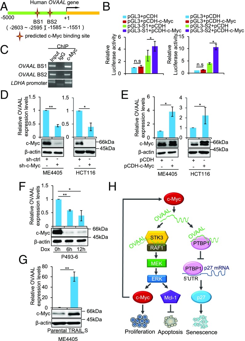
Long noncoding RNAs (lncRNAs) function through a diverse array of mechanisms that are not presently fully understood. Here, we sought to find lncRNAs differentially regulated in cancer cells resistant to either TNF-related apoptosis-inducing ligand (TRAIL) or the Mcl-1 inhibitor UMI-77, agents that act through the extrinsic and intrinsic apoptotic pathways, respectively. This work identified a commonly up-regulated lncRNA, ovarian adenocarcinoma-amplified lncRNA (OVAAL), that conferred apoptotic resistance in multiple cancer types. Analysis of clinical samples revealed OVAAL expression was significantly increased in colorectal cancers and melanoma in comparison to the corresponding normal tissues. Functional investigations showed that OVAAL depletion significantly inhibited cancer cell proliferation and retarded tumor xenograft growth. Mechanically, OVAAL physically interacted with serine/threonine-protein kinase 3 (STK3), which, in turn, enhanced the binding between STK3 and Raf-1. The ternary complex OVAAL/STK3/Raf-1 enhanced the activation of the RAF protooncogene serine/threonine-protein kinase (RAF)/mitogen-activated protein kinase kinase 1 (MEK)/ERK signaling cascade, thus promoting c-Myc-mediated cell proliferation and Mcl-1-mediated cell survival. On the other hand, depletion of OVAAL triggered cellular senescence through polypyrimidine tract-binding protein 1 (PTBP1)-mediated p27 expression, which was regulated by competitive binding between OVAAL and p27 mRNA to PTBP1. Additionally, c-Myc was demonstrated to drive OVAAL transcription, indicating a positive feedback loop between c-Myc and OVAAL in controlling tumor growth. Taken together, these results reveal that OVAAL contributes to the survival of cancer cells through dual mechanisms controlling RAF/MEK/ERK signaling and p27-mediated cell senescence.

Keywords: OVAAL; c-Myc; p27; proliferation; senescence.


Conflict of interest statement

The authors declare no conflict of interest.

Figures

Fig. 1.
Fig. 1.
OVAAL is selectively up-regulated and confers resistance against cancer cells to TRAIL and the Mcl-1 inhibitor UMI-77. (A) ME4405 TRAIL.S and ME4405 UMI-77.S cell sublines display resistance to TRAIL (25 ng/mL) and the Mcl-1 inhibitor UMI-77 (4 μM) as shown in colony formation assays (2D) and hanging drop assays (3D) (n = 3). (Scale bars: 2D, 1 cm; 3D, 25 μm.) (B) Thirty-seven lncRNAs were up-regulated in common between selected cells in comparison to ME4405 cells. The heat map illustrates the five most up-regulated lncRNAs. (C) OVAAL was up-regulated in TRAIL.S and UMI-77.S cells as shown in Northern blotting. β-Actin mRNA was used as a loading control (n = 3). Depletion of OVAAL in resistant TRAIL.S and UMI-77.S cells using three independent shRNA targeting vectors (D) results in increased apoptosis as measured by annexin V-FITC staining in cells treated with vehicle control (ctrl), TRAIL, or UMI-77 for 24 h (E) and accompanying activation of caspase-3 and cleavage of PARP visualized by Western blotting (F) (n = 3; mean ± SD; Student’s t test). OVAAL overexpression in parental ME4405 cells (G) treated with vehicle ctrl, TRAIL (25 ng/mL), or UMI-77 (4 μM) for 24 h promotes resistance to both TRAIL- and UMI-77–induced apoptosis as shown in annexin V-FITC staining assays (H) and reduced activation of caspase-3 and cleavage of PARP as shown in Western blotting (I) (n = 3; mean ± SD; Student’s t test). **P < 0.01; ***P < 0.001.
Fig. 2.
Fig. 2.
OVAAL is frequently overexpressed in colon cancer and melanoma, and functions to promote cancer cell survival and proliferation. (A, Top) As shown in Western blotting, OVAAL shRNA caused enhanced activation of caspase-3 and cleavage of PARP in ME4405 and HCT116 cells treated with vehicle control (ctrl) or TRAIL (200 ng/mL) for 24 h. (A, Bottom) OVAAL depletion was confirmed by qPCR. β-actin was used as a loading ctrl throughout (n = 3; mean ± SD; Student’s t test). (B, Top) As shown in Western blotting, OVAAL shRNA did not cause activation of caspase-3 and cleavage of PARP in HAFF cells treated with vehicle ctrl or TRAIL (200 ng/mL) for 24 h. (B, Bottom) OVAAL depletion was confirmed by qPCR (n = 3; mean ± SD; Student’s t test). (C) OVAAL shRNA inhibited cell proliferation in ME4405 and HCT116 cells (n = 3; mean ± SD; Student’s t test). (D) OVAAL shRNA caused cell cycle arrest in G0/G1 phase in ME4405 and HCT116 cells measured by propidium iodide staining, followed by flow cytometry (n = 3; mean ± SD; Student’s t test). (E) OVAAL shRNA decreased the levels of p-Rb, cyclin A, and cyclin B levels and increased cyclin D and cyclin E levels in ME4405 and HCT116 cells as shown by Western blotting (n = 3). (F) OVAAL shRNA inhibited cell proliferation in ME4405 and HCT116 cells as shown in colony formation assays (n = 3). (Scale bar: 1 cm.) (G and H) OVAAL depletion by shRNA inhibited HCT116 xenograft growth in nu/nu mice (n = 7; mean ± SD; Student’s t test). (I) Measurement of expression using qPCR shows higher levels of OVAAL in CRC compared with paired adjacent normal tissues (n = 33; mean ± SD; Student’s t test). (J) Assessment of OVAAL expression using in situ hybridization assays demonstrates significantly increased expressed in CRC in comparison to adjacent normal tissues (n = 75; mean ± SD; Student’s t test) RS, reactive score. (K) Representative photomicrographs of OVAAL ISH staining in FFPE tissue samples from J showing high differential expression between cancer and normal tissue. **P < 0.01; ***P < 0.001; ****P < 0.0001.
Fig. 3.
Fig. 3.
OVAAL interacts with STK3. (A) Visualization of protein bands stained with Coomassie Brilliant Blue pulled down by biotin-labeled antisense probes against OVAAL in total protein extracts of ME4405 and HCT116 cells. Protein identities with high probabilities as determined using mass spectrometry are labeled. (B) Biotin-labeled antisense probes used to capture OVAAL from total HCT116 cellular extracts also pulled down by STK3 as shown by qPCR analysis (Left) and Western blotting (Right) (n = 3; mean ± SD; Student’s t test). *P < 0.05. (C) Antibodies against STK3 coprecipitated OVAAL from total cellular extracts of ME4405 and HCT116 cells as shown in Western blotting (Top) and PCR analysis (Bottom) (n = 3). (D) Anti-Flag (M2) antibodies precipitated OVAAL, along with Flag-STK3, from total ME4405 and HCT116 cellular extracts as shown in Western blotting (Top) and PCR analysis (Bottom) (n = 3). (E) Subcellular fractionation studies conducted in ME4405 and HCT116 cells showed that approximately half of OVAAL RNA was located to the cytoplasm, along with the STK3 protein, as shown by qPCR analysis (Left) and Western blotting (Right) (n = 3, mean ± SD). Cyto, cytoplasmic; Nuc, nuclear. (F) Subcellular localization of OVAAL was examined by RNA FISH (red) in ME4405 and HCT116 cells. Nuclear DNA was visualized with DAPI (blue). (Scale bar: 20 μm.) (G) Schematic representation of OVAAL RNA and the design of exon-deletion constructs used for the analyses shown in H. E3 was further was subdivided into four regions for the analyses shown in I. (H and I) Anti-Flag (M2) antibody precipitates from whole-cell lysates of Flag-STK3–transfected HCT116 cells were co-incubated with the indicated in vitro-transcribed OVAAL constructs. RT-PCR analyses (Top) were used to detect binding of OVAAL RNA, and Western blotting (Bottom) was used to validate input and capture of Flag-STK3 (n = 3).
Fig. 4.
Fig. 4.
OVAAL activates RAF/MEK/ERK cascade through facilitating the binding between STK3 and Raf-1. (A) STK3 shRNA decreased levels of p-ERK, p-MEK, p-MSK1, and p-RSK1 in ME4405 and HCT116 cells as shown in Western blotting. Analysis of total ERK, MEK, MSK1, and RSK1, along with a GAPDH loading control (ctrl), is shown for comparison (n = 3). (B) OVAAL shRNA decreased levels of p-ERK, p-MEK, p-MSK1, and p-RSK1 in ME4405 and HCT116 cells as shown in Western blotting (n = 3). (C) Co-silencing of OVAAL with STK3 in HCT116 cells did not impart further reductions in the phosphorylated levels of ERK, MEK, MSK1, or RSK1 levels in comparison to STK3 knockdown alone as measured by Western blotting (n = 3). (D) Biotin-labeled antisense probes used to capture OVAAL from total cellular lysates of ME4405 and HCT116 cells also pulled down STK3, along with Raf-1, as shown by Western blotting. β-Actin was used as a negative ctrl here and in the following experiments (n = 3). (E) Flag-STK3, Raf-1, and OVAAL were coprecipitated with an anti-Flag antibody in whole-cell lysates of HCT116 cells transfected with Flag-STK3 (Left), and after elution with Flag peptide, all were further coprecipitated with an anti–Raf-1 antibody in the resultant precipitates (Right). IP, immunoprecipitation. (F) OVAAL depletion strongly diminished the levels of Raf-1 associating with STK3. Endogenous STK3 was immunoprecipitated from sh-ctrl– or sh-OVAAL–treated ME4405 and HCT116 cells, with results shown by Western blotting (n = 3). (G) Presence of in vitro-transcribed OVAAL, but not an antisense RNA, enhanced the binding between recombinant HA–Raf-1 and Flag-STK3. Assay inputs were visualized by Coomassie Brilliant Blue (CBB) and ethidium bromide staining (n = 3). OVAAL shRNA (H) and STK3 shRNA (I) increased levels of p-Raf-1 (Ser259) in ME4405 and HCT116 cells as shown in Western blotting (n = 3).
Fig. 5.
Fig. 5.
OVAAL promotes cell proliferation via stabilization of c-Myc. (A) OVAAL shRNA reduced c-Myc and increased p21 and p27 protein expression levels in ME4405 and HCT116 cells as shown in qPCR analyses (Top) and Western blotting (Bottom). GAPDH or β-actin is used throughout as a loading control (ctrl) (n = 3; mean ± SD; Student’s t test). (B) STK3 shRNA reduced c-Myc and increased p21 and p27 protein expression levels in ME4405 and HCT116 cells as shown in Western blotting (n = 3). (C) Reductions in c-Myc expression following OVAAL shRNA are rescued by overexpression of OVAAL in ME4405 and HCT116 cells as shown in Western blotting (n = 3). (D, Bottom) As shown in Western blotting, overexpression of OVAAL increased c-Myc protein expression levels in ME4405 and HCT116 cells. (D, Top) Analysis of total RNA by qPCR analysis confirmed the increase in OVAAL expression in cells transduced with pCDH-OVAAL compared with the control pCDH vector (n = 3; mean ± SD; Student’s t test). (E) Treatment of ME4405 and HCT116 cells with the proteasome inhibitor MG132 (50 μM) reversed the decrease in c-Myc protein levels resulting from silencing of OVAAL as shown in Western blotting (n = 3). (F) shRNA silencing of either OVAAL (Top) or STK3 (Bottom) shortened the half-life of c-Myc protein in cycloheximide chase assays (n = 3). (G) OVAAL shRNA increased c-Myc polyubiquitination levels in the presence of MG132 (50 μM) (n = 3). IP, immunoprecipitation. (H) OVAAL shRNA reduced phosphorylated c-Myc (Ser62) levels in ME4405 and HCT116 cells as shown in Western blotting (n = 3). **P < 0.01; ***P < 0.001.
Fig. 6.
Fig. 6.
OVAAL counteracts TRAIL- and UMI-77–induced apoptosis through up-regulation of Mcl-1. (A) OVAAL shRNA decreased levels of Mcl-1 in ME4405 and HCT116 cells as shown in Western blotting (n = 3). ctrl, control. (B) MEK inhibitor U0126 (10 μM) caused the reduction of Mcl-1 expression in TRAIL.S cells as shown in Western blotting (n = 3). (C) STK3 shRNA reduced Mcl-1 expression and increased activation of caspase-3 and cleavage of PARP in ME4405 and HCT116 cells as shown in Western blotting (n = 3). (D) Mcl-1 shRNA enhanced activation of caspase-3 and cleavage of PARP in ME4405, HCT116, TRAIL.S, and UMI-77.S cells treated with vehicle ctrl, TRAIL, or UMI-77 for 24 h as shown in Western blotting (n = 3). (E) Overexpression of Mcl-1 diminished OVAAL depletion-increased cleavage of caspase-3 and PARP under TRAIL (50 ng/mL) treatment in HCT116 cells (n = 3). (F) Proteasome inhibitor MG132 (50 μM) reversed the decrease in Mcl-1 protein levels by OVAAL silencing in HCT116 cells as shown in Western blotting (n = 3). (G) OVAAL shRNA shortened the half-life of Mcl-1 protein in HCT116 cells as shown in Western blotting (n = 3). (H) OVAAL shRNA reduced p-Mcl-1 (Thr163) levels in ME4405 and HCT116 cells as shown in Western blotting (n = 3).
Fig. 7.
Fig. 7.
OVAAL blocks cellular senescence via regulation of p27 translation. (A) OVAAL shRNA triggered cellular senescence as shown by induction of SA–β-gal staining in ME4405 and HCT116 cells. Representative photographs (Left) and quantification of staining (Right) are shown (n = 3; mean ± SD; Student’s t test). ctrl, control. (Scale bar: 200 μm.) (B) OVAAL shRNA increased the levels of p27 protein in ME4405 and HCT116 cells as shown in Western blotting (n = 3). (C) OVAAL shRNA did not affect p27 mRNA levels in ME4405 and HCT116 cells as shown in qPCR analysis (n = 3; mean ± SD). (D) Biotin-labeled antisense probes against OVAAL pulled down OVAAL, along with PTBP1, from total ME4405 and HCT116 cellular extracts as shown in qPCR analysis (Left) and Western blotting (Right) (n = 3; mean ± SD; Student’s t test). (E) Antibody against PTBP1 precipitated PTBP1, along with OVAAL, from total ME4405 and HCT116 cellular extracts as shown in Western blotting (Left) and qPCR analysis (Right) (n = 3; mean ± SD; Student’s t test). (F) Myc-tag antibody precipitated Myc-PTBP1 from HCT116 whole-cell lysates, along with co-incubated in vitro-transcribed mutant OVAAL, as shown in PCR analysis (Top) and Western blotting (Bottom) (n = 3). (G) Cosilencing of PTBP1 abolished the up-regulation of p27 protein following OVAAL knockdown in parental ME4405 and HCT116 cells bearing an inducible knockdown system (i-sh-OVAAL) activated with doxycycline (Dox; 100 ng/mL). Results are shown in Western blotting (Left) and qPCR analysis (Right) (n = 3; mean ± SD; Student’s t test). (H) Depletion of OVAAL following Dox (100 ng/mL) treatment increased the association between PTBP1 and p27 mRNA in OVAAL-inducible knockdown UMI-77.S cells (UMI-77.S i-sh-OVAAL). PTBP1 immunoprecipitates were assessed using Western blotting (Left) and qPCR analysis (Right) (n = 3; mean ± SD; Student’s t test). (I) Overexpression of OVAAL in a dose-dependent manner decreased the association between PTBP1 and p27 mRNA in ME4405 cells. Analysis as per H is shown using Western blotting (Left) and qPCR (Right) (n = 3, mean ± SD; Student’s t test). *P < 0.05; **P < 0.01; ***P < 0.001; ****P < 0.0001.
Fig. 8.
Fig. 8.
c-Myc transcriptionally regulates OVAAL in cancer cells. (A) Two potential c-Myc–binding sites in the OVAAL promoter region were predicted in the high-quality transcription factor binding profile database (JASPAR). (B) Both predicted regions were transcriptionally responsive to c-Myc overexpression as shown in luciferase reporter assays (n = 3; mean ± SD; Student’s t test). (C) c-Myc bound to both predicted c-Myc–binding sites in OVAAL promoter as shown in ChIP assays. Lactate dehydrogenase A (LDHA) promoter was used as a positive control (n = 3). (D) c-Myc shRNA reduced the levels of OVAAL in ME4405 and HCT116 cells as shown in qPCR analysis (Top) and Western blotting (Bottom) (n = 3; mean ± SD; Student’s t test). ctrl, control. (E) Overexpression of c-Myc increased the expression levels of OVAAL in ME4405 and HCT116 cells as shown in qPCR analysis (Top) and Western blotting (Bottom) (n = 3; mean ± SD; Student’s t test). (F) Expression levels of OVAAL in P493-6 cells carrying a c-Myc tet-off system were decreased when cells were treated with doxycycline (Dox; 1 μM) as shown in qPCR analysis (Top) and Western blotting (Bottom) (n = 3; mean ± SD; Student’s t test). (G) c-Myc, along with OVAAL, was up-regulated in TRAIL.S cells in comparison to ME4405 cells as shown in qPCR analysis (Top) and Western blotting (Bottom) (n = 3; mean ± SD; Student’s t test). (H) Schematic illustration of proposed model depicting dual roles of lncRNA OVAAL in regulating cell proliferation/apoptosis and senescence. *P < 0.05; **P < 0.01.

Similar articles

Cited by

References

    1. Kung JT, Colognori D, Lee JT. Long noncoding RNAs: Past, present, and future. Genetics. 2013;193:651–669. - PMC - PubMed
    1. Huarte M. The emerging role of lncRNAs in cancer. Nat Med. 2015;21:1253–1261. - PubMed
    1. Huang JZ, et al. A peptide encoded by a putative lncRNA HOXB-AS3 suppresses colon cancer growth. Mol Cell. 2017;68:171–184.e6. - PubMed
    1. Ruiz-Orera J, Messeguer X, Subirana JA, Alba MM. Long non-coding RNAs as a source of new peptides. eLife. 2014;3:e03523. - PMC - PubMed
    1. Wang JZ, Xu CL, Wu H, Shen SJ. LncRNA SNHG12 promotes cell growth and inhibits cell apoptosis in colorectal cancer cells. Braz J Med Biol Res. 2017;50:e6079. - PMC - PubMed

Publication types

MeSH terms