. 2019 Jan 28;201(4):e00583-18.
doi: 10.1128/JB.00583-18. Print 2019 Feb 15.

The Core and Holoenzyme Forms of RNA Polymerase from Mycobacterium smegmatis

Affiliations

The Core and Holoenzyme Forms of RNA Polymerase from Mycobacterium smegmatis

Tomáš Kouba et al. J Bacteriol. .

Abstract

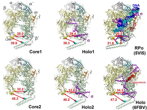
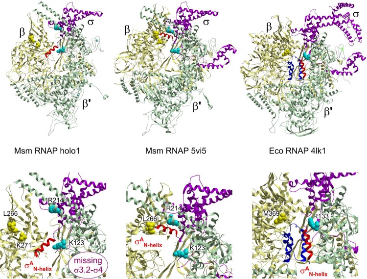
Bacterial RNA polymerase (RNAP) is essential for gene expression and as such is a valid drug target. Hence, it is imperative to know its structure and dynamics. Here, we present two as-yet-unreported forms of Mycobacterium smegmatis RNAP: core and holoenzyme containing σA but no other factors. Each form was detected by cryo-electron microscopy in two major conformations. Comparisons of these structures with known structures of other RNAPs reveal a high degree of conformational flexibility of the mycobacterial enzyme and confirm that region 1.1 of σA is directed into the primary channel of RNAP. Taken together, we describe the conformational changes of unrestrained mycobacterial RNAP.IMPORTANCE We describe here three-dimensional structures of core and holoenzyme forms of mycobacterial RNA polymerase (RNAP) solved by cryo-electron microscopy. These structures fill the thus-far-empty spots in the gallery of the pivotal forms of mycobacterial RNAP and illuminate the extent of conformational dynamics of this enzyme. The presented findings may facilitate future designs of antimycobacterial drugs targeting RNAP.

Keywords: RNA polymerase; bacterial transcription; conformational change; cryo-electron microscopy; mycobacteria; protein structure; transcription initiation factor.


Figures

FIG 1
FIG 1
Cryo-EM structure determinations of M. smegmatis RNAP complexes. (Top row) Secondary structures of two forms of RNAP from M. smegmatis as observed in the experimental cryo-EM densities: core (left) and holoenzyme (right, RNAP in complex with primary sigma factor, σA). Subunits are colored as follows: β, yellow; β′, green; α, gray; α′, cyan; ω, orange; σA, magenta. (Middle row) Cryo-EM maps are filtered and colored according to local resolution calculated with the RELION software package (see Fig. S3 for more detail). (Bottom row) Cryo-EM maps, together with fit of secondary structure models of M. smegmatis RNAP complexes. Maps are visualized as 0.5 transparent at 0.08 volume threshold in Chimera (see Fig. S4 for more detail). Color coding is as described for the top row.
FIG 2
FIG 2
M. smegmatis RNAP exhibits conformational dynamics. Conformational dynamics are illustrated by distances (arrows) between the β and β′ subunits at the narrowest point of the dwDNA entrance (distance between βGly271Cα and β′Lys123Cα) and by positioning of the nonconserved domain β’i1 (distances between βLeu266Cα and β′Arg214Cα) in the main conformation of M. smegmatis RNAP. Core1/Core2 and Holo1/Holo2 represent the two most represented conformations of respective forms of M. smegmatis RNAP. Closed RNAP conformations of M. smegmatis RPo (PDB entry 5VI5) and M. tuberculosis holoenzyme in complex with the antibacterial compound fidaxomicin (PDB entry 6FBV) that traps the open conformation of the pincer are shown for comparison. RNAP subunits and nucleic acids chains are colored as follows: β, yellow; β′, green; α, gray; α′, cyan; ω, orange; σA, magenta; DNA duplex, blue and cyan.
FIG 3
FIG 3
The σAN-helix changes position within the RNAP. (Top left) The N-terminal α-helix (red) of σA (amino acids [aa] 149 to 162) is located within the primary channel of the M. smegmatis open-conformation holoenzyme RNAP. The σAN-helix is positioned between the β′ coiled-coil and the inner edge of β domain 2 (Table S2) and oriented perpendicular to the nonconserved domain (NCD) β′i1 finger. (Top middle) The N-terminal α-helix (red) of σA (aa 149 to 162) in the closed-conformation mycobacterial RNAP structures (12) is positioned on the periphery in between the β and σ′ subunit claws, parallel to the NCD β′i1 finger. (Top right) σ701.1 from E. coli in complex with RNAP positioned in the primary channel of RNAP (8). RNAP subunits are colored as follows: β, yellow; β′, green; α, gray; α′, cyan; ω, orange; σA/70, magenta; the N-terminal part of σA/70, dark blue; σAN-helix, red. (Bottom row) Detailed views of the respective σA/70N helix regions from the structures in the upper row.

References

    1. Molodtsov V, Scharf NT, Stefan MA, Garcia GA, Murakami KS. 2017. Structural basis for rifamycin resistance of bacterial RNA polymerase by the three most clinically important RpoB mutations found in Mycobacterium tuberculosis. Mol Microbiol 103:1034–1045. doi: 10.1111/mmi.13606. - DOI - PMC - PubMed
    1. Ma C, Yang X, Lewis PJ. 2016. Bacterial transcription as a target for antibacterial drug development. Microbiol Mol Biol Rev 80:139–160. doi: 10.1128/MMBR.00055-15. - DOI - PMC - PubMed
    1. Sutherland C, Murakami KS. 2018. An introduction to the structure and function of the catalytic core enzyme of Escherichia coli RNA polymerase. EcoSal Plus 8. doi: 10.1128/ecosalplus.ESP-0004-2018. - DOI - PMC - PubMed
    1. Paget MS. 2015. Bacterial sigma factors and anti-sigma factors: structure, function and distribution. Biomolecules 5:1245–1265. doi: 10.3390/biom5031245. - DOI - PMC - PubMed
    1. Bae B, Chen J, Davis E, Leon K, Darst SA, Campbell EA. 2015. CarD uses a minor groove wedge mechanism to stabilize the RNA polymerase open promoter complex. Elife 4:e08505. doi: 10.7554/eLife.08505.001. - DOI - PMC - PubMed

Publication types

MeSH terms

LinkOut - more resources