. 2018 Nov 27;2(22):3163-3176.
doi: 10.1182/bloodadvances.2017015172.

CD19-positive antibody-secreting cells provide immune memory

Affiliations

CD19-positive antibody-secreting cells provide immune memory

C J Groves et al. Blood Adv. .

Abstract

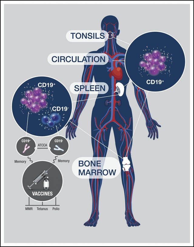
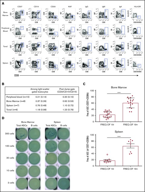
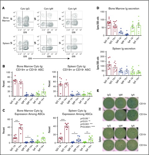
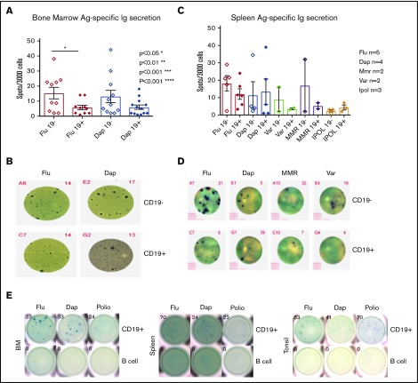
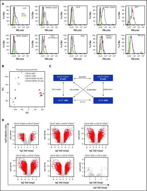
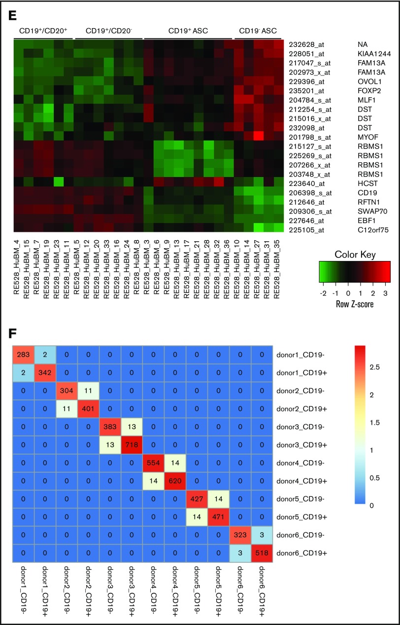
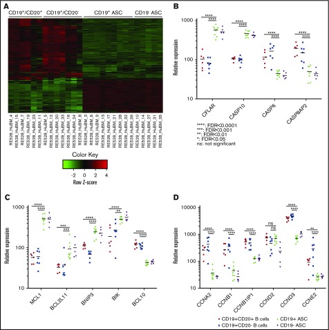
Long-lived antibody-secreting cells (ASCs) are critical for the maintenance of humoral immunity through the continued production of antibodies specific for previously encountered pathogen or vaccine antigens. Recent reports describing humoral immune memory have suggested the importance of long-lived CD19- bone marrow (BM) ASCs, which secrete antibodies recognizing previously encountered vaccine antigens. However, these reports do not agree upon the unique contribution of the CD19+ BM ASC subset toward humoral immunity. Here, we found both CD19+ and negative ASCs from human BM were similar in functional capacity to react to a number of vaccine antigens via ELISpot assays. The CD19+ cells were the predominant ASC population found in lymphoid tissues, and unlike the CD19- ASCs, which were found only in spleen and BM, the CD19+ ASCs were found in tonsil and blood. CD19+ ASCs from the BM, spleen, and tonsil were capable of recognizing polio vaccine antigens, indicating the CD19+ ASC cells play a novel role in long-lasting immune defense. Comparative gene expression analysis indicated CD19+ and negative BM ASCs differed significantly by only 14 distinct messenger RNAs and exhibited similar gene expression for cell cycle, autophagy, and apoptosis control necessary for long life. In addition, we show identical CDR-H3 sequences found on both BM ASC subsets, indicating a shared developmental path. Together, these results provide novel insight for the distribution, function, genetic regulation, and development of long-lived ASCs and may not only impact improved cell therapies but also enhance strategies for vaccine development.


Conflict of interest statement

Conflict-of-interest disclosure: C.J.G., J.C., B.R., C.A.M., R.G., J. Wang, J. Wu, R. Halpin, Y.S., R.R., R.K., Y.W., and R. Herbst are/were full-time employees and shareholders of MedImmune/AstraZeneca.

Figures

None
Graphical abstract
Figure 1.
Figure 1.
Most CD38highCD27+ASCs in human tissues express CD19. (A) Contour plots revealed a rare cell subset with high expression of CD38 and positive for CD27, which showed a clear cytoplasmic IgG, IgM, or IgA positive expression and was found in all human tissues (BM, PB, tonsil, and spleen) indicative of ASCs. (B) ASC (CD38highCD27+) frequencies in human tissues: mean, %, and standard deviation (SD). (C) CD19+ ASCs were statistically more abundant that CD19 ASCs in the BM (upper graph, unpaired Student t test, ****P < .0001, mean ± standard error of the mean (SEM) 23.14 ± 2.477 and 60.74 ± 3.994, both n = 16, median ± SD age 32 ± 7.713) and spleen (lower graph, unpaired Student t test, ****P < .0001, mean ± SEM 17.70 ± 2.983 and 64.7 ± 2.983, both n = 8, median ± SD age 51 ± 14.125). (D) CD38highCD27+ ASCs or CD19+CD20+ non-ASC B cells from BM were sorted directly onto total IgG/IgM/IgA precoated ELISpot plates. BCs, B cells; Freq., frequency.
Figure 2.
Figure 2.
CD19+ASCs are the most common immunoglobulin-secreting cells in BM and spleen. (A) Cytoplasmic immunoglobulin immunophenotyping gated on CD38highCD27+ ASCs. Representative plots showed CD19+ and CD19 vs cytoplasmic immunoglobulin in BM and spleen. (B) Relative frequency of immunoglobulin-secreting isotypes among ASC CD19+ or CD19 from BM (n = 8) and spleen (n = 7). (C) The frequency between CD19+ and CD19 ASCs for immunoglobulin isotype secretion via cytoplasmic immunoglobulin FCM in BM (upper, unpaired Student t test, IgG P < .0002, CD19+ mean ± SEM 39.58 ± 3.952, CD19 mean ± SEM 13.18 ± 2.473, n = 6, IgM P < .043, CD19+ mean ± SEM 4.393 ± 1.359, CD19 mean ± SEM 1.095 ± 0.4234, n = 6) and spleen (lower, Student unpaired Student t test, IgG P < .0001, CD19+ mean ± SEM 40.78 ± 2.48, CD19 mean ± SEM 11.54 ± 2.515, n = 6, IgM P < .0179, CD19+ mean ± SEM 8.665 ± 2.491, CD19 mean ± SEM 1.515 ± 0.4246, n = 6, IgA P < .0035, CD19+ mean ± SEM 10.3 ± 2.242, CD19 mean ± SEM 1.686 ± 0.3156, n = 6) immunoglobulin isotype testing via cytoplasmic immunoglobulin FCM. (D) Immunoglobulin secretion by ELISpot showed no significance between CD19+ and negative ASCs in BM (upper, IgG n = 13, IgM n = 6, IgA n = 6) or spleen (lower, IgG n = 8, IgM n = 6, IgA n = 7). (E) Representative ELISpots are shown for both BM (upper) and spleen (lower). Cyto, cytoplasmic; Ig, immunoglobulin.
Figure 3.
Figure 3.
Vaccine-specific IgG are produced by both the CD19+and the CD19ASC subsets. (A) BD CD19+ and CD19 ASC antigen-specific ELISpot results for Daptacel and Fluzone (unpaired Student t test, P < .0327, CD19+ mean ± SEM 5.810 ± 1.498, n = 10, CD19 mean ± SEM 15.29 ± 3.814, n = 10). (B) Representative ELISpot wells from 3000 CD19+ and CD19 FACS sorted ASCs from BM. (C) 3000 CD19+ and CD19 ASCs from spleen were sorted onto Daptacel (n = 4), Fluzone (n = 5), MMR (n = 2), Varicella (n = 2), or IPOL (n = 3) vaccine antigen precoated ELISpot plates and detected for total IgG. (D) Representative ELISpot wells showing sorted ASCs from either CD19+ or negative spleen subsets. (E) 10 000 (Flu/Dap) and 30 000 (polio) CD19+ ASCs or B cells were FACS sorted from BM, spleen, and tonsil, which indicated antigen-specific CD19+ ASCs existed in all tissues. Ag., antigen; Dap., Daptacel; Flu., Fluzone; MMR, measles, mumps, and rubella; Var., Varicella.
Figure 4.
Figure 4.
CD19+and negative BM ASC gene expression and sequence identity indicates a common differentiation path. (A) Known ASC genes (XBP-1, PRDM-1, SDC-1), B-cell genes (IL-4R, CD19, MS4A1), and phenotype genes (TNFRSF7, CD38) were assayed from freshly isolated BM. CD38highCD27+ gated cells showed increased expression of ASC genes and low levels of expression of B-cell genes by PrimeFlow. (B) Principal component (PC) analysis was performed on all genes based on relative expression from Affymetrix microarray (U133) for all 4 sorted cell populations from human BM. (C) Graphical representation showing the number of differentially regulated probes (upregulated/downregulated) for each cell population combination. (D) A volcano plot showing log2 (fold change) vs −log10 (adjusted P value) for each paired BM population comparison. Red dots indicate genes with significant expression change; gray dots indicate genes with no significant expression change. (E) Six genes with increased expression on CD19+ ASC and 8 uncharacterized genes with increased expression specific to CD19 ASCs. (F) Heat map of a pairwise comparison of common CDR-H3 sequences between donors. CD19+ and CD19 ASCs were profiled for 6 donors, and the sequences with identical CDR-H3 were considered as identical clones. The number in the heat map describes the number of common clones between 2 samples, and the number in the diagonal line showed the total number of CDR-H3 sequences in the sample. Max., maximum.
Figure 4.
Figure 4.
CD19+and negative BM ASC gene expression and sequence identity indicates a common differentiation path. (A) Known ASC genes (XBP-1, PRDM-1, SDC-1), B-cell genes (IL-4R, CD19, MS4A1), and phenotype genes (TNFRSF7, CD38) were assayed from freshly isolated BM. CD38highCD27+ gated cells showed increased expression of ASC genes and low levels of expression of B-cell genes by PrimeFlow. (B) Principal component (PC) analysis was performed on all genes based on relative expression from Affymetrix microarray (U133) for all 4 sorted cell populations from human BM. (C) Graphical representation showing the number of differentially regulated probes (upregulated/downregulated) for each cell population combination. (D) A volcano plot showing log2 (fold change) vs −log10 (adjusted P value) for each paired BM population comparison. Red dots indicate genes with significant expression change; gray dots indicate genes with no significant expression change. (E) Six genes with increased expression on CD19+ ASC and 8 uncharacterized genes with increased expression specific to CD19 ASCs. (F) Heat map of a pairwise comparison of common CDR-H3 sequences between donors. CD19+ and CD19 ASCs were profiled for 6 donors, and the sequences with identical CDR-H3 were considered as identical clones. The number in the heat map describes the number of common clones between 2 samples, and the number in the diagonal line showed the total number of CDR-H3 sequences in the sample. Max., maximum.
Figure 5.
Figure 5.
CD19+and CD19BM ASCs exhibit similar genetic mechanisms for survival. (A) A heat map showing expression of genes with differential expression between 1 of any 2 BM cell populations. Most gene expression appears similar between ASCs and distinct from BM B-cell populations. (B) Scatter plot of 4 caspase-family relative gene expression between BM CD19+CD20+ B cells (circle), CD19+CD20 B cells (square), CD19+ ASCs (triangle), and CD19 ASCs (inverted triangle). (C) Relative gene expression of bcl2-family genes on BM cell populations. (D) Relative expression of cyclin genes on sorted BM cell populations. None of the scatter plots showed a significant difference between the CD19+ and CD19 ASCs.

References

    1. Ahmed R, Gray D.. Immunological memory and protective immunity: understanding their relation. Science. 1996;272(5258):54-60. - PubMed
    1. Manz RA, Thiel A, Radbruch A. Lifetime of plasma cells in the bone marrow. Nature. 1997;388(6638):133-134. - PubMed
    1. Slifka MK, Antia R, Whitmire JK, Ahmed R. Humoral immunity due to long-lived plasma cells. Immunity. 1998;8(3):363-372. - PubMed
    1. Nutt SL, Hodgkin PD, Tarlinton DM, Corcoran LM. The generation of antibody-secreting plasma cells. Nat Rev Immunol. 2015;15(3):160-171. - PubMed
    1. Shaffer AL, Lin KI, Kuo TC, et al. . Blimp-1 orchestrates plasma cell differentiation by extinguishing the mature B cell gene expression program. Immunity. 2002;17(1):51-62. - PubMed