. 2019 Jan 18;294(3):953-967.
doi: 10.1074/jbc.RA118.004883. Epub 2018 Nov 26.

Structures of AMP-activated protein kinase bound to novel pharmacological activators in phosphorylated, non-phosphorylated, and nucleotide-free states

Affiliations

Structures of AMP-activated protein kinase bound to novel pharmacological activators in phosphorylated, non-phosphorylated, and nucleotide-free states

Yan Yan et al. J Biol Chem. .

Abstract

AMP-activated protein kinase (AMPK) is an attractive therapeutic target for managing metabolic diseases. A class of pharmacological activators, including Merck 991, binds the AMPK ADaM site, which forms the interaction surface between the kinase domain (KD) of the α-subunit and the carbohydrate-binding module (CBM) of the β-subunit. Here, we report the development of two new 991-derivative compounds, R734 and R739, which potently activate AMPK in a variety of cell types, including β2-specific skeletal muscle cells. Surprisingly, we found that they have only minor effects on direct kinase activity of the recombinant α1β2γ1 isoform yet robustly enhance protection against activation loop dephosphorylation. This mode of activation is reminiscent of that of ADP, which activates AMPK by binding to the nucleotide-binding sites in the γ-subunit, more than 60 Å away from the ADaM site. To understand the mechanisms of full and partial AMPK activation, we determined the crystal structures of fully active phosphorylated AMPK α1β1γ1 bound to AMP and R734/R739 as well as partially active nonphosphorylated AMPK bound to R734 and AMP and phosphorylated AMPK bound to R734 in the absence of added nucleotides at <3-Å resolution. These structures and associated analyses identified a novel conformational state of the AMPK autoinhibitory domain associated with partial kinase activity and provide new insights into phosphorylation-dependent activation loop stabilization in AMPK.

Keywords: ADaM site; AMP; AMP-activated kinase (AMPK); CBS3; R734; R739; X-ray crystallography; activation loop phosphorylation; energy sensor; hydrogen exchange mass spectrometry; metabolic disorder; phosphorylation.


Conflict of interest statement

S. J. S., Y. L., and Y. H. are or have been employees and shareholders of Rigel Pharmaceuticals, Inc

Figures
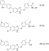
Figure 1.
Figure 1.
Chemical structures of R734, R739, and Merck 991.
Figure 2.
Figure 2.
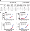
AMPK activation in cells. A, summary of AMPK activity data. Activation was determined by ELISA-based pACC assay. AC2X, compound concentration at which activity is 2-fold above background. 991, PF249, and GSK621 activation data were included as references. nd, not determined. B, R734 and R739 dose-response curves in pACC cell-based ELISAs. n = 2; error bars, S.D.
Figure 3.
Figure 3.
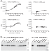
Activation of purified, recombinant AMPK. A, activity dose-response curves determined by radioactive kinase assays using the ACC-based SAMS peptide as substrate. n = 3; error bars, S.D. B, effects of 20 μm R734, R739, or A769662 or 200 μm AMP on protection against activation loop dephosphorylation. The indicated AMPK proteins were incubated with protein phosphatase 2Cα (PP2C; left blot) or with λ-phosphatase (λ-pp; right blot) for the indicated length of time. Protein phosphatase 2Cα was chosen for AMPK α1β2γ1 as the activation loop phosphate of this isoform was inefficiently removed by λ-phosphatase. Phospho-AMPK was visualized by immunoblotting. ΔST, deletion of the ST loop in the α-subunit (amino acids 476–529).
Figure 4.
Figure 4.
R734, R739, 991, and A769662 increase phosphorylation of AMPK, Raptor, and ACC in cells. A–D, dose-dependent phosphorylation increases in the presence of R734 (A), R739 (B), 991 (C), and A769662 (D). HepG2 cells were treated with increasing doses of R734, R739, 991, and A769662 for 1 h. Phosphorylated and total proteins were visualized by immunoblotting with the indicated antibodies. Target bands are indicated by arrows. Higher order phosphorylation of AMPKα due to autophosphorylation is marked by asterisks. Bands above the phosphorylated Raptor bands are cross-reacting bands. β-Actin is shown as loading control.
Figure 5.
Figure 5.
Structures of phosphorylated AMPK α1β1γ1 in complex with R734/R739, AMP, and staurosporine. A and B, structure overviews. C–E, Close-ups of R734 (C)-, R739 (D)-, and 991-occupied (E) ADaM sites. Dashed lines, polar interactions. The CBM is colored cyan, the KD is green, and the ATP-binding G-loop of the KD is dark green.
Figure 6.
Figure 6.
Structure of nonphosphorylated AMPK α1β1γ1–R734–AMP–staurosporine. A, R734 weakly synergizes with AMP for AMPK activation. Kinase assays were performed with recombinant, E. coli–expressed AMPK. The kinase assay with phosphorylated AMPK was performed on ice to compensate for the much higher activity compared with nonphosphorylated AMPK. n = 3; error bars, S.D. B, structure overview.
Figure 7.
Figure 7.
The AID of nonphosphorylated AMPK α1β1γ1–R734–AMP adopts a unique γ-subunit–bound conformation. A, cartoon representation of the AID (brown), its immediate flanking residues (yellow), and adjacent β- (cyan) and γ-subunit (magenta) regions with AID side chains shown in thin stick presentation. Key interface residues are shown in the inset. B, 2FoFc composite omit map of the AID contoured at 1.0 σ in the same orientation as in A. A subset of AID residues with well-resolved densities is labeled. Residues 298–313 were not resolved. C, structural overlay of nonphosphorylated AMPK α1β1γ1 (AID, green; γ-subunit, magenta) and phosphorylated AMPK α1β2γ1 (PDB code 4RER) (yellow) to highlight the extent of the AID rotation indicated by the arrow. D, AID mutational analysis. Met-289 and Glu-293 of the AID α1 helix interact with the γ-subunit in the structure of nonphosphorylated AMPK α1β1γ1–R734–AMP but make no interactions in any structure of phosphorylated AMPK (n = 3; error bars, S.D.). ns, not significant (p > 0.05); **, p < 0.01; ***, p < 0.001; ****, p < 0.0001 (two-way analysis of variance with Bonferroni post-test).
Figure 8.
Figure 8.
The activation loop of nonphosphorylated AMPK α1β1γ1–R734–AMP adopts an active conformation stabilized by binding to the AMPK core. The activation loop (dark green) is stabilized by interaction with the AMPK core complex (green, α-subunit; cyan, β-subunit). Key interface residues are shown in stick representation (yellow, phosphorylated AMPK–R734–AMP residues in overlay panel).
Figure 9.
Figure 9.
Phosphorylation stabilizes the activation loop in solution. A, kinase domain structure of AMPK α1β1γ1–R734–AMP with activation loop highlighted in red. B–D, perturbation (difference) HDX-MS heat maps overlaid on the structure shown in A. Relative changes in protection against hydrogen/deuterium exchange are indicated by the color bar in the center. Gray indicates no change or unresolved regions. The full maps are shown in Fig. S8–S10.
Figure 10.
Figure 10.
AMP is bound at CBS4 in the structure of phosphorylated AMPK α1β1γ1–R734 in the absence of added nucleotides. A, structure overview. B, 2FoFc composite omit map contoured at 1 σ around the three AMP-binding sites. The omit map is overlaid with the transparent cartoon structure of the heteromeric complex and a stick model of AMP at CBS4. For orientation, it is also overlaid with stick models (white) of the three AMP molecules of pAMPK in the presence of added AMP (PDB code 6C9F; see Fig. 5A).

References

    1. Cheung P. C., Salt I. P., Davies S. P., Hardie D. G., and Carling D. (2000) Characterization of AMP-activated protein kinase γ-subunit isoforms and their role in AMP binding. Biochem. J. 346, 659–669 - PMC - PubMed
    1. Stapleton D., Gao G., Michell B. J., Widmer J., Mitchelhill K., Teh T., House C. M., Witters L. A., and Kemp B. E. (1994) Mammalian 5′-AMP-activated protein kinase non-catalytic subunits are homologs of proteins that interact with yeast Snf1 protein kinase. J. Biol. Chem. 269, 29343–29346 - PubMed
    1. Thornton C., Snowden M. A., and Carling D. (1998) Identification of a novel AMP-activated protein kinase β subunit isoform that is highly expressed in skeletal muscle. J. Biol. Chem. 273, 12443–12450 10.1074/jbc.RA117.001068 - DOI - PubMed
    1. Birk J. B., and Wojtaszewski J. F. (2006) Predominant α2/β2/γ3 AMPK activation during exercise in human skeletal muscle. J. Physiol. 577, 1021–1032 10.1074/jbc.RA117.001068 - DOI - PMC - PubMed
    1. Cokorinos E. C., Delmore J., Reyes A. R., Albuquerque B., Kjøbsted R., Jørgensen N. O., Tran J. L., Jatkar A., Cialdea K., Esquejo R. M., Meissen J., Calabrese M. F., Cordes J., Moccia R., Tess D., et al. (2017) Activation of skeletal muscle AMPK promotes glucose disposal and glucose lowering in non-human primates and mice. Cell Metab. 25, 1147–1159.e10 10.1074/jbc.RA117.001068 - DOI - PubMed

Publication types

MeSH terms

LinkOut - more resources