. 2018 Dec;25(12):1103-1110.
doi: 10.1038/s41594-018-0154-1. Epub 2018 Nov 26.

Architecture of the CBF3-centromere complex of the budding yeast kinetochore

Affiliations

Architecture of the CBF3-centromere complex of the budding yeast kinetochore

Kaige Yan et al. Nat Struct Mol Biol. 2018 Dec.

Abstract

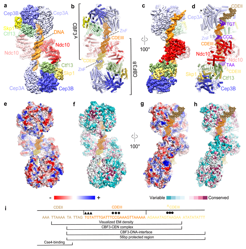
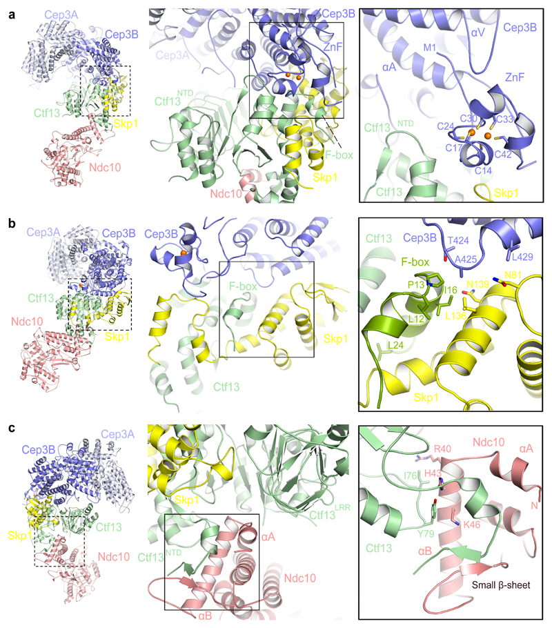
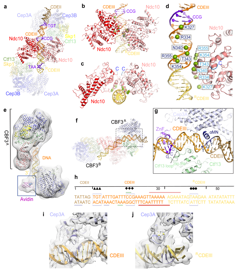
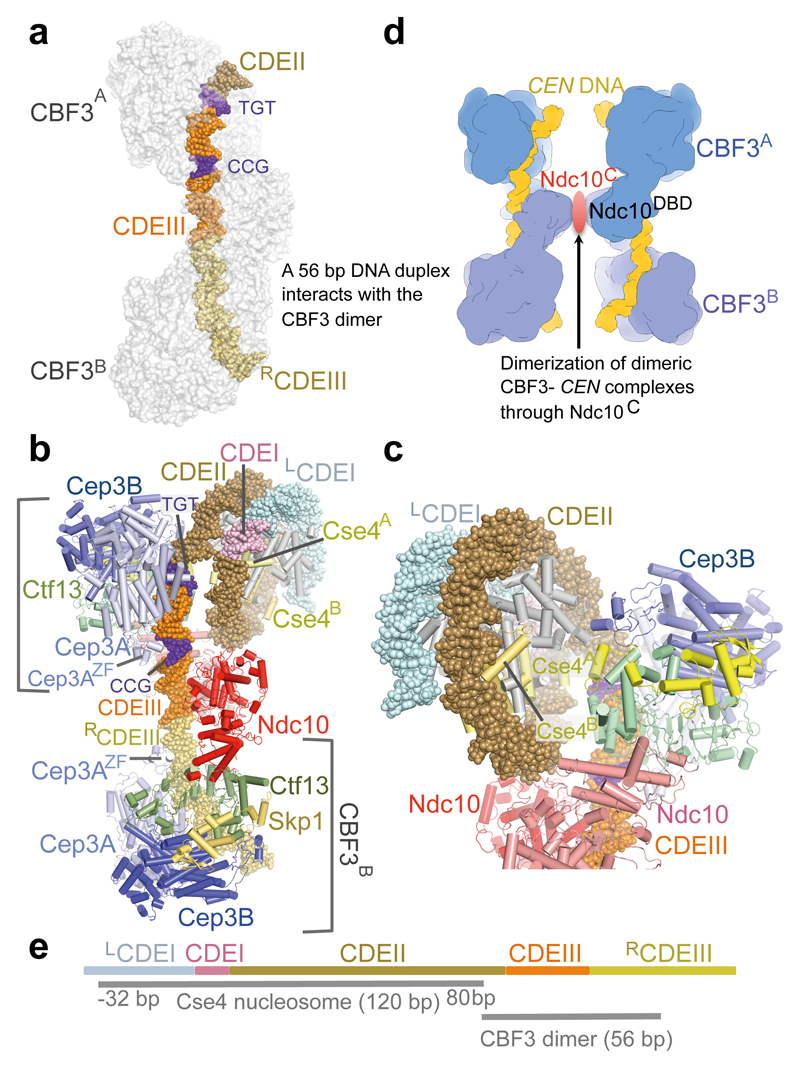
Kinetochores are multicomponent complexes responsible for coordinating the attachment of centromeric DNA to mitotic-spindle microtubules. The point centromeres of budding yeast are organized into three centromeric determining elements (CDEs), and are associated with the centromere-specific nucleosome Cse4. Deposition of Cse4 at CEN loci is dependent on the CBF3 complex that engages CDEIII to direct Cse4 nucleosomes to CDEII. To understand how CBF3 recognizes CDEIII and positions Cse4, we determined a cryo-EM structure of a CBF3-CEN complex. CBF3 interacts with CEN DNA as a head-to-head dimer that includes the whole of CDEIII and immediate 3' regions. Specific CEN-binding of CBF3 is mediated by a Cep3 subunit of one of the CBF3 protomers that forms major groove interactions with the conserved and essential CCG and TGT motifs of CDEIII. We propose a model for a CBF3-Cse4-CEN complex with implications for understanding CBF3-directed deposition of the Cse4 nucleosome at CEN loci.


Conflict of interest statement

Competing Interests. The authors declare that there are no competing interests.

Figures

Figure 1
Figure 1. Overall structure of the dimeric−CEN3 complex.
(a) and (c) Two orthogonal views of the EM density map of the complex, (b) and (d) Corresponding cartoon representations. The TGT and CCG motifs of CDEIII are colored blue. (e) and (g) Electrostatic surface representation of the dimeric CBF3−CEN3 complex. Color code is displayed below. (f) and (h) Surface conservation of the CBF3−CEN3 dimer. Conservation score is indicated below. (i) Region of CEN3 sequence and 3’ flanking region recognized by the dimeric CBF3-CEN3 complex. Sequences indicated with diamonds and circles are the CCG and TAA motifs recognized by the Zn2Cys6 cluster of CBF3A and CBF3B, respectively. The TGT motif contacted by αMN helix of Cep3A is indicated with arrowheads.
Figure 2
Figure 2. CBF3 inter-subunit interfaces.
(a) Interactions of the Cep3B Zn2Cys6 cluster at the Skp1 and Ctf13 interface. (b) Contacts of the Ctf13 F-box with Skp1 and Cep3B. L12, P13 and L24 are essential for Ctf13−Skp1 interactions. (c) The N-terminus of Ndc10 contacts Ctf13. Mutating I76 and Y79 of Ctf13 disrupts Ctf13-Ndc10 interactions (Supplementary Fig. 1c).
Figure 3
Figure 3. Basis for CBF3−CEN3 recognition.
(a) Overview of the dimeric CBF3−CEN3 complex. (b) and (c) Two views of the Ndc10DBD-DNA interface. DNA induces dimerization of Ndc10DBD. (d) Details of Ndc10DBD−DNA contacts. Residues in contact with DNA are shown as green spheres. Cartoon representation of only one Ndc10 subunit is shown. (e) EM density map of the CBF3−CEN3-biotin avidin complex orients and positions the DNA duplex. (f) Overview of Cep3B-Ctf13 contacts with DNA in CBF3A. (g) Details of Cep3A−Ctf13−CDEIII interface, showing Zn2Cys6 cluster contacting the CCG motif and αMN helix contacting the TGT motif. Ctf13 contacts the minor groove midway between the CCG and TGT motifs. (h) Schematic of CBF3-DNA interactions. Sequence of the nuclease resistant 56 bp segment protected by CBF3 is shown . Arrow, diamond and circle symbols as in Fig. 1i. Colored horizontal lines indicate contacts to CBF3 subunits. (i) and (j) EM density of the Zn2Cys6 clusters interacting with CEN3-DNA for (i) Cep3A of CBF3A and (j) Cep3A of CBF3B.
Figure 4
Figure 4. Model of CBF3−Cse4 nucleosome complex.
(a). Model of dimeric CBF3 with nuclease resistant 56 bp DNA segment. CBF3 is represented with a transparent molecular surface. (b) CBF3−Cse4−CEN structure modeled on the cryo-EM CBF3−CEN complex and the CENP-A crystal structure (PDB 3AN2). The two CENP-A subunits (labeled as Cse4A and Cse4B) are highlighted. (c) Close up of the view of the CBF3−Cse4−CEN model showing close proximity of the N-terminus of Cse4A and Ctf13. (d) Cartoon showing possible pairing of CBF3−CEN complexes mediated by dimerization of Ndc10C domains. (e) Schematic of the proposed CBF3−Cse4 nucleosome complex at the CEN3 locus. CDEI, CDEII and CDEIII, and the DNA contact regions of Cse4 and CBF3 are drawn to scale. For Cse4, the start and end of the binding site are shown, -32 bp: 32 bp to the left of the LCDEI-CDEI junction, 80 bp: 80 bp to the right of the CDEI-CDEII junction.

References

    1. Bloom K, Costanzo V. Centromere Structure and Function. Prog Mol Subcell Biol. 2017;56:515–539. - PMC - PubMed
    1. Musacchio A, Desai A. A Molecular View of Kinetochore Assembly and Function. Biology (Basel) 2017;6 - PMC - PubMed
    1. Jenni S, Dimitrova YN, Valverde R, Hinshaw SM, Harrison SC. Molecular Structures of Yeast Kinetochore Subcomplexes and Their Roles in Chromosome Segregation. Cold Spring Harb Symp Quant Biol. 2017 - PubMed
    1. Musacchio A. The Molecular Biology of Spindle Assembly Checkpoint Signaling Dynamics. Curr Biol. 2015;25:R1002–18. - PubMed
    1. Joglekar AP. A Cell Biological Perspective on Past, Present and Future Investigations of the Spindle Assembly Checkpoint. Biology (Basel) 2016;5 - PMC - PubMed

Publication types

MeSH terms

Substances