. 2018 Nov 26;9(1):4976.
doi: 10.1038/s41467-018-07309-4.

VDAC2 enables BAX to mediate apoptosis and limit tumor development

Affiliations

VDAC2 enables BAX to mediate apoptosis and limit tumor development

Hui San Chin et al. Nat Commun. .

Abstract

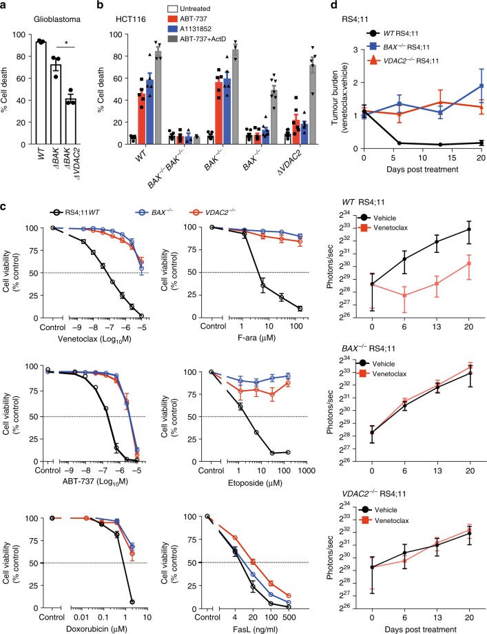
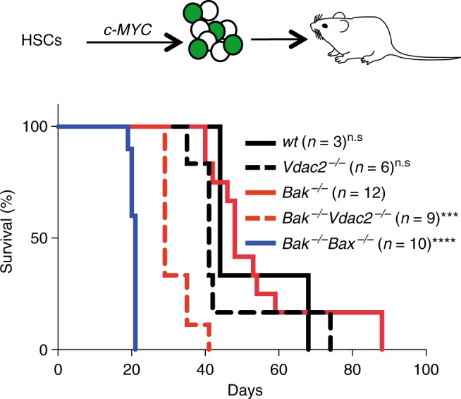
Intrinsic apoptosis is critical to prevent tumor formation and is engaged by many anti-cancer agents to eliminate tumor cells. BAX and BAK, the two essential mediators of apoptosis, are thought to be regulated through similar mechanisms and act redundantly to drive apoptotic cell death. From an unbiased genome-wide CRISPR/Cas9 screen, we identified VDAC2 (voltage-dependent anion channel 2) as important for BAX, but not BAK, to function. Genetic deletion of VDAC2 abrogated the association of BAX and BAK with mitochondrial complexes containing VDAC1, VDAC2, and VDAC3, but only inhibited BAX apoptotic function. Deleting VDAC2 phenocopied the loss of BAX in impairing both the killing of tumor cells by anti-cancer agents and the ability to suppress tumor formation. Together, our studies show that efficient BAX-mediated apoptosis depends on VDAC2, and reveal a striking difference in how BAX and BAK are functionally impacted by their interactions with VDAC2.


Conflict of interest statement

M.Fv.D., D.H.D.G., C.C., C.M.H., G.L.K., I.K.L.T., L.F.D., A.W., J.J.S., A.J.K., M.J.H., K.B., A.L.S., B.R., P.B., R.M.K., D.C.S.H., and G.D. are employees of the Walter and Eliza Hall Institute of Medical Research, which receives milestone payments for venetoclax. All other authors declare no competing interests.

Figures

Fig. 1
Fig. 1
CRISPR/Cas9 screen identifies VDAC2 as a promoter of BAX-mediated apoptosis. a Outline of the genome-wide CRISPR/Cas9 library screens to identify mediators of intrinsic apoptosis. b VDAC2 promotes BAX apoptotic function. MEFs expressing Cas9 and a whole-genome sgRNA library were treated with ABT-737 (LC90, 250 nM, see Supplementary Fig. 1b) for 48 h. Resistant cells were recovered after 5 days and enriched sgRNAs identified by deep-sequencing. Plots show independent sgRNAs in ABT-737 treated versus untreated controls cells collated and normalized from 2 to 4 independent experiments. Dashed lines represent LOWESS regression curves fitted to the data. c Deletion of Vdac2 protects from BAX-mediated apoptosis in response to ABT-737. Clones (Mcl1−/−Bax−/−Vdac2−/− and Mcl1−/−Bak−/−Vdac2−/−) or polyclonal populations (Mcl1−/−, Bax−/− and, Bak−/−) of MEFs were treated with escalating doses of ABT-737 for 24 h and cell viability assessed by PI exclusion. Data are mean+/− SEM of at least three independent experiments with 3–4 independent clones. d Deletion of Vdac2 provides long-term protection from BAX-mediated cell death. MEFs of the indicated genotype were treated with the indicated concentration of ABT-737 and colony formation was assessed after 5 days. e Deletion of Bak protects Vdac2−/− MEFs from etoposide-induced apoptosis. Polyclonal populations or three independent MEF clones of the indicated genotype (all on 129sv;C57BL/6 background) were treated with etoposide (10 μM for 24 h) and cell viability assessed by PI exclusion. Data are mean+/− SEM shown for three independent experiments
Fig. 2
Fig. 2
VDAC2 promotes the association of BAX and BAK with a VDAC complex. a Endogenous BAX and BAK associate with independent complexes in mitochondria. Mitochondria-enriched fractions from HeLa or HCT116 cells were solubilized in 1% digitonin prior to incubation with a control antibody or an antibody that binds inactive human BAK (7D10), prior to BN-PAGE and immunoblotting for BAK or BAX. * likely cross-reactivity of anti-rat secondary antibody with rat IgG used for gel-shift. Importantly, whilst all of the BAK:VDAC2 complex was gel-shifted by the BAK antibody, the BAX:VDAC2 complex was unaffected. b Mass spectrometry analysis of the native BAX complex. Mitochondria from MEFs expressing FLAG-BAXS184L or untagged BAXS184L were solubilized in 1% digitonin prior to anti-FLAG affinity purification and proteins identified by quantitative mass spectrometry analysis. Volcano plot illustrating the log2 protein ratios of proteins enriched in the native complex following quantitative pipeline analysis. Proteins were deemed differentially regulated if the log2 fold change in protein expression was greater than two-fold (red) or four-fold (green) and a –log10 p value ≥ 1.3, equivalent to a p value ≤ 0.05. c Mass spectrometry of the native BAK complex. Mitochondria from MEFs expressing FLAG-BAK or untagged BAK harvested and analyzed as in (b). d Deletion of VDAC2 impacts mitochondrial localization of BAX and BAK. Clonal populations of Bax−/− and Bak−/− MEFs with deleted Vdac1, Vdac2 or Vdac3 (denoted ∆) were fractionated into cytosol and membrane and immunoblotted for BAX, BAK or TIM44 as a mitochondrial control. e VDAC2 plays the major role in BAX and BAK complex stability. Mitochondria isolated from clonal populations of Bax−/− and Bak−/− MEFs with deleted Vdac1, Vdac2 or Vdac3 were analyzed on BN-PAGE. Data are representative of two independent clones (see Supplementary Fig. 2e). Intermediate complexes indicated (arrows). f BAX-mediated apoptosis is impaired in the absence of VDAC2 and to a lesser extent by VDAC3. Polyclonal populations were treated with etoposide (10 μM) and cell death was assessed by PI uptake. Data are mean+/ SEM of three independent experiments. ***p < 0.001; **p < 0.01; n.s, not significant; based on unpaired Student’s t-test
Fig. 3
Fig. 3
Interaction with VDAC2 is important for BAX apoptotic function. a BAX mitochondrial complex formation specifically relies on VDAC2. Mitochondria-enriched fractions from Mcl1−/−Bak−/−Vdac2−/− MEFs reconstituted with FLAG-mVDAC1 or FLAG-hVDAC2 were analyzed by BN-PAGE and immunoblotted for BAX (left) or FLAG to detect ectopically-expressed VDACs (right). b BAX apoptotic function relies on VDAC2. Cells as in (a) were treated with ABT-737 and cell viability was assessed. ce Rescue of BAX apoptotic function correlates with interaction with a specific region of VDAC2. Mcl1−/−Bak−/−Vdac2−/− MEFs stably expressing FLAG-mVDAC1/hVDAC2 chimeras (c) were analyzed for expression by immunoblotting for FLAG (or GAPDH as a loading control), cell viability following treatment with ABT-737 (d), and complex formation by BN-PAGE and immunoblotting for BAX or FLAG (e). f BAX interacts with a defined region of VDAC2. Interaction of BAX requires aa109-171 of hVDAC2 (salmon) mapped onto the structure of zebrafish VDAC2 (PDB 4BUM). MOM mitochondrial outer membrane, IMS intermembrane space. g VDAC2 is essential for BAX, but not BAK to target mitochondria and mediate apoptosis. In wild-type cells, cytosolic BAX (BAXcyto) relies on VDAC2 to associate with mitochondria (BAXmito) and activate (BAX*). In the absence of VDAC2, BAX cannot drive cell death. Although the ability of BAK to associate with mitochondria is also perturbed in VDAC2−/− cells, , sufficient BAK can still target mitochondria through a VDAC2-independent mechanism to drive apoptosis. Data presented in (b) and (d) is mean+/ SEM of three independent experiments
Fig. 4
Fig. 4
Vdac2−/− mice do not show evidence of excessive BAK activity. a Bak deletion is not essential for early embryonic development of Vdac2−/− mice. C57BL/6J zygotes were injected with DNA encoding Cas9 and sgRNAs targeting Bak and Vdac2 (or Rosa as a control) and transplanted into pseudo-pregnant mothers. The percentage embryos at E14.5 with homozygous null alleles are indicated (number of mice in parentheses). (b) Gene targeting strategy to generate Vdac2−/− mice. Targeting by both sgRNAs will result in a deletion of 500 bp whereas targeting by the 3’ sgRNA alone results in indels (see Supplementary Fig. 3a). c, d Vdac2−/− mice are runted die post-natally. Kaplan–Meier survival curve of Vdac2+/− and Vdac2−/− F0 mice. e Vdac2−/− mitochondria are resistant to MOM permeabilization. Liver mitochondria isolated from age-matched WT and Vdac2−/− mice were treated with cBID prior to fractionation into supernatant (S) and membrane (P) and immunoblotting for cytochrome c. Data are representative of N = 2 mice (see Supplementary Fig. 3d). f Vdac2−/− mice show defects in the hematopoietic system. Blood counts for individual age-matched wild-type (WT, N = 6) or Vdac2−/− (N = 9) mice shown with mean+/SD. WBC white blood cells, RBC red blood cells. P values calculated by two-tailed Student’s t-test. n.s, not significant. g Early passage primary Bak−/− (BR1.4, triangles; BR2.1, circles), Vdac2−/− (BV1.1, triangles; BV2.4, circles), Bak−/−Vdac2−/− (BV2.1, triangles, BV2.3, circles) or wild-type (wt, circles) MEFs (see Supplementary Figure 8) were treated with etoposide (10 μM), ABT-737 (1 μΜ) and S63485 (1 μM), staurosporine (STS, 1 μM) or actinomycin D (ActD, 1 μM) and cell death assessed by PI uptake after 24 h. Data is mean+/SD of three independent experiments. **p < 0.01; ****p < 0.0001; n.s, not significant based on Students unpaired t-test
Fig. 5
Fig. 5
VDAC2 enables BAX-mediated killing of cancer cells in vitro and in vivo. a Deletion of VDAC2 inhibits BAX-mediated apoptosis in glioblastoma cells. Glioblastoma cells (U-251) were treated with ABT-737 (1 μM) and S63845 (1 μM) and cell death assessed after 24 h. Data are mean+/SEM of three independent experiments. *p < 0.05 based on Student’s unpaired t-test. b Deletion of VDAC2 protects HCT116 colorectal cancer cells from apoptosis. HCT116 cells were treated with ABT-737 (5 μM), A1331852 (5 μM) or ABT-737 (5 μM) + actinomycin D (1 μM) for 24 h and cell death assessed. Data are mean+/SEM of five independent experiments. c BAX or VDAC2 deletion renders RS4;11 acute lymphoblastic leukemia cells resistant to venetoclax or other chemotherapeutic agents. WT, BAX−/− or VDAC2−/− RS4;11 cells were treated with venetoclax, ABT-737 and standard-of-care chemotherapies (F-ara, etoposide, doxorubicin) or the BAX/BAK-independent stimulus Fas ligand (FasL) and cell viability assessed by PI exclusion. Data are mean+/SEM of at least 3 independent experiments. d Deletion of VDAC2 renders RS4;11 cells resistant to venetoclax in vivo. RS4;11 cells were subcutaneously engrafted into NOD/SCID/IL-2Rγnull mice, treated with venetoclax and tumor growth monitored by IVIS imaging. The collated data (top panel) is normalized to represent relative tumor burden. Data are mean+/ SEM collated from four independent experiments, N = 12 mice engrafted with each genotype of RS4;11 cells
Fig. 6
Fig. 6
VDAC2 enables BAX to limit tumor development. Vdac2 deletion accelerates the development of MYC-driven AML. Kaplan–Meier survival plot of mice transplanted with wild-type (wt), Vdac2−/−, Bak−/−, Bak−/−Bax−/− or Bak−/−Vdac2−/− fetal liver-derived hematopoietic stem cells (HSCs) (2 livers per genotype, see Supplementary Figure 7a) infected with a c-MYC–expressing retrovirus. P values (Log-rank analysis) of mice injected with Bak−/− hematopoietic precursors compared with Bak−/−Vdac2−/− or Bak−/−Bax−/− precursors are <0.001 (***) and <0.0001 (****) respectively. n.s, not significant

References

    1. Wei MC, et al. Proapoptotic BAX and BAK: a requisite gateway to mitochondrial dysfunction and death. Science. 2001;292:727–730. doi: 10.1126/science.1059108. - DOI - PMC - PubMed
    1. Czabotar PE, Lessene G, Strasser A, Adams JM. Control of apoptosis by the BCL-2 protein family: implications for physiology and therapy. Nat. Rev. Mol. Cell Biol. 2014;15:49–63. doi: 10.1038/nrm3722. - DOI - PubMed
    1. Lindsten T, et al. The combined functions of proapoptotic Bcl-2 family members Bak and Bax are essential for normal development of multiple tissues. Mol. Cell. 2000;6:1389–1399. doi: 10.1016/S1097-2765(00)00136-2. - DOI - PMC - PubMed
    1. Edlich F, et al. Bcl-x(L) Retrotranslocates bax from the mitochondria into the cytosol. Cell. 2011;145:104–116. doi: 10.1016/j.cell.2011.02.034. - DOI - PMC - PubMed
    1. Schellenberg B, et al. Bax exists in a dynamic equilibrium between the cytosol and mitochondria to control apoptotic priming. Mol. Cell. 2013;49:959–971. doi: 10.1016/j.molcel.2012.12.022. - DOI - PMC - PubMed

Publication types

Substances