. 2018 Dec;3(12):1369-1376.
doi: 10.1038/s41564-018-0273-9. Epub 2018 Nov 26.

The interferon-inducible isoform of NCOA7 inhibits endosome-mediated viral entry

Affiliations

The interferon-inducible isoform of NCOA7 inhibits endosome-mediated viral entry

Tomas Doyle et al. Nat Microbiol. 2018 Dec.

Erratum in

Abstract

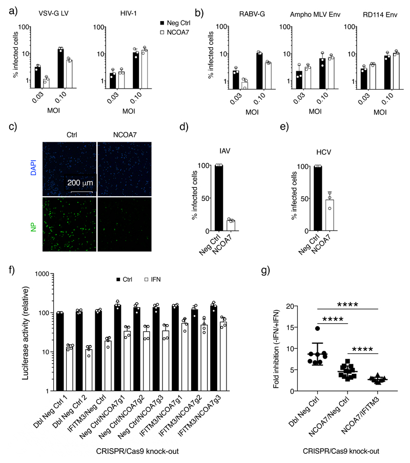
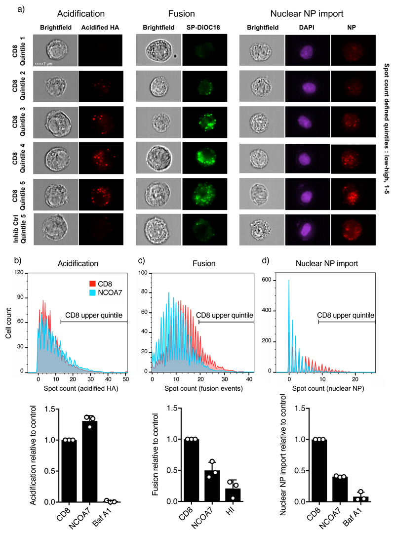
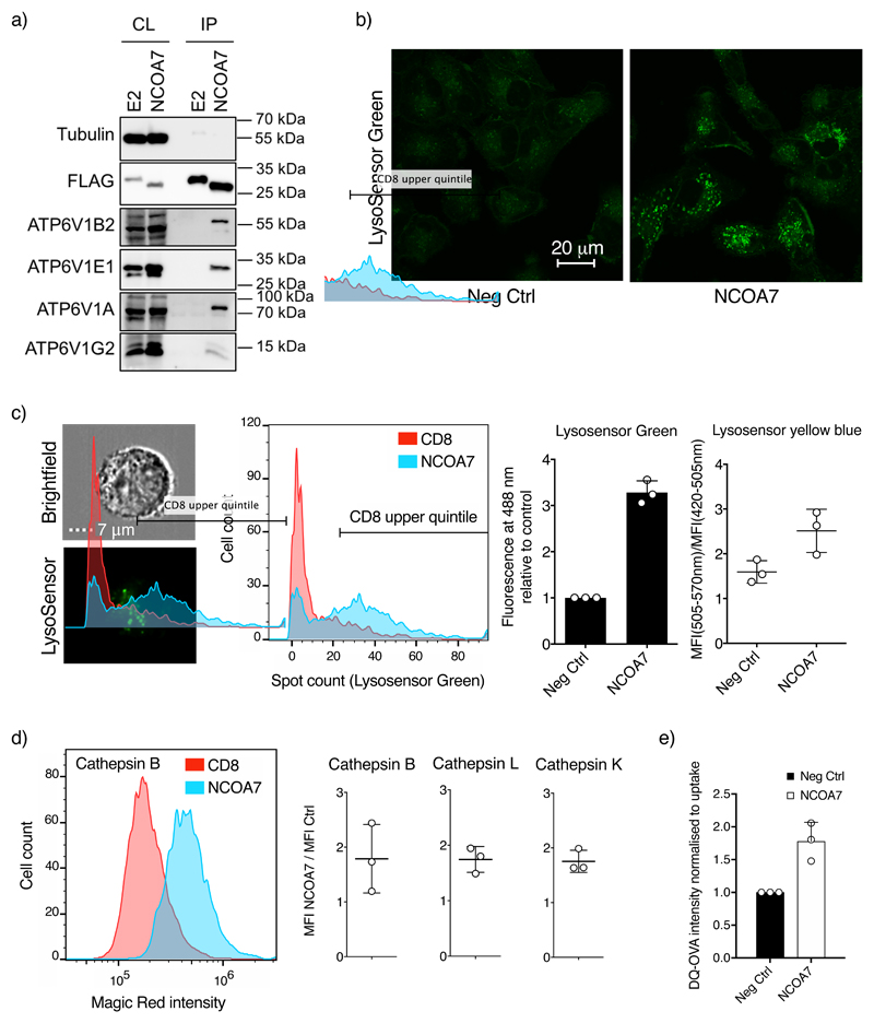
Interferons (IFNs) mediate cellular defence against viral pathogens by upregulation of IFN-stimulated genes whose products interact with viral components or alter cellular physiology to suppress viral replication1-3. Among the IFN-stimulated genes that can inhibit influenza A virus (IAV)4 are the myxovirus resistance 1 GTPase5 and IFN-induced transmembrane protein 3 (refs 6,7). Here, we use ectopic expression and gene knockout to demonstrate that the IFN-inducible 219-amino acid short isoform of human nuclear receptor coactivator 7 (NCOA7) is an inhibitor of IAV as well as other viruses that enter the cell by endocytosis, including hepatitis C virus. NCOA7 interacts with the vacuolar H+-ATPase (V-ATPase) and its expression promotes cytoplasmic vesicle acidification, lysosomal protease activity and the degradation of endocytosed antigen. Step-wise dissection of the IAV entry pathway demonstrates that NCOA7 inhibits fusion of the viral and endosomal membranes and subsequent nuclear translocation of viral ribonucleoproteins. Therefore, NCOA7 provides a mechanism for immune regulation of endolysosomal physiology that not only suppresses viral entry into the cytosol from this compartment but may also regulate other V-ATPase-associated cellular processes, such as physiological adjustments to nutritional status, or the maturation and function of antigen-presenting cells.


Conflict of interest statement

Conflicts of interest statement:

The authors have no conflicts of interest to declare in relation to this manuscript.

Figures

Figure 1
Figure 1. NCOA7 inhibits infection by viruses entering through the endocytic pathway.
U87MG CD4+ CXCR4+ cells were transduced with EasiLV expressing NCOA7 or a control (CD8 or luciferase) and treated with doxycycline for 48 hr. Cells were challenged with a) VSV-G-pseudotyped HIV-1 based, GFP-expressing lentiviral vector (VSV-G LV) or HIV-1 (NL4.3/Nef-IRES-GFP) or b) lentiviral vectors pseudotyped with: rabies virus GP (RABV-G), or amphotropic MLV Env or RD114 Env. 48 hr later, E2 crimson-positive cells were gated and the percentage of infected, GFP-positive cells determined by flow cytometry. c) A549 cells expressing NCOA7 or CD8 were infected with A/Eng/195/2009, fixed at 5 hr, stained with anti-NP antibody (green) and visualized by confocal microscopy, and d) shows the percentage of infected cells in c) as quantitated by ImageJ. e) Huh-7.5 cells were transduced with lentiviral vectors expressing NCOA7 or GFP, and infected with HCV. After 48 hr, cells were fixed, stained with anti-NS5A antibody and the percentage of infected cells was determined by flow cytometry. f) NCOA7 requirement for effective IFNα-induced inhibition of IAV. A549-Cas9 cells were transduced with lentiviral vectors expressing CRISPR non-targeting guide RNAs (g1/g2-CTRL) or guide RNAs targeting IFITM3 or NCOA7, singly or in combination. Cells were antibiotic selected for at least 15 days, seeded and treated for 24 hr with IFNα prior to challenge with 5 x 10⁴ PFU/well of IAV NanoLuc. The cells were lysed 7 hr post infection and luciferase activity measured. Relative luminescence results for IFNα-treated and untreated conditions are shown. g) Statistical analysis of infectivity in the absence (-) or presence (+) of IFNα from (f) combining different guides from control (n=8), NCOA7 knock-out (n=12) and NCOA7/IFITM3 double knock-out (n=12) cell populations, ****p-value (95% Confidence Interval) was 6.351e-05 (2.2-5.3), 1.588e-05 (4.3-6.3) and 4.955e-05 (0.8-2.9) for Dbl Neg Ctrl vs NCOA7/Neg Ctrl, Dbl Neg Ctrl vs NCOA7/IFITM3 and NCOA7/Neg Ctrl vs NCOA7/IFITM3 respectively. Mann Whitney test, unadjusted, two-sided. Charts show the mean of 3 independent experiments for a), b), d) and e) and 4 for f). Error bars indicate one standard deviation from the mean.
Figure 2
Figure 2. NCOA7 function inhibits IAV membrane fusion.
a) Images from HA-acidification, IAV membrane fusion and nuclear vRNP import assays performed by imaging flow cytometry are shown. For the acidification assay, A549 cells expressing NCOA7 or CD8, or treated for 1 hr with bafilomycin A1, were challenged with A/Victoria/3/75 in serum free medium. At 1 hr, cells were fixed, and stained with the A1 antibody that recognizes the acidified form of HA. For the fusion assay, cells were challenged with SP-DiOC18-labelled A/Victoria/3/75 and fixed at 1.5 hr post infection. The control virus was heat-inactivated (HI) prior to labelling and infection. For the nuclear vRNP import assay, cells were challenged with A/Eng/195/2009, the medium changed at 1 and 3 hr while maintained in 1 mM cycloheximide. Cells were fixed at 5 hr and stained with anti-NP antibody; nuclei were demarcated with DAPI. Acidified HA staining, fluorescent SP-DiOC18, and nuclear NP spot counts were quantitated by imaging flow cytometry. Representative images (from 3 independent experiments) from spot count defined quintiles for the CD8 control population are shown in addition to the highest quintile for the bafilomycin A1 and heat inactivation (inhibition) controls. b-d) Histograms show spot count distributions across the CD8 control and NCOA7 populations (equal numbers of cells displayed, >1000 per condition) from a representative experiment for each assay (n=3 independent experiments). Populations are gated on the upper quintile of the CD8 control, with the bar graphs indicating the mean relative proportion of the gated populations from 3 independent experiments. Error bars represent one standard deviation from the mean.
Figure 3
Figure 3. NCOA7 interacts with the V-ATPase and promotes cytoplasmic vesicle acidification and lysosomal protease activation.
a) U87MG cells were transduced with lentiviral vectors constitutively expressing Flag-tagged E2-crimson (E2) or NCOA7. Cells were lysed, and the Flag-tagged proteins recovered by immunoprecipitation and analyzed by immunoblotting using anti-tubulin (loading control), anti-Flag, anti-ATP6V1B2, anti-ATP6V1A, anti-ATP6V1E or anti-ATP6V1G2 antibodies. CL, whole cell lysate; IP, immunoprecipitate. One representative immunoblot from 3 independent experiments is shown. b) A549 cells expressing NCOA7 or CD8 were plated on glass-bottomed dishes and treated for 1 hr with 1 mM LysoSensor Green. Cells were subsequently washed, incubated in clear medium and were visualized (live) by confocal microscopy. Representative images from 3 independent experiments are shown. c) A549 cells (as in panel b) were seeded, treated with 0.1 μM LysoSensor Green DND-189 for 1 hr, or with 10 μM Lysosensor Yellow/Blue DND-160 for 30 min, trypsinized and washed. For LysoSensor Green, fluorescent spot counts were determined by imaging flow cytometry and the data are represented as in Fig 2. For Lysosensor Yellow/Blue the ratio of the mean fluorescence intensity of the cells in channels 420-505 nm and 505-570 nm was measured for the analysis (>2,000 cells per condition) d) Arranged as in c) but the cells were trypsinized prior to treatment with Magic Red cathepsin B, L or K reagents for 1.5 hr. Cells were fixed and analyzed by imaging flow cytometry (>3,000 cells per condition are displayed). e) Arranged as in c) but cells were incubated in serum-free media containing 250 μg/ml of DQ Ovalbumin (Ova-DQ) or Alexa Fluor 647 Ovalbumin (Ova-647) for 1 hr followed by washing. Ova-647 containing cells were subsequently fixed whereas Ova-DQ containing samples were incubated for a further 4 hr before washing and fixation. Ova-DQ fluorescence was enumerated by flow cytometry and Ova-647 was used to normalize for uptake. The mean of 3 independent experiments is shown with error bars representing one standard deviation from the mean.

References

    1. Doyle T, Goujon C, Malim MH. HIV-1 and interferons: who's interfering with whom? Nature reviews Microbiology. 2015;13(7):403–13. - PMC - PubMed
    1. McNab F, Mayer-Barber K, Sher A, Wack A, O'Garra A. Type I interferons in infectious disease. Nature reviews Immunology. 2015;15(2):87–103. - PMC - PubMed
    1. Randall RE, Goodbourn S. Interferons and viruses: an interplay between induction, signalling, antiviral responses and virus countermeasures. The Journal of general virology. 2008;89(Pt 1):1–47. - PubMed
    1. Iwasaki A, Pillai PS. Innate immunity to influenza virus infection. Nature reviews Immunology. 2014;14(5):315–28. - PMC - PubMed
    1. Haller O, Kochs G. Human MxA protein: an interferon-induced dynamin-like GTPase with broad antiviral activity. Journal of interferon & cytokine research : the official journal of the International Society for Interferon and Cytokine Research. 2011;31(1):79–87. - PubMed

Publication types

MeSH terms