Review
. 2019 Jun;140(3):355-369.
doi: 10.1007/s11120-018-0607-8. Epub 2018 Nov 26.

Time-resolved fluorescence measurements on leaves: principles and recent developments

Affiliations
Review

Time-resolved fluorescence measurements on leaves: principles and recent developments

Volha U Chukhutsina et al. Photosynth Res. 2019 Jun.

Abstract

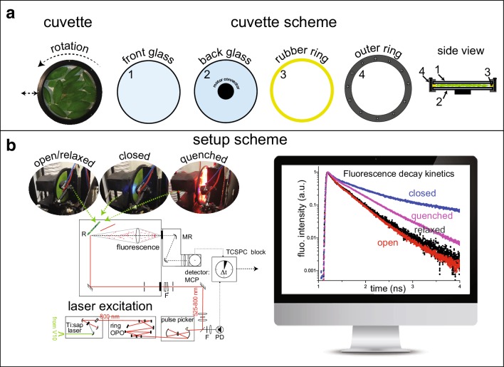
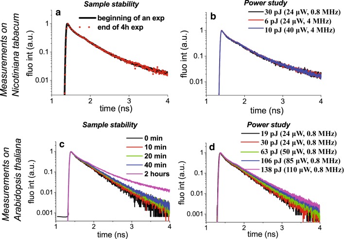
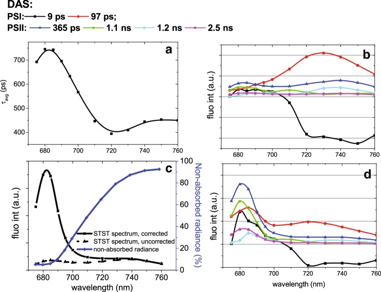
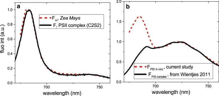
Photosynthesis starts when a pigment in the photosynthetic antennae absorbs a photon. The electronic excitation energy is then transferred through the network of light-harvesting pigments to special chlorophyll (Chl) molecules in the reaction centres, where electron transfer is initiated. Energy transfer and primary electron transfer processes take place on timescales ranging from femtoseconds to nanoseconds, and can be monitored in real time via time-resolved fluorescence spectroscopy. This method is widely used for measurements on unicellular photosynthetic organisms, isolated photosynthetic membranes, and individual complexes. Measurements on intact leaves remain a challenge due to their high structural heterogeneity, high scattering, and high optical density, which can lead to optical artefacts. However, detailed information on the dynamics of these early steps, and the underlying structure-function relationships, is highly informative and urgently required in order to get deeper insights into the physiological regulation mechanisms of primary photosynthesis. Here, we describe a current methodology of time-resolved fluorescence measurements on intact leaves in the picosecond to nanosecond time range. Principles of fluorescence measurements on intact leaves, possible sources of alterations of fluorescence kinetics and the ways to overcome them are addressed. We also describe how our understanding of the organisation and function of photosynthetic proteins and energy flow dynamics in intact leaves can be enriched through the application of time-resolved fluorescence spectroscopy on leaves. For that, an example of a measurement on Zea mays leaves is presented.

Keywords: Fluorescence; Leaf; Re-absorption; Time-resolved spectroscopy.


Conflict of interest statement

The authors declare that they have no conflict of interest.

Figures

Fig. 1
Fig. 1
a Details of the rotation cuvette specially designed to measure heterogeneous, scattering samples such as intact leaves (Miloslavina et al. ; Holzwarth et al. 2009). It is also very useful for measuring isolated photosynthetic complexes or other liquid samples when multiple excitation effects within the same sample volume are undesirable (Miloslavina et al. ; Slavov et al. , ; Szczepaniak et al. 2008, 2009). Dimensions of the cuvette: diameter 10 cm, sample thickness variable between 0.5 and 2 mm. b Scheme of the experimental setup that allows measurement of fluorescence decay traces on intact leaves in different physiological states: open (Fo) or closed (Fm) photosystem II (corresponding to minimum and maximum fluorescence levels of PSII, which occur when the electron transport carriers from PSII are in oxidised or reduced state, respectively), quenched (in the presence of NPQ, induced by illumination with strong red LEDs) and relaxed (40 min recovery in darkness after high light illumination, quenched state)
Fig. 2
Fig. 2
Sample stability study and power study should always be performed during time-resolved fluorescence measurements on intact leaves and isolated samples. The checks are performed at a characteristic wavelength, 686 nm, which is close to PSII and free Chl emission maxima. a, b Measurements on Nicotiana tabacum leaves in Fo state. Rotation speed was 1200 rpm. Side movement was 80 mpm. a Sample stability was confirmed by measuring fluorescence decay traces at the beginning and end of the experiment. b The power study demonstrated that the fluorescence kinetics are not affected in the range between 20 and 60 µW (corresponding to the pulse energy of 6–30 pJ). c, dArabidopsis thaliana leaves require different measuring conditions than N. tabacum to achieve Fo state. c Rotation speed was 1200 rpm. Side movement was 80 mpm. Sample stability was tested by measuring fluorescence decay traces every 10 min during the experiment. The stability study demonstrated that A. thaliana leaves cannot withstand the same rpm as N. tabacum: irreversible damage to the leaves occurs after only 40 min of rotation. Experimentally, a slower speed of 700 rpm was determined to be optimal for A. thaliana leaves. d Power study of A. thaliana leaves upon 700 rpm rotation: no more than 25 µE should be used to achieve Fo state at speeds of 700 rpm
Fig. 3
Fig. 3
Steady-state emission spectrum obtained from an intact leaf. The fluorescence was dominated by two emission bands: 682 nm, characteristic for PSII (dotted line) and Light-harvesting complex II, and 720–740 nm, characteristic for PSI emission (dashed line)
Fig. 4
Fig. 4
a, b Test of the presence of re-emission in time-resolved decay traces of intact leaves. a The decay traces were measured at various detection wavelengths in two extreme positions of the focusing lens: (1) the surface of the leaf; (2) into the leaf tissue. b Time-resolved fluorescence traces obtained from: (1) N. tabacum leaf (black line), (2) free Chl (blue line), and (3) the same N. tabacum leaf together with a Chl filter paper, when the latter is positioned on the back side of the cuvette (red line). Fluorescence kinetics measured from intact leaves (with/without Chl in the back) are indistinguishable. c The presence of time-of-flight dispersion is tested: time-resolved decay traces were measured at different angles of incidence. The angle of incidence does not affect fluorescence kinetics, excluding the possible contribution of time-of-flight dispersion
Fig. 5
Fig. 5
a Average lifetime of Z. mays leaves in Fm state at different detection wavelengths; b, d DAS obtained from a 6-component fit of fluorescence decay traces. Original DAS (b) and the DAS corrected for re-absorption (d). c Correction of Chl re-absorption in fluorescence measurements on intact leaves. Spectra of the measured chlorophyll fluorescence emission (black dotted line, Fm) are corrected for re-absorption according to Formula 1 of the main text. The radiation not absorbed by Z. mays leaves (transmitted and reflected light, parameter r + t in Formula 1) is indicated with a blue line
Fig. 6
Fig. 6
a Reconstructed steady-state emission spectra obtained from time-resolved measurements on intact leaves of Z. mays in Fm state. The spectra are corrected for re-absorption. The resulting spectra were compared to that of isolated Photosystem II [C2S2 from (Xu et al. 2015)]. b Spectrum of PSI in vivo compared to the spectrum of the isolated PSI [from (Wientjes et al. 2011b)]

References

    1. Agati G, Fusi F, Mazzinghi P, Dipaolo ML. A simple approach to the evaluation of the reabsorption of chlorophyll fluorescence-spectra in intact leaves. J Photochem Photobiol B. 1993;17(2):163–171.
    1. Ahmad P, Prasad MNV. Environmental adaptations and stress tolerance of plants in the era of climate change. New York: Springer; 2012.
    1. Akimoto S, Yokono M, Hamada F, Teshigahara A, Aikawa S, Kondo A. Adaptation of light-harvesting systems of Arthrospira platensis to light conditions, probed by time-resolved fluorescence spectroscopy. Biochim Biophys Acta. 2012;1817(8):1483–1489. - PubMed
    1. Amarnath K, Zaks J, Park SD, Niyogi KK, Fleming GR. Fluorescence lifetime snapshots reveal two rapidly reversible mechanisms of photoprotection in live cells of Chlamydomonas reinhardtii. Proc Natl Acad Sci USA. 2012;109(22):8405–8410. - PMC - PubMed
    1. Andrizhiyevskaya EG, Chojnicka A, Bautista JA, Diner BA, van Grondelle R, Dekker JP. Origin of the F685 and F695 fluorescence in photosystem II. Photosynth Res. 2005;84(1–3):173–180. - PubMed

Grants and funding

LinkOut - more resources