. 2018 Nov 27;8(1):17412.
doi: 10.1038/s41598-018-35817-2.

Flavonoids are identified from the extract of Scutellariae Radix to suppress inflammatory-induced angiogenic responses in cultured RAW 264.7 macrophages

Affiliations

Flavonoids are identified from the extract of Scutellariae Radix to suppress inflammatory-induced angiogenic responses in cultured RAW 264.7 macrophages

Guowei Gong et al. Sci Rep. .

Abstract

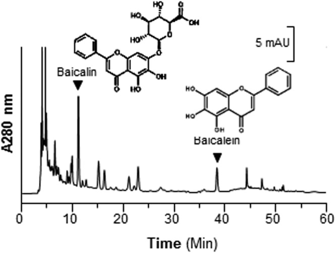
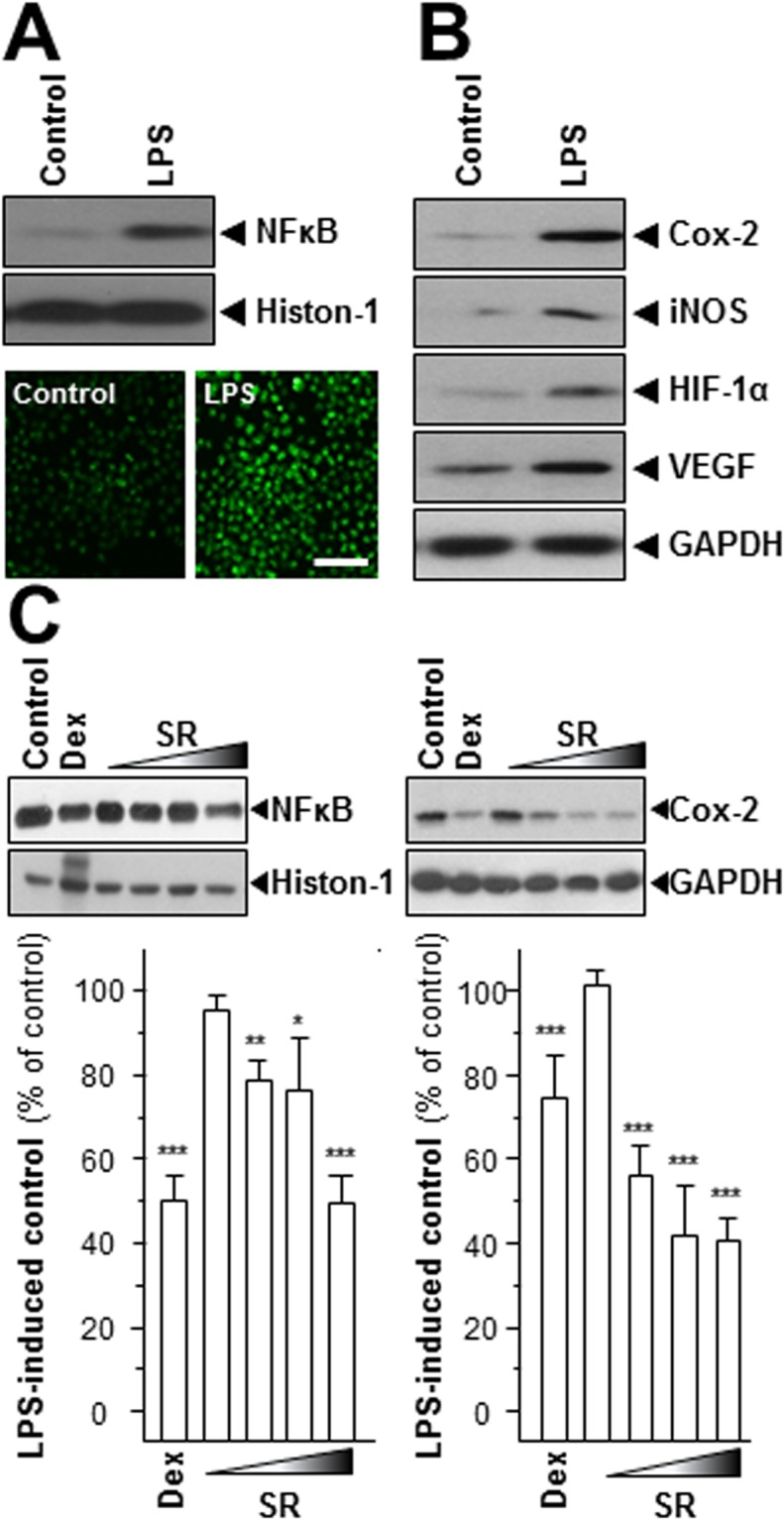
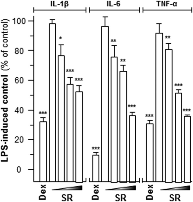
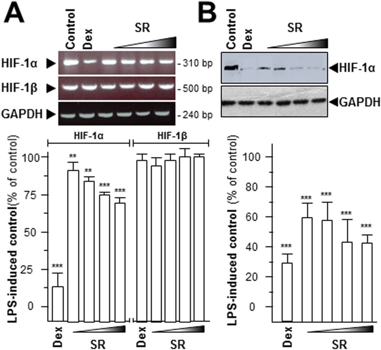
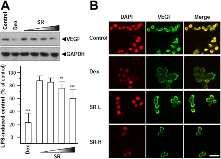
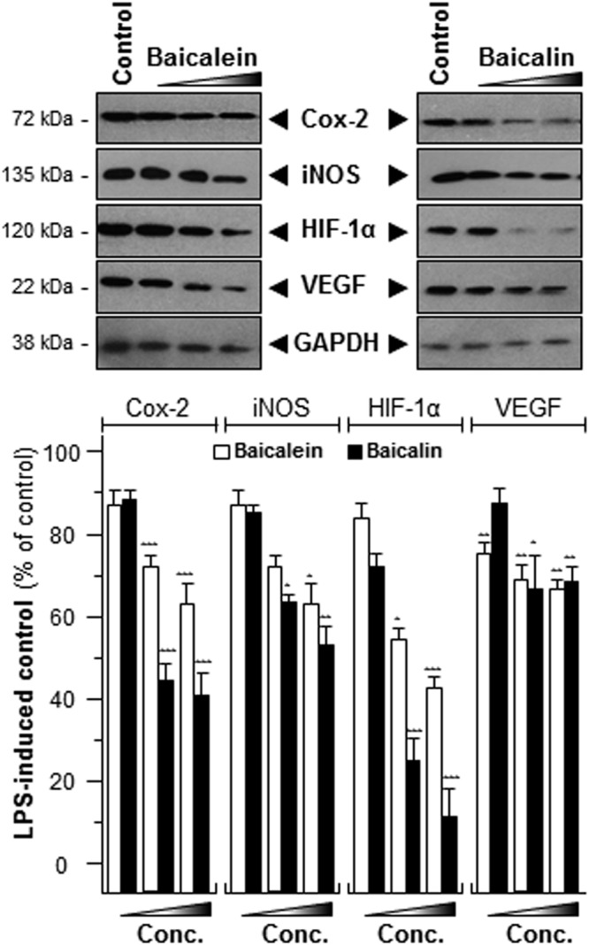
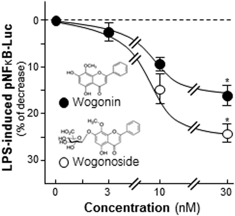
Scutellariae Radix (SR), also named Huangqin in China, is the dried root of Scutellaria baicalensis Georgi. Historically, the usage of SR was targeted to against inflammation. In fact, chronic inflammation has a close relationship with hypoxia and abnormal angiogenesis in tumor cells. Hence, we would like to probe the water extract of SR in suppressing the inflammation-induced angiogenesis. Prior to determine the pharmaceutical values of SR, the first step is to analysis the chemical compositions of SR according to China Pharmacopeia (2015). From the results, the amount of baicalin was 12.6% by weight. Furthermore, the anti-angiogenic properties of SR water extract were evaluated in lipopolysaccharide (LPS) pre-treated cultured macrophage RAW 264.7 cells by detecting the inflammatory markers, i.e. Cox-2, cytokine and iNOS, as well as the translocation activity of NFκB and angiogenic biomarker, i.e. VEGF. This herbal extract was capable of declining both inflammatory and angiogenic hallmarks in a concentration-dependent manner. Moreover, the SR-derived flavonoids, i.e. baicalin, baicalein, wogonin and wogonoside, were shown to be active chemicals in the anti-inflammatory-induced angiogenesis. Therefore, the inflammation-induced angiogenesis is believed to be suppressed by SR water extract, or its major ingredients. These results shed light in the benefiting role of SR in the inflammation-induced angiogenesis in vitro.


Conflict of interest statement

The authors declare no competing interests.

Figures

Figure 1
Figure 1
Fingerprint of water extract of SR. Ten μL of 100 mg/mL of SR water extract was subjected to HPLC-DAD analysis, and the chemical fingerprint was revealed at the wavelength 280 nm. The identification and chemical structures of baicalin and baicalein were shown here. Representative chromatograms were shown, n = 3.
Figure 2
Figure 2
SR suppresses the expressions of NFκB and Cox-2. LPS (1 μg/mL) were utilized for 24 hours as mimicking inflammatory condition. (A,B) The translational levels of Cox-2 (~72 kDa), iNOS (~130 kDa), HIF-1α (~90 kDa) and VEGF (~27 kDa) and nuclear protein of NFκB (~60 kDa) were detected by immunoblot analysis. Histon-1 (~27 kDa) acted as nuclear internal control and GAPDH (~38 kDa) served as an cytosolic internal control. Cells were labeled with fluorescent NO indicator DAF-FM DA for 30 min. The amounts of NO were evaluated by measuring the fluorescence intensity. Micrographs were taken by a confocal microscope (lower panel), Bar = 100 µm. (C) Cells were treated with various concentrations of SR extracts (0.03, 0.1, 0.3, 1.0 mg/mL) for 48 hours. The nuclear protein of NFκB was isolated and detected by immunoblot analysis using specific antibodies. The translational level of Cox-2 was detected by specific antibodies (upper panel), and GAPDH served as an internal control. Here, dexamethasone (Dex; 10 μM) served as positive control. All data were exhibited as the percentage of LPS-induced maximum blank reading (lower panel), in Mean ± SEM, where n = 3. Statistically significant changes were classified as significant (*) where p < 0.05 more significant (**) where p < 0.01 and highly significant (***) where p < 0.001.
Figure 3
Figure 3
SR modulates cytokine mRNA levels. LPS-stimulated cells were treated with various concentrations of SR extracts (0.03, 0.1, 0.3, 1.0 mg/mL) for 48 hours. Total RNAs were isolated and reverse transcribed to cDNA for PCR analysis. The mRNA levels were determined by the Ct-value method and normalized by the house keeping gene GAPDH rRNA. Here, dexamethasone (Dex; 10 μM) served as positive control. Values were in the percentage of LPS-induced maximum reading, in Mean ± SEM, where n = 3. Statistically significant changes were classified as significant (*) where p < 0.05 more significant (**) where p < 0.01 and highly significant (***) where p < 0.001 as compared with control group.
Figure 4
Figure 4
SR declines iNOS and NO productions. (A) Various concentrations of SR extracts (0.03, 0.1, 0.3, 1.0 mg/mL) were applied onto LPS-stimulated RAW 264.7 for 48 hours, and cytosolic protein of iNOS (~130 kDa) was detected. GAPDH (~38 kDa) served as an internal control. (B) The LPS-stimulated cells were treated with various concentrations of SR (SR-L at 0.03 mg/mL and SR-H at 1.0 mg/mL) for 48 hours, and then labeled with fluorescent NO indicator DAF-FM DA for 30 min. The amounts of NO were evaluated by measuring the fluorescence intensity. Micrographs were taken by a confocal microscope (upper panel), Bar = 100 µm. Here, dexamethasone (Dex; 10 μM) served as positive control. Values were at the percentage of LPS-induced maximum reading (lower panel), in Mean ± SEM, where n = 3. Statistically significant changes were classified as significant (*) where p < 0.05 more significant (**) where p < 0.01 and highly significant (***) where p < 0.001 as compared with control group.
Figure 5
Figure 5
SR decrease HIF-1α mRNA and protein levels. Series dilutions of SR extracts (0.03, 0.1, 0.3, 1.0 mg/mL) were used onto LPS-stimulated RAW 264.7 cells for 48 hours. (A) Total RNAs were isolated and reverse transcribed to cDNA for PCR analysis and normalized by the house keeping gene GAPDH. (B) The protein level of HIF-1α (~90 kDa) was detected by immunoblot analysis, and GAPDH (~38 kDa) served as an internal control. Here, dexamethasone (Dex; 10 μM) served as positive control. Values were at the percentage of LPS-induced maximum blank reading, in Mean ± SEM, where n = 3. Statistically significant changes were classified as significant more significant (**) where p < 0.01 and highly significant (***) where p < 0.001 as compared with control group.
Figure 6
Figure 6
SR reduces the expression of angiogenic marker. Different dilutions of SR extracts (0.03, 0.1, 0.3, 1.0 mg/mL) were used onto LPS-stimulated RAW 264.7 cells for 48 hours. (A) The protein expression level of VEGF (~27 kDa) was detected by immunoblot analysis using specific antibodies (upper panel), and GAPDH (~38 kDa) served as an internal control. Here, dexamethasone (Dex; 10 μM) served as positive control. Values were shown as the percentage of LPS-induced maximum reading (lower panel), in Mean ± SEM, where n = 3. Statistically significant changes were classified as significant more significant (**) where p < 0.01 and highly significant (***) where p < 0.001 as compared with control group. (B) VEGF expression level was revealed by immunochemical staining. Bar = 10 µm.
Figure 7
Figure 7
Baicalein and baicalin suppress the expressions of Cox-2, iNOS, HIF-1α and VEGF. The LPS-stimulated cells were treated with LPS (1 μg/mL) for 24 hours and then challenging with various concentrations of baicalin or baicalein (3, 10, 30 nM) for another 48 hours. The protein levels of Cox-2 (~72 kDa), iNOS (~130 kDa), HIF-1α (~90 kDa) and VEGF (~27 kDa) were detected by immunoblot analysis (upper panel), and GAPDH (~38 kDa) served as an internal control. Values were shown as the percentage of LPS-induced maximum reading (lower panel), in Mean ± SEM, where n = 3. Statistically significant changes were classified as significant (*) where p < 0.05 more significant (**) where p < 0.01 and highly significant (***) where p < 0.001 as compared with control group.
Figure 8
Figure 8
Wogonin and wogonoside inhibit the activity of pNFκB-Luc. A luciferase reporter contains 5 repeat NFκB response elements, named pNFκB-Luc, was applied here. Transfected cells were treated with LPS (1 μg/mL) for 24 hours and then application with wogonin or wogonoside at different concentrations for another 2 days. The cell lysates were subjected to luciferase assay. Chemical structures of wogonin and wogonoside were shown. Data were expressed as percentage of control and in Mean ± SEM, where n = 3. *p < 0.05 as compared to the control.

References

    1. Turner MD, Nedjai B, Hurst T, Pennington DJ. Cytokines and chemokines: at the crossroads of cell signaling and inflammatory disease. Biochim Biophys Acta. 2014;1843:2563–2582. doi: 10.1016/j.bbamcr.2014.05.014. - DOI - PubMed
    1. Newton K, Dixit VM. Signaling in innate immunity and inflammation. Cold Spring Harb Perspect Biol. 2012;4:a006049. doi: 10.1101/cshperspect.a006049. - DOI - PMC - PubMed
    1. Navab KD, et al. Chronic inflammatory disorders and accelerated atherosclerosis: chronic kidney disease. Curr Pharm Des. 2011;17:17–20. doi: 10.2174/138161211795049787. - DOI - PubMed
    1. Nonnenmacher Y, Hiller K. Biochemistry of proinflammatory macrophage activation. Cell Mol Life Sci. 2018;75:2093–2109. doi: 10.1007/s00018-018-2784-1. - DOI - PMC - PubMed
    1. Coussens LM, Werb Z. Inflammation and cancer. Nature. 2002;420:860–867. doi: 10.1038/nature01322. - DOI - PMC - PubMed

Publication types

MeSH terms