. 2018 Nov 27;9(1):5014.
doi: 10.1038/s41467-018-07452-y.

NF1 heterozygosity fosters de novo tumorigenesis but impairs malignant transformation

Affiliations

NF1 heterozygosity fosters de novo tumorigenesis but impairs malignant transformation

Jean-Philippe Brosseau et al. Nat Commun. .

Abstract

Neurofibromatosis type 1 (NF1) is an autosomal genetic disorder. Patients with NF1 are associated with mono-allelic loss of the tumor suppressor gene NF1 in their germline, which predisposes them to develop a wide array of benign lesions. Intriguingly, recent sequencing efforts revealed that the NF1 gene is frequently mutated in multiple malignant tumors not typically associated with NF1 patients, suggesting that NF1 heterozygosity is refractory to at least some cancer types. In two orthogonal mouse models representing NF1- and non-NF1-related tumors, we discover that an Nf1+/- microenvironment accelerates the formation of benign tumors but impairs further progression to malignancy. Analysis of benign and malignant tumors commonly associated with NF1 patients, as well as those with high NF1 gene mutation frequency, reveals an antagonistic role for NF1 heterozygosity in tumor initiation and malignant transformation and helps to reconciliate the role of the NF1 gene in both NF1 and non-NF1 patient contexts.


Conflict of interest statement

The authors declare no competing interests.

Figures

Fig. 1
Fig. 1
Loss of Nf1 in keratinocytes does not enhance DMBA/TPA-induced tumors. a Schematic of the DMBA/TPA treatment schedule used. Pictures of DMBA/TPA-induced tumors from b Nf1f/f and c K14Cre; Nf1f/f mice. White scale bar is equal to 5 mm. Representative d, e H&E-stained and fi IHC using K1 antibodies of histological slides of DMBA/TPA-induced tumor from d, f Nf1f/f and e, g K14Cre; Nf1f/f and their normal margin from h Nf1f/f and i K14Cre; Nf1f/f. Black scale bar is equal to 50 μm. j Tumor incidence in relation to mice genotype. Fischer exact test statistic was performed and is not significant (n.s.). A fraction of Nf1f/f (3 out of 22 (14%) and K14Cre; Nf1f/f (3 out of 28 (11%) developed at least one tumor (papilloma or SCC)
Fig. 2
Fig. 2
Loss of Nf1 in keratinocytes does not trigger obvious skin phenotype. Mice genotyping with specific PCR primers for a Cre recombinase, b Nf1 flox allele and c Nf1 deleted. Lane 1 = no template control, lane 2 = DNA from Nf1f/f mice’s skin tail, lane 3 = DNA from K14Cre; Nf1f/f mice’s skin tail, lane 4 = DNA from wild-type mice’s skin tail. d Western blot of skin protein extract quantified using anti-NF1, anti-LUMICAN (dermal marker), and anti-K1 (epidermal marker). Lane A = epidermis from Nf1f/f mice; Lane B = dermis from Nf1f/f mice; Lane C = epidermis from K14Cre; Nf1f/f mice; Lane D = dermis from K14Cre; Nf1f/f mice. e, f Representative IHC using anti-NF1 antibodies of dissected normal dorsal skin from e Nf1f/f and f K14Cre; Nf1f/f (arrow head pointing to NF1 expression in brown). g, h Representative X-gal staining counterstained with Nuclear Fast Red of dissected normal dorsal skin from g Nf1f/f; ROSA-LacZ and h K14Cre; Nf1f/f; ROSA-LacZ mice. i, j Representative H&E of dissected normal dorsal skin from i Nf1f/f and j K14Cre; Nf1f/f. Scale bar is equal to 50 μm
Fig. 3
Fig. 3
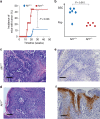
Antagonistic role of Nf1+/− microenvironment in DMBA/TPA-induced tumors. a Incidence of mice developing tumor under DMBA/TPA treatment as a function of time. A fraction of mice with Nf1+/+ (6 out of 50; 12%) and Nf1+/− (4 out of 9; 44%) microenvironment developed at least one tumor (papilloma or SCC). Fischer exact test statistic was performed. b Tumors from six mice with Nf1+/+ and five Nf1+/−microenvironment were reviewed by a pathologist and classified as papilloma (benign) or SCC (malignant) based on cf. Fischer exact test statistic was performed. Representative c, d H&E-stained and e, f IHC using K1 antibodies in histological sections of dissected DMBA/TPA-treated dorsal skin from c, e Nf1+/+ and d, f Nf1+/−. Scale bar is equal to 50 μm
Fig. 4
Fig. 4
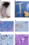
A novel mouse model to study the sequential progression from plexiform neurofibroma to MPNST. a, b Representative gross image of a PLPCreERT2; Nf1f/f mouse that develops MPNST in the right sciatic nerve and b its dissected spinal cord along with peripheral nerves. cf Representative c X-Gal staining (PLPCreERT2; Nf1f/f; ROSA-LacZ), IHC using d S100 and e H3K27 (arrow head) antibodies and f H&E in histological sections of dissected MPNST from PLPCreERT2; Nf1f/f. Scale bar is equal to 50 μm
Fig. 5
Fig. 5
Antagonistic role of Nf1+/− microenvironment in plexiform neurofibroma and MPNST formation. a Incidence of mice with plexiform neurofibroma (pNF). b Tumor were classified as pNF (benign) or MPNST (malignant) based on histology. Fischer exact test statistic was performed. cj Representative c, d H&E; e, f IHC using S100 g, h trichrome and i, j toluidine blue (arrow head pointing toward mast cells) staining in histological sections of dissected pNF from PLPCreERT2; Nf1f/f (c, e, g, i) and PLPCreERT2; Nf1f/− (d, f, h, j). Scale bar is equal to 50 μm
Fig. 6
Fig. 6
Impact of Nf1 heterozygosity on T cells proliferation and function. a, b Proliferation of peripheral T cells from Nf1+/+ and Nf1+/− mice stimulated by various concentrations of a anti-CD3e antibody alone or with b CD28 costimulation. Proliferation was assessed in biological duplicate and technical triplicate by 3H-thymidine incorporation during the last 20 h of culture. c Delayed-type hypersensitivity (DTH) assay: On day 0, Nf1+/+ and Nf1+/− mice were sensitized on abdominal skin by topical application of oxazolone. On day 6, Nf1+/+ and Nf1+/− mice were challenged by topical application on the left and right ear with oxazolone and control solvent, respectively. DTH was assessed daily through by measuring ear thickness (Δ). Experiments were performed in biological triplicates. d Two days after challenge, cellularity of draining lymph nodes (DLNs) of Nf1+/+ and Nf1+/− mice were determined. e Spontaneous activation of cells from DLNs (T cells) was measured by 3H-thymidine incorporation after culturing for 3 days without stimuli. f Fraction of CD4+ cells that are activated. g Fraction of CD8+ cells that are activated. h Fraction of B cells+ that are activated. i Fraction of Treg+ cells in total CD4+ subset. Panels di were performed in biological triplicates. Statistical paired t-test performed; * P< 0.05. Variation was estimated using standard deviation with a 95% confidence level
Fig. 7
Fig. 7
NF1 heterozygosity fosters de novo tumorigenesis but impairs malignant transformation. Schematic summarizing the antagonistic role of NF1+/− microenvironment on the sequential cancer progression from normal to benign to malignant tumor

References

    1. Le LQ, Parada LF. Tumor microenvironment and neurofibromatosis type I: connecting the GAPs. Oncogene. 2007;26:4609–4616. doi: 10.1038/sj.onc.1210261. - DOI - PMC - PubMed
    1. Friedman, J. M. in GeneReviews(R) (eds. Pagon, R. A., et al.) (University of Washington, Seattle, WA,1993). - PubMed
    1. Brosseau JP, et al. The biology of cutaneous neurofibromas: consensus recommendations for setting research priorities. Neurology. 2018;91:S14–S20. doi: 10.1212/WNL.0000000000005788. - DOI - PMC - PubMed
    1. Upadhyaya M, et al. An absence of cutaneous neurofibromas associated with a 3-bp inframe deletion in exon 17 of the NF1 gene (c.2970-2972 delAAT): evidence of a clinically significant NF1 genotype-phenotype correlation. Am. J. Hum. Genet. 2007;80:140–151. doi: 10.1086/510781. - DOI - PMC - PubMed
    1. Rojnueangnit K, et al. High incidence of Noonan syndrome features including short stature and pulmonic stenosis in patients carrying NF1 missense mutations affecting p.Arg1809: genotype-phenotype correlation. Human. Mutat. 2015;36:1052–1063. doi: 10.1002/humu.22832. - DOI - PMC - PubMed

Publication types

MeSH terms

Substances