An Update on Optical Coherence Tomography Angiography in Diabetic Retinopathy
- PMID: 30479720
- PMCID: PMC6210870
- DOI: 10.4103/jovr.jovr_57_18
An Update on Optical Coherence Tomography Angiography in Diabetic Retinopathy
Abstract
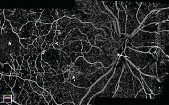
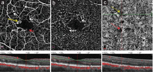
Optical coherence tomography angiography (OCTA) is a novel non-invasive imaging modality for 3-dimensional visualization of retinal and optic nerve capillary networks. In this article, a comprehensive review of relevant original articles in the PubMed database was performed using the search terms "diabetic retinopathy," "diabetic macular edema," "diabetes mellitus," and "optical coherence tomography angiography." OCTA was found to detect microvascular changes early in diabetes mellitus, even before they become clinically evident. Morphological and qualitative assessment of vascular changes can help to determine the pathophysiological processes, activity, treatment, and follow-up of diabetic retinopathy (DR). Vessel density and foveal avascular zone are the most investigated quantified indices shown to be early predictors of DR, correlated to DR severity and visual function, and useful in predicting response to treatment. OCTA has shown to be a promising alternative to fluorescein angiography in the management of DR. Further studies are warranted to determine the role of OCTA in the routine clinical management of DR.
Keywords: Deep Capillary Plexus; Diabetic Retinopathy; Foveal Avascular Zone; Ischemia; Optical Coherence Tomography Angiography; Superficial Capillary Plexus; Vessel Density.
Conflict of interest statement
There are no conflicts of interest.
Figures
References
-
- Soares M, Neves C, Marques IP, Pires I, Schwartz C, Costa MÂ, et al. Comparison of diabetic retinopathy classification using fluorescein angiography and optical coherence tomography angiography. Br J Ophthalmol. 2017;101:62–68. - PubMed
Publication types
LinkOut - more resources
Full Text Sources