Subclinical oral involvement in patients with endemic pemphigus foliaceus
- PMID: 30479852
- PMCID: PMC6246068
- DOI: 10.5826/dpc.0804a02
Subclinical oral involvement in patients with endemic pemphigus foliaceus
Abstract
Background: We have described a variant of endemic pemphigus foliaceus (EPF) in El Bagre area known as pemphigus Abreu-Manu. Our previous study suggested that Colombian EPF seemed to react with various plakin family proteins, such as desmoplakins, envoplakin, periplakin BP230, MYZAP, ARVCF, p0071 as well as desmoglein 1.
Objectives: To explore whether patients affected by a new variant of endemic pemphigus foliaceus (El Bagre-EPF) demonstrated oral involvement.
Materials and methods: A case-control study was done by searching for oral changes in 45 patients affected by El Bagre-EPF, as well as 45 epidemiologically matched controls from the endemic area matched by demographics, oral hygiene habits, comorbidities, smoking habits, place of residence, age, sex, and work activity. Oral biopsies were taken and evaluated via hematoxylin and eosin staining, direct immunofluorescence, indirect immunofluorescence, confocal microscopy, and immunohistochemistry.
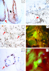
Results: Radicular pieces and loss of teeth were seen in in 43 of the 45 El Bagre-EPF patients and 20 of the 45 controls (P < 0.001) (confidence interval [CI] 98%). Hematoxylin and eosin staining showed 23 of 45 El Bagre-EPF patients had corneal/subcorneal blistering and lymphohistiocytic infiltrates under the basement membrane zone and around the salivary glands, the periodontal ligament, and the neurovascular bundles in all cell junction structures in the oral cavity; these findings were not seen in the controls (P < 0.001) (CI 98%). The direct immunofluorescence, indirect immunofluorescence, confocal microscopy, and microarray staining displayed autoantibodies to the salivary glands, including their serous acini and the excretory duct cell junctions, the periodontal ligament, the neurovascular bundles and their cell junctions, striated muscle and their cell junctions, neuroreceptors, and connective tissue cell junctions. The autoantibodies were polyclonal. IgA autoantibodies were found in neuroreceptors in the glands and were positive in 41 of 45 patients and 3 of 45 controls.
Conclusions: Patients affected by El Bagre-EPF have some oral anomalies and an immune response, primarily to cell junctions. The intrinsic oral mucosal immune system, including IgA and secretory IgA, play an important role in this autoimmunity. Our data contradict the hypothesis that pemphigus foliaceus does not affect the oral mucosa due to the desmoglein 1-desmoglein 3 compensation.
Keywords: IgA; cell junctions; endemic pemphigus foliaceus; oral mucosa; salivary glands; secretory immunoglobulin A.
Conflict of interest statement
Competing interests: The authors have no conflicts of interest to disclose.
Figures


References
-
- Abreu Velez AM, Hashimoto T, Bollag W, et al. A unique form of endemic pemphigus in Northern Colombia. J Am Acad Dermatol. 2003;49(4):599–608. - PubMed
-
- Abreu Velez AM, Beutner EH, Montoya F, Hashimoto T. Analyses of autoantigens in a new form of endemic pemphigus foliaceus in Colombia. J Am Acad Dermatol. 2003;49(4):609–614. - PubMed
-
- Hisamatsu Y, Abreu Velez AM, Amagai M, et al. Comparative study of autoantigen profile between Colombian and Brazilian types of endemic pemphigus foliaceus by various biochemical and molecular biological techniques. J Dermatol Sci. 2003;32(1):33–41. - PubMed
-
- Abreu Velez AM, Javier Patiño P, Montoya F, et al. The tryptic cleavage product of the mature form of the bovine desmoglein 1 ectodomain is one of the antigen moieties immunoprecipitated by all sera from symptomatic patients affected by a new variant of endemic pemphigus. Eur J Dermatol. 2003;13(4):359–366. - PubMed
-
- Abréu Vélez AM, Yepes MM, Patiño PJ, Bollag WB, Montoya F., Sr A cost-effective, sensitive and specific enzyme linked immunosorbent assay useful for detecting a heterogeneous antibody population in sera from people suffering a new variant of endemic pemphigus. Arch Dermatol Res. 2004;295(10):434–441. - PubMed
LinkOut - more resources
Full Text Sources
Molecular Biology Databases
Miscellaneous
